SLIT3 Antibody - #DF9909


 and mouse anti-beta tubulin Ab(T0023 1:200) for 1 hour at 37°C. An AlexaFluor594 conjugated goat anti-rabbit IgG(H+L) Ab(Red) and an AlexaFluor488 conjugated goat anti-mouse IgG(H+L) Ab(Green) were used as the secondary antibody.
The nuclear counter stain is DAPI(blue).")
| Product: | SLIT3 Antibody |
| Catalog: | DF9909 |
| Description: | Rabbit polyclonal antibody to SLIT3 |
| Application: | WB IHC IF/ICC |
| Cited expt.: | WB, IHC, IF/ICC |
| Reactivity: | Human, Mouse, Rat |
| Prediction: | Pig, Bovine, Horse, Sheep, Rabbit, Dog, Chicken |
| Mol.Wt.: | 168 kDa; 168kD(Calculated). |
| Uniprot: | O75094 |
| RRID: | AB_2843103 |
Related Downloads
Protocols
Product Info
*The optimal dilutions should be determined by the end user. For optimal experimental results, antibody reuse is not recommended.
*Tips:
WB: For western blot detection of denatured protein samples. IHC: For immunohistochemical detection of paraffin sections (IHC-p) or frozen sections (IHC-f) of tissue samples. IF/ICC: For immunofluorescence detection of cell samples. ELISA(peptide): For ELISA detection of antigenic peptide.
Cite Format: Affinity Biosciences Cat# DF9909, RRID:AB_2843103.
Fold/Unfold
FLJ10764; KIAA0814; MEGF 5; MEGF5; Multiple EGF-like domains protein 5; Multiple epidermal growth factor like domains 5; Multiple epidermal growth factor-like domains protein 5; SLIL 2; SLIL2; SLIT 1; Slit 2; Slit 3; Slit homolog 3 (Drosophila); Slit homolog 3; Slit homolog 3 protein; Slit-3; SLIT1; Slit2; Slit3; SLIT3_HUMAN;
Immunogens
A synthesized peptide derived from human SLIT3, corresponding to a region within C-terminal amino acids.
- O75094 SLIT3_HUMAN:
- Protein BLAST With
- NCBI/
- ExPASy/
- Uniprot
MAPGWAGVGAAVRARLALALALASVLSGPPAVACPTKCTCSAASVDCHGLGLRAVPRGIPRNAERLDLDRNNITRITKMDFAGLKNLRVLHLEDNQVSVIERGAFQDLKQLERLRLNKNKLQVLPELLFQSTPKLTRLDLSENQIQGIPRKAFRGITDVKNLQLDNNHISCIEDGAFRALRDLEILTLNNNNISRILVTSFNHMPKIRTLRLHSNHLYCDCHLAWLSDWLRQRRTVGQFTLCMAPVHLRGFNVADVQKKEYVCPAPHSEPPSCNANSISCPSPCTCSNNIVDCRGKGLMEIPANLPEGIVEIRLEQNSIKAIPAGAFTQYKKLKRIDISKNQISDIAPDAFQGLKSLTSLVLYGNKITEIVKGLFDGLVSLQLLLLNANKINCLRVNTFQDLQNLNLLSLYDNKLQTISKGLFAPLQSIQTLHLAQNPFVCDCHLKWLADYLQDNPIETSGARCSSPRRLANKRISQIKSKKFRCSGSEDYRSRFSSECFMDLVCPEKCRCEGTIVDCSNQKLVRIPSHLPEYVTDLRLNDNEVSVLEATGIFKKLPNLRKINLSNNKIKEVREGAFDGAASVQELMLTGNQLETVHGRVFRGLSGLKTLMLRSNLIGCVSNDTFAGLSSVRLLSLYDNRITTITPGAFTTLVSLSTINLLSNPFNCNCHLAWLGKWLRKRRIVSGNPRCQKPFFLKEIPIQDVAIQDFTCDGNEESSCQLSPRCPEQCTCMETVVRCSNKGLRALPRGMPKDVTELYLEGNHLTAVPRELSALRHLTLIDLSNNSISMLTNYTFSNMSHLSTLILSYNRLRCIPVHAFNGLRSLRVLTLHGNDISSVPEGSFNDLTSLSHLALGTNPLHCDCSLRWLSEWVKAGYKEPGIARCSSPEPMADRLLLTTPTHRFQCKGPVDINIVAKCNACLSSPCKNNGTCTQDPVELYRCACPYSYKGKDCTVPINTCIQNPCQHGGTCHLSDSHKDGFSCSCPLGFEGQRCEINPDDCEDNDCENNATCVDGINNYVCICPPNYTGELCDEVIDHCVPELNLCQHEAKCIPLDKGFSCECVPGYSGKLCETDNDDCVAHKCRHGAQCVDTINGYTCTCPQGFSGPFCEHPPPMVLLQTSPCDQYECQNGAQCIVVQQEPTCRCPPGFAGPRCEKLITVNFVGKDSYVELASAKVRPQANISLQVATDKDNGILLYKGDNDPLALELYQGHVRLVYDSLSSPPTTVYSVETVNDGQFHSVELVTLNQTLNLVVDKGTPKSLGKLQKQPAVGINSPLYLGGIPTSTGLSALRQGTDRPLGGFHGCIHEVRINNELQDFKALPPQSLGVSPGCKSCTVCKHGLCRSVEKDSVVCECRPGWTGPLCDQEARDPCLGHRCHHGKCVATGTSYMCKCAEGYGGDLCDNKNDSANACSAFKCHHGQCHISDQGEPYCLCQPGFSGEHCQQENPCLGQVVREVIRRQKGYASCATASKVPIMECRGGCGPQCCQPTRSKRRKYVFQCTDGSSFVEEVERHLECGCLACS
Predictions
Score>80(red) has high confidence and is suggested to be used for WB detection. *The prediction model is mainly based on the alignment of immunogen sequences, the results are for reference only, not as the basis of quality assurance.
High(score>80) Medium(80>score>50) Low(score<50) No confidence
Research Backgrounds
May act as molecular guidance cue in cellular migration, and function may be mediated by interaction with roundabout homolog receptors.
Secreted.
Predominantly expressed in thyroid.
Research Fields
· Organismal Systems > Development > Axon guidance. (View pathway)
References

Application: IF/ICC Species: mouse Sample: BMSCs
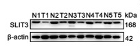
Application: WB Species: Human Sample: LUAD tumor
Application: IHC Species: Human Sample:
Restrictive clause
Affinity Biosciences tests all products strictly. Citations are provided as a resource for additional applications that have not been validated by Affinity Biosciences. Please choose the appropriate format for each application and consult Materials and Methods sections for additional details about the use of any product in these publications.
For Research Use Only.
Not for use in diagnostic or therapeutic procedures. Not for resale. Not for distribution without written consent. Affinity Biosciences will not be held responsible for patent infringement or other violations that may occur with the use of our products. Affinity Biosciences, Affinity Biosciences Logo and all other trademarks are the property of Affinity Biosciences LTD.