
, using Cytokeratin 10 Antibody at 1/1000 dilution.
5ug/NC membrane strip.
Exposure for 10s with Affinity™ ECL Kit(#KF8001).
Bands result from membrane strip incubation.")


| Product: | Cytokeratin 10 Antibody |
| Catalog: | AF0197 |
| Description: | Rabbit polyclonal antibody to Cytokeratin 10 |
| Application: | WB IHC IF/ICC |
| Cited expt.: | IHC |
| Reactivity: | Human, Mouse, Rat |
| Prediction: | Pig, Bovine, Sheep, Rabbit, Dog |
| Mol.Wt.: | 59kDa(Observed); 59kD(Calculated). |
| Uniprot: | P13645 |
| RRID: | AB_2833390 |
Control Products

Related Downloads
Protocols
Product Info
*The optimal dilutions should be determined by the end user. For optimal experimental results, antibody reuse is not recommended.
*Tips:
WB: For western blot detection of denatured protein samples. IHC: For immunohistochemical detection of paraffin sections (IHC-p) or frozen sections (IHC-f) of tissue samples. IF/ICC: For immunofluorescence detection of cell samples. ELISA(peptide): For ELISA detection of antigenic peptide.
Cite Format: Affinity Biosciences Cat# AF0197, RRID:AB_2833390.
Fold/Unfold
BCIE; BIE; CK 10; CK-10; Cytokeratin-10; EHK; K10; K1C10_HUMAN; Keratin 10; Keratin 10 type I; Keratin; Keratin type i cytoskeletal 10; Keratin type I cytoskeletal 59 kDa; Keratin-10; Keratin10; KPP; KRT10; type I cytoskeletal 10;
Immunogens
A synthesized peptide derived from human Cytokeratin 10, corresponding to a region within the internal amino acids.
Seen in all suprabasal cell layers including stratum corneum. Expressed on the surface of lung cell lines (PubMed:19627498).
- P13645 K1C10_HUMAN:
- Protein BLAST With
- NCBI/
- ExPASy/
- Uniprot
MSVRYSSSKHYSSSRSGGGGGGGGCGGGGGVSSLRISSSKGSLGGGFSSGGFSGGSFSRGSSGGGCFGGSSGGYGGLGGFGGGSFRGSYGSSSFGGSYGGIFGGGSFGGGSFGGGSFGGGGFGGGGFGGGFGGGFGGDGGLLSGNEKVTMQNLNDRLASYLDKVRALEESNYELEGKIKEWYEKHGNSHQGEPRDYSKYYKTIDDLKNQILNLTTDNANILLQIDNARLAADDFRLKYENEVALRQSVEADINGLRRVLDELTLTKADLEMQIESLTEELAYLKKNHEEEMKDLRNVSTGDVNVEMNAAPGVDLTQLLNNMRSQYEQLAEQNRKDAEAWFNEKSKELTTEIDNNIEQISSYKSEITELRRNVQALEIELQSQLALKQSLEASLAETEGRYCVQLSQIQAQISALEEQLQQIRAETECQNTEYQQLLDIKIRLENEIQTYRSLLEGEGSSGGGGRGGGSFGGGYGGGSSGGGSSGGGHGGGHGGSSGGGYGGGSSGGGSSGGGYGGGSSSGGHGGSSSGGYGGGSSGGGGGGYGGGSSGGGSSSGGGYGGGSSSGGHKSSSSGSVGESSSKGPRY
Predictions
Score>80(red) has high confidence and is suggested to be used for WB detection. *The prediction model is mainly based on the alignment of immunogen sequences, the results are for reference only, not as the basis of quality assurance.
High(score>80) Medium(80>score>50) Low(score<50) No confidence
Research Backgrounds
Plays a role in the establishment of the epidermal barrier on plantar skin.
(Microbial infection) Acts as a mediator of S.aureus adherence to desquamated nasal epithelial cells via clfB, and hence may play a role in nasal colonization.
(Microbial infection) Binds S.pneumoniae PsrP, mediating adherence of the bacteria to lung cell lines. Reduction of levels of KRT10 keratin decrease adherence, overexpression increases adherence. Neither protein has to be glycosylated for the interaction to occur.
Secreted>Extracellular space. Cell surface.
Note: Localized on the surface of desquamated nasal epithelial cells (PubMed:12427098). Localized on the surface of lung cell lines (PubMed:19627498).
Seen in all suprabasal cell layers including stratum corneum. Expressed on the surface of lung cell lines.
Belongs to the intermediate filament family.
Research Fields
· Human Diseases > Infectious diseases: Bacterial > Staphylococcus aureus infection.
· Organismal Systems > Endocrine system > Estrogen signaling pathway. (View pathway)
References
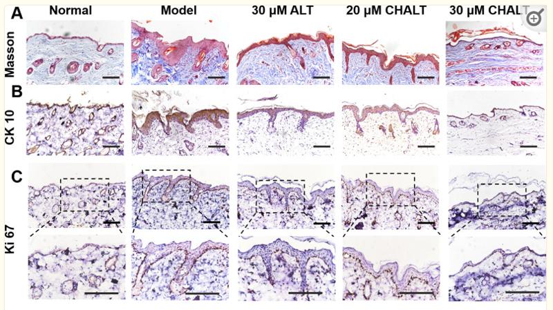
Application: IHC Species: Mice Sample: skin tissue
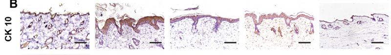
Application: IHC Species: mouse Sample: skin
Restrictive clause
Affinity Biosciences tests all products strictly. Citations are provided as a resource for additional applications that have not been validated by Affinity Biosciences. Please choose the appropriate format for each application and consult Materials and Methods sections for additional details about the use of any product in these publications.
For Research Use Only.
Not for use in diagnostic or therapeutic procedures. Not for resale. Not for distribution without written consent. Affinity Biosciences will not be held responsible for patent infringement or other violations that may occur with the use of our products. Affinity Biosciences, Affinity Biosciences Logo and all other trademarks are the property of Affinity Biosciences LTD.