
.
Bands result from membrane strip incubation.")
 and mouse anti-beta tubulin Ab(T0023 1:200) for 1 hour at 37°C. An AlexaFluor594 conjugated goat anti-rabbit IgG(H+L) Ab(Red) and an AlexaFluor488 conjugated goat anti-mouse IgG(H+L) Ab(Green) were used as the secondary antibody.
The nuclear counter stain is DAPI(blue).")
| Product: | FOLR2 Antibody |
| Catalog: | DF9518 |
| Description: | Rabbit polyclonal antibody to FOLR2 |
| Application: | WB IF/ICC |
| Cited expt.: | WB, IF/ICC |
| Reactivity: | Human, Mouse |
| Prediction: | Bovine, Horse, Sheep, Rabbit, Dog |
| Mol.Wt.: | 29 kDa(Observed); 29kD(Calculated). |
| Uniprot: | P14207 |
| RRID: | AB_2842714 |
Control Products

Related Downloads
Protocols
Product Info
*The optimal dilutions should be determined by the end user. For optimal experimental results, antibody reuse is not recommended.
*Tips:
WB: For western blot detection of denatured protein samples. IHC: For immunohistochemical detection of paraffin sections (IHC-p) or frozen sections (IHC-f) of tissue samples. IF/ICC: For immunofluorescence detection of cell samples. ELISA(peptide): For ELISA detection of antigenic peptide.
Cite Format: Affinity Biosciences Cat# DF9518, RRID:AB_2842714.
Fold/Unfold
beta hFR; FBP; FBP/PL 1; FBP2; fetal/placental; folate receptor 2 (fetal); Folate receptor 2; Folate receptor; Folate receptor beta; Folate receptor, fetal/placental; Folbp 2; Folbp2; Folr2; FOLR2_HUMAN; FR beta; FR P3; FR-beta; Placental folate binding protein; Placental folate-binding protein;
Immunogens
A synthesized peptide derived from human FOLR2, corresponding to a region within the internal amino acids.
Expressed in placenta and hematopoietic cells. Expression is increased in malignant tissues.
- P14207 FOLR2_HUMAN:
- Protein BLAST With
- NCBI/
- ExPASy/
- Uniprot
MVWKWMPLLLLLVCVATMCSAQDRTDLLNVCMDAKHHKTKPGPEDKLHDQCSPWKKNACCTASTSQELHKDTSRLYNFNWDHCGKMEPACKRHFIQDTCLYECSPNLGPWIQQVNQSWRKERFLDVPLCKEDCQRWWEDCHTSHTCKSNWHRGWDWTSGVNKCPAGALCRTFESYFPTPAALCEGLWSHSYKVSNYSRGSGRCIQMWFDSAQGNPNEEVARFYAAAMHVNAGEMLHGTGGLLLSLALMLQLWLLG
Predictions
Score>80(red) has high confidence and is suggested to be used for WB detection. *The prediction model is mainly based on the alignment of immunogen sequences, the results are for reference only, not as the basis of quality assurance.
High(score>80) Medium(80>score>50) Low(score<50) No confidence
Research Backgrounds
Binds to folate and reduced folic acid derivatives and mediates delivery of 5-methyltetrahydrofolate and folate analogs into the interior of cells. Has high affinity for folate and folic acid analogs at neutral pH. Exposure to slightly acidic pH after receptor endocytosis triggers a conformation change that strongly reduces its affinity for folates and mediates their release.
N-glycosylated.
Cell membrane>Lipid-anchor. Secreted.
Expressed in placenta and hematopoietic cells. Expression is increased in malignant tissues.
Belongs to the folate receptor family.
Research Fields
· Cellular Processes > Transport and catabolism > Endocytosis. (View pathway)
· Human Diseases > Drug resistance: Antineoplastic > Antifolate resistance.
References

Application: IF/ICC Species: Mouse Sample: RAW264.7 cells
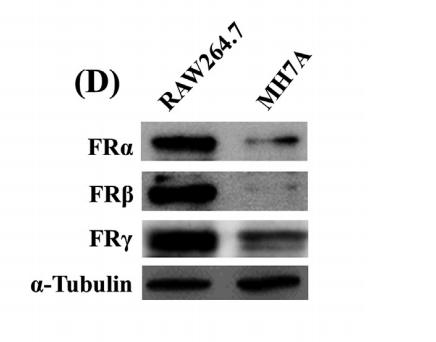
Application: WB Species: Mouse Sample: Raw264.7 and MH7A cells
Restrictive clause
Affinity Biosciences tests all products strictly. Citations are provided as a resource for additional applications that have not been validated by Affinity Biosciences. Please choose the appropriate format for each application and consult Materials and Methods sections for additional details about the use of any product in these publications.
For Research Use Only.
Not for use in diagnostic or therapeutic procedures. Not for resale. Not for distribution without written consent. Affinity Biosciences will not be held responsible for patent infringement or other violations that may occur with the use of our products. Affinity Biosciences, Affinity Biosciences Logo and all other trademarks are the property of Affinity Biosciences LTD.