Product: Cytokeratin 18 Antibody
Catalog: AF0191
Description: Rabbit polyclonal antibody to Cytokeratin 18
Application: WB IHC IF/ICC
Reactivity: Human, Mouse, Rat
Prediction: Pig, Sheep, Rabbit
Mol.Wt.: 46kDa; 48kD(Calculated).
Uniprot: P05783
RRID: AB_2833384

View similar products>>

   Size Price Inventory
 100ul $280 In stock
 200ul $350 In stock

Lead Time: Same day delivery

For pricing and ordering contact:
Local distributors

Product Info

Source:
Rabbit
Application:
WB 1:500-1:3000, IHC 1:50-1:200, IF/ICC: 1:100-1:500
*The optimal dilutions should be determined by the end user.
*Tips:

WB: For western blot detection of denatured protein samples. IHC: For immunohistochemical detection of paraffin sections (IHC-p) or frozen sections (IHC-f) of tissue samples. IF/ICC: For immunofluorescence detection of cell samples. ELISA(peptide): For ELISA detection of antigenic peptide.

Reactivity:
Human,Mouse,Rat
Prediction:
Pig(88%), Sheep(82%), Rabbit(94%)
Clonality:
Polyclonal
Specificity:
Cytokeratin 18 Antibody detects endogenous levels of total Cytokeratin 18.
RRID:
AB_2833384
Cite Format: Affinity Biosciences Cat# AF0191, RRID:AB_2833384.
Conjugate:
Unconjugated.
Purification:
The antiserum was purified by peptide affinity chromatography using SulfoLink™ Coupling Resin (Thermo Fisher Scientific).
Storage:
Rabbit IgG in phosphate buffered saline , pH 7.4, 150mM NaCl, 0.02% sodium azide and 50% glycerol. Store at -20 °C. Stable for 12 months from date of receipt.
Alias:

Fold/Unfold

Cell proliferation inducing gene 46 protein; Cell proliferation inducing protein 46; Cell proliferation-inducing gene 46 protein; CK 18; CK-18; CK18; CYK 18; CYK18; Cytokeratin 18; Cytokeratin endo B; Cytokeratin-18; K 18; K18; K1C18_HUMAN; KA18; Keratin 18; Keratin 18, type I; Keratin D; keratin, type I cytoskeletal 18; Keratin-18; Krt18;

Immunogens

Immunogen:
Uniprot:
Gene(ID):
Expression:
P05783 K1C18_HUMAN:

Expressed in colon, placenta, liver and very weakly in exocervix. Increased expression observed in lymph nodes of breast carcinoma.

Description:
K18 a type I cytoskeletal keratin. The keratins are intermediate filament proteins responsible for the structural integrity of epithelial cells and are subdivided into cytokeratins and hair keratins. Keratin 18 and its filament partner keratin 8 are perhaps the most commonly found members of the intermediate filament gene family. They are expressed in single layer epithelial tissues of the body.
Sequence:
MSFTTRSTFSTNYRSLGSVQAPSYGARPVSSAASVYAGAGGSGSRISVSRSTSFRGGMGSGGLATGIAGGLAGMGGIQNEKETMQSLNDRLASYLDRVRSLETENRRLESKIREHLEKKGPQVRDWSHYFKIIEDLRAQIFANTVDNARIVLQIDNARLAADDFRVKYETELAMRQSVENDIHGLRKVIDDTNITRLQLETEIEALKEELLFMKKNHEEEVKGLQAQIASSGLTVEVDAPKSQDLAKIMADIRAQYDELARKNREELDKYWSQQIEESTTVVTTQSAEVGAAETTLTELRRTVQSLEIDLDSMRNLKASLENSLREVEARYALQMEQLNGILLHLESELAQTRAEGQRQAQEYEALLNIKVKLEAEIATYRRLLEDGEDFNLGDALDSSNSMQTIQKTTTRRIVDGKVVSETNDTKVLRH

Predictions

Predictions:

Score>80(red) has high confidence and is suggested to be used for WB detection. *The prediction model is mainly based on the alignment of immunogen sequences, the results are for reference only, not as the basis of quality assurance.

Species
Results
Score
Rabbit
94
Pig
88
Sheep
82
Zebrafish
64
Horse
0
Bovine
0
Dog
0
Xenopus
0
Chicken
0
Model Confidence:
High(score>80) Medium(80>score>50) Low(score<50) No confidence

PTMs - P05783 As Substrate

Site PTM Type Enzyme
M1 Acetylation
S2 Acetylation
S2 Phosphorylation
T4 Phosphorylation
S7 Phosphorylation
T8 Phosphorylation
S10 Phosphorylation
T11 Phosphorylation
Y13 Phosphorylation
S15 Phosphorylation
S18 Phosphorylation
S23 Phosphorylation
Y24 Phosphorylation
R27 Methylation
S30 O-Glycosylation
S30 Phosphorylation
S31 O-Glycosylation
S31 Phosphorylation
S34 Phosphorylation P06493 (CDK1)
Y36 Phosphorylation
S42 Phosphorylation
S44 Phosphorylation
R45 Methylation
S47 Phosphorylation
S49 O-Glycosylation
S49 Phosphorylation
R50 Methylation
S51 Phosphorylation
T52 Phosphorylation
S53 Phosphorylation P17252 (PRKCA) , Q9UBS0 (RPS6KB2) , P49137 (MAPKAPK2) , Q02156 (PRKCE) , P51812 (RPS6KA3) , Q14012 (CAMK1) , Q9UQM7 (CAMK2A)
R55 Methylation
S60 Phosphorylation
T65 Phosphorylation
K81 Acetylation
K81 Sumoylation
K81 Ubiquitination
T83 Phosphorylation
S86 Phosphorylation
R90 Methylation
S93 Phosphorylation
Y94 Phosphorylation
R97 Methylation
S100 Phosphorylation
S110 Phosphorylation
K111 Acetylation
K118 Methylation
K118 Ubiquitination
S127 Phosphorylation
Y129 Phosphorylation
K131 Acetylation
K131 Methylation
K131 Ubiquitination
K167 Acetylation
K167 Methylation
K167 Sumoylation
K167 Ubiquitination
Y168 Phosphorylation
S177 Phosphorylation
K187 Acetylation
K187 Methylation
K187 Sumoylation
K187 Ubiquitination
T192 Phosphorylation
T195 Phosphorylation
T201 Phosphorylation
K207 Sumoylation
K207 Ubiquitination
K214 Acetylation
K214 Ubiquitination
K215 Ubiquitination
K222 Acetylation
S230 Phosphorylation
K241 Ubiquitination
S242 Phosphorylation
K247 Acetylation
K247 Sumoylation
K247 Ubiquitination
Y256 Phosphorylation
K262 Ubiquitination
K269 Sumoylation
Y270 Phosphorylation
S272 Phosphorylation
T302 Phosphorylation
S305 Phosphorylation
S312 Phosphorylation
K317 Acetylation
K317 Sumoylation
K317 Ubiquitination
S319 Phosphorylation
S323 Phosphorylation
Y331 Phosphorylation
S347 Phosphorylation
Y363 Phosphorylation
K370 Ubiquitination
K372 Sumoylation
K372 Ubiquitination
T379 Phosphorylation
Y380 Phosphorylation
S398 Phosphorylation
S399 Phosphorylation
S401 Phosphorylation
T404 Phosphorylation
K407 Ubiquitination
T409 Phosphorylation
K417 Acetylation
K417 Sumoylation
K417 Ubiquitination
S420 Phosphorylation
T422 Phosphorylation
T425 Phosphorylation
K426 Acetylation
K426 Methylation
K426 Sumoylation
K426 Ubiquitination

Research Backgrounds

Function:

Involved in the uptake of thrombin-antithrombin complexes by hepatic cells (By similarity). When phosphorylated, plays a role in filament reorganization. Involved in the delivery of mutated CFTR to the plasma membrane. Together with KRT8, is involved in interleukin-6 (IL-6)-mediated barrier protection.

PTMs:

Phosphorylation at Ser-34 increases during mitosis. Hyperphosphorylated at Ser-53 in diseased cirrhosis liver. Phosphorylation increases by IL-6.

Proteolytically cleaved by caspases during epithelial cell apoptosis. Cleavage occurs at Asp-238 by either caspase-3, caspase-6 or caspase-7.

O-GlcNAcylation increases solubility, and decreases stability by inducing proteasomal degradation.

Subcellular Location:

Cytoplasm>Perinuclear region. Nucleus>Nucleolus.

Extracellular region or secreted Cytosol Plasma membrane Cytoskeleton Lysosome Endosome Peroxisome ER Golgi apparatus Nucleus Mitochondrion Manual annotation Automatic computational assertionSubcellular location
Tissue Specificity:

Expressed in colon, placenta, liver and very weakly in exocervix. Increased expression observed in lymph nodes of breast carcinoma.

Subunit Structure:

Heterotetramer of two type I and two type II keratins. KRT18 associates with KRT8. Interacts with the thrombin-antithrombin complex (By similarity). Interacts with PNN and mutated CFTR. Interacts with YWHAE, YWHAH and YWHAZ only when phosphorylated. Interacts with DNAJB6, TCHP and TRADD. Interacts with FAM83H. Interacts with EPPK1 (By similarity).

(Microbial infection) Interacts with hepatitis C virus/HCV core protein.

Family&Domains:

Belongs to the intermediate filament family.

Research Fields

· Human Diseases > Infectious diseases: Bacterial > Pathogenic Escherichia coli infection.

· Organismal Systems > Endocrine system > Estrogen signaling pathway.   (View pathway)

References

1). Establishment and Characterization of a New Cell Line from Enzootic Nasal Adenocarcinoma in Goats: ENA-1. Veterinary sciences, 2024 (PubMed: 38922007) [IF=2.4]

Application: IF/ICC    Species: Mouse    Sample: ENA-1 cells

Figure 5 Immunofluorescence staining of ENA-1 cells for CK7 and CK18. ENA-1 cells expressed CK7 and CK18 in the cytoplasm, with CK18 most strongly expressed. (A) Negative control; (B) ENA-1 cells expressing CK7; and (C) ENA-1 cells strongly expressing CK18. (A–C), bar—100 μm.

2). TGFβ2 mediates oxidative stress-induced epithelial-to-mesenchymal transition of bladder smooth muscle. In vitro cellular & developmental biology. Animal, 2024 (PubMed: 38409639) [IF=2.1]

Application: WB    Species: Rat    Sample:

Figure 4. Expression of E-cadherin, CK-18, N-cadherin, vimentin, and α-SMA in bladder tissue of control and BOO rats. The mRNA expression levels of (a) E-cadherin, (b) CK-18, (c) N-cadherin, (d) vimentin, and (e) α-SMA were detected using RT-qPCR. ( f ) The protein expression levels of E-cadherin, N-cadherin, vimentin, CK-18, and α-SMA were detected using western blotting. The data are presented as the mean ± SD; **P 

3). Comparative study of the external auditory canal in humans and large mammals. The Anatomical Record, 2022 (PubMed: 34050614)

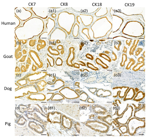
Application: IHC    Species: Human,Goat,Dog,Pig    Sample: EAC glandular cells

FIGURE 4 Immunostaining of cytoskeletal proteins of the ceruminous gland of a (a) human, (b) goat, (c) dog, and (d) pig. CK7, CK8, CK18, and CK19 are expressed in the EAC glandular cells of humans, goats, dogs, and pig. The positive expressions appeared to be brown or dark brown. Scale bar = 50 μm

4). Isolation and characterization of small extracellular vesicles from murine primary mammary tumor. STAR protocols, 2024 (PubMed: 38103192)

Application: WB    Species: Mouse    Sample: liver, adipose, and tumor tissue

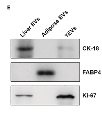
Figure 3 Characterization of EVs isolated from liver and adipose tissue and tissue immune markers for EVs (A) Particle concentration (particles/mL) of isolated liver EVs acquired by nano-flow cytometry. Data are represented as mean ± SD of three independent biological replicates (n = 3). P value (P): ∗P < 0.05, ∗∗∗P < 0.001, ∗∗∗∗P < 0.0001, unpaired two-tailed Student’s t test. (B) Particle concentration (particles/mL) of isolated adipose tissue EVs acquired by nano-flow cytometry. Data are represented as mean ± SD of three independent biological replicates (n = 3). P value (P): ∗∗∗P < 0.001, ∗∗∗∗P < 0.0001, unpaired two-tailed Student’s t test. (C) Transmission electron microscopy (TEM) images of EVs derived from liver. Scale bar, 100 nm. (D) Transmission electron microscopy (TEM) images of EVs derived from adipose tissue. Scale bar, 100 nm. (E) Immunoblotting analysis of samples from liver, adipose, and tumor tissue derived EVs.

Restrictive clause


Affinity Biosciences tests all products strictly. Citations are provided as a resource for additional applications that have not been validated by Affinity Biosciences. Please choose the appropriate format for each application and consult Materials and Methods sections for additional details about the use of any product in these publications.

For Research Use Only.
Not for use in diagnostic or therapeutic procedures. Not for resale. Not for distribution without written consent. Affinity Biosciences will not be held responsible for patent infringement or other violations that may occur with the use of our products. Affinity Biosciences, Affinity Biosciences Logo and all other trademarks are the property of Affinity Biosciences LTD.