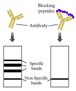
Product: Cytochrome c Oxidase 1 Antibody
Catalog: DF8920
Description: Rabbit polyclonal antibody to Cytochrome c Oxidase 1
Application: WB IHC IF/ICC
Cited expt.: WB
Reactivity: Human, Mouse, Rat
Prediction: Pig, Zebrafish, Bovine, Horse, Sheep, Rabbit, Dog, Chicken, Xenopus
Mol.Wt.: 57 kDa, 73 kDa; 57kD(Calculated).
Uniprot: P00395
RRID: AB_2842116

View similar products>>

   Size Price Inventory
 100ul $280 In stock
 200ul $350 In stock

Lead Time: Same day delivery

For pricing and ordering contact:
Local distributors

Product Info

Source:
Rabbit
Application:
WB 1:1000-3000, IF/ICC 1:100-1:500, IHC 1:50-1:200
*The optimal dilutions should be determined by the end user. For optimal experimental results, antibody reuse is not recommended.
*Tips:

WB: For western blot detection of denatured protein samples. IHC: For immunohistochemical detection of paraffin sections (IHC-p) or frozen sections (IHC-f) of tissue samples. IF/ICC: For immunofluorescence detection of cell samples. ELISA(peptide): For ELISA detection of antigenic peptide.

Reactivity:
Human,Mouse,Rat
Prediction:
Pig(100%), Zebrafish(90%), Bovine(100%), Horse(100%), Sheep(100%), Rabbit(100%), Dog(100%), Chicken(90%), Xenopus(90%)
Clonality:
Polyclonal
Specificity:
Cytochrome c Oxidase 1 Antibody detects endogenous levels of total Cytochrome c Oxidase 1.
RRID:
AB_2842116
Cite Format: Affinity Biosciences Cat# DF8920, RRID:AB_2842116.
Conjugate:
Unconjugated.
Purification:
The antiserum was purified by peptide affinity chromatography using SulfoLink™ Coupling Resin (Thermo Fisher Scientific).
Storage:
Rabbit IgG in phosphate buffered saline , pH 7.4, 150mM NaCl, 0.02% sodium azide and 50% glycerol. Store at -20 °C. Stable for 12 months from date of receipt.
Alias:

Fold/Unfold

COI; COX I; COX1; COX1_HUMAN; COXI; Cytochrome c oxidase polypeptide I; Cytochrome c oxidase subunit 1; Cytochrome C Oxidase subunit I; Mitochondrially encoded cytochrome c oxidase I; MT CO1; MT-CO1; MTCO 1; MTCO1;

Immunogens

Immunogen:

A synthesized peptide derived from human Cytochrome c Oxidase 1, corresponding to a region within C-terminal amino acids.

Uniprot:
Gene(ID):
Sequence:
MFADRWLFSTNHKDIGTLYLLFGAWAGVLGTALSLLIRAELGQPGNLLGNDHIYNVIVTAHAFVMIFFMVMPIMIGGFGNWLVPLMIGAPDMAFPRMNNMSFWLLPPSLLLLLASAMVEAGAGTGWTVYPPLAGNYSHPGASVDLTIFSLHLAGVSSILGAINFITTIINMKPPAMTQYQTPLFVWSVLITAVLLLLSLPVLAAGITMLLTDRNLNTTFFDPAGGGDPILYQHLFWFFGHPEVYILILPGFGMISHIVTYYSGKKEPFGYMGMVWAMMSIGFLGFIVWAHHMFTVGMDVDTRAYFTSATMIIAIPTGVKVFSWLATLHGSNMKWSAAVLWALGFIFLFTVGGLTGIVLANSSLDIVLHDTYYVVAHFHYVLSMGAVFAIMGGFIHWFPLFSGYTLDQTYAKIHFTIMFIGVNLTFFPQHFLGLSGMPRRYSDYPDAYTTWNILSSVGSFISLTAVMLMIFMIWEAFASKRKVLMVEEPSMNLEWLYGCPPPYHTFEEPVYMKS

Predictions

Predictions:

Score>80(red) has high confidence and is suggested to be used for WB detection. *The prediction model is mainly based on the alignment of immunogen sequences, the results are for reference only, not as the basis of quality assurance.

Species
Results
Score
Pig
100
Horse
100
Bovine
100
Sheep
100
Dog
100
Rabbit
100
Xenopus
90
Zebrafish
90
Chicken
90
Model Confidence:
High(score>80) Medium(80>score>50) Low(score<50) No confidence

Research Backgrounds

Function:

Component of the cytochrome c oxidase, the last enzyme in the mitochondrial electron transport chain which drives oxidative phosphorylation. The respiratory chain contains 3 multisubunit complexes succinate dehydrogenase (complex II, CII), ubiquinol-cytochrome c oxidoreductase (cytochrome b-c1 complex, complex III, CIII) and cytochrome c oxidase (complex IV, CIV), that cooperate to transfer electrons derived from NADH and succinate to molecular oxygen, creating an electrochemical gradient over the inner membrane that drives transmembrane transport and the ATP synthase. Cytochrome c oxidase is the component of the respiratory chain that catalyzes the reduction of oxygen to water. Electrons originating from reduced cytochrome c in the intermembrane space (IMS) are transferred via the dinuclear copper A center (CU(A)) of subunit 2 and heme A of subunit 1 to the active site in subunit 1, a binuclear center (BNC) formed by heme A3 and copper B (CU(B)). The BNC reduces molecular oxygen to 2 water molecules using 4 electrons from cytochrome c in the IMS and 4 protons from the mitochondrial matrix.

Subcellular Location:

Mitochondrion inner membrane>Multi-pass membrane protein.

Extracellular region or secreted Cytosol Plasma membrane Cytoskeleton Lysosome Endosome Peroxisome ER Golgi apparatus Nucleus Mitochondrion Manual annotation Automatic computational assertionSubcellular location
Family&Domains:

Belongs to the heme-copper respiratory oxidase family.

Research Fields

· Human Diseases > Endocrine and metabolic diseases > Non-alcoholic fatty liver disease (NAFLD).

· Human Diseases > Neurodegenerative diseases > Alzheimer's disease.

· Human Diseases > Neurodegenerative diseases > Parkinson's disease.

· Human Diseases > Neurodegenerative diseases > Huntington's disease.

· Metabolism > Energy metabolism > Oxidative phosphorylation.

· Metabolism > Global and overview maps > Metabolic pathways.

· Organismal Systems > Circulatory system > Cardiac muscle contraction.   (View pathway)

References

1). Mitochondrial-cytochrome c oxidase II promotes glutaminolysis to sustain tumor cell survival upon glucose deprivation. Nature communications, 2025 (PubMed: 39747079) [IF=16.6]

2). Multimodal analysis stratifies genetic susceptibility and reveals the pathogenic mechanism of kidney injury in diabetic nephropathy. Cell reports. Medicine, 2025 (PubMed: 40712574) [IF=11.7]

3). NAD+-boosting agent nicotinamide mononucleotide potently improves mitochondria stress response in Alzheimer's disease via ATF4-dependent mitochondrial UPR. Cell death & disease, 2024 (PubMed: 39394148) [IF=8.1]

Application: WB    Species: Mouse    Sample: N2a APPswe cells
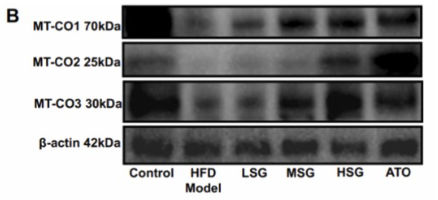
Fig. 5: Cross-species MSR signature attenuates mitochondrial dysfunction. a, c Effects of NMN on the expression level of proteins involved in UPRmt and mitophagy in the 5xFAD mice hippocampi of individuals with and without NMN (n = 3 biologically independent samples; two-sided unpaired t-test). b, d Corresponding quantification of western blot data of (a, c) (n = 3 mice in each group). Data were analyzed by two-sided one-way ANOVA followed by Tukey’s multiple comparisons test. e, f Representative immunostained images (e) and quantification (f) of ATF4 in the hippocampi of AD (VEH) and AD (NMN) tissues (n = 3 mice; two-sided unpaired t-test). Scale bar, 100 μm. g The mRNA expression measurement of MSR signature after supplementing with NMN in 5xFAD mice (n = 3; two-way ANOVA). h Synaptic proteins from the 6-month hippocampi were analyzed by Western blotting (n = 3 mice in each group). Data were analyzed by two-sided one-way ANOVA followed by Tukey’s multiple comparisons test. i–k we obtained T2-weighted anatomical images to analyze the structure difference in the ventricle system using a 9.4 Tesla magnetic resonance imaging (MRI) scanner. Data were pooled from at least 3 biological replicates. Data were analyzed by two-sided one-way ANOVA followed by Tukey’s multiple comparisons test. l Mito-nuclear protein imbalance evaluated by the ratio of mitochondrial DNA (mtDNA)-encoded protein (MTCO1) and nuclear DNA (nDNA)-encoded protein (ATP5a) in three groups (n = 3 mice; one-way ANOVA). m, n Representative immunostaining and quantification of the MitoSOX in the cultured primary astrocytes (n = 6 mice; two-sided unpaired t-test). Scale bar, 20 μm. o, p Representative electron microscopic images, and multiple quantifications, showing the effects of NMN on mitochondrial morphology in mouse hippocampal brain tissues. The quantification was obtained in two independent experiments performed in duplicates in at least 20 ROIs. q ATP Assay was performed in 6-month-old AD mice brain (n = 5 mice, two-sided unpaired t-test). All experiments were performed independently with at least three biological replicates with similar results. Data are shown as mean ± s.e.m. The p-values are indicated on the graphs. ns not significant.

4). A classical herbal formula alleviates high-fat diet induced nonalcoholic steatohepatitis (NASH) via targeting mitophagy to rehabilitate dysfunctional mitochondria, validated by UPLC-HRMS identification combined with in vivo experiment. Biomedicine & pharmacotherapy = Biomedecine & pharmacotherapie, 2023 (PubMed: 37939615) [IF=6.9]

Application: WB    Species: Mouse    Sample:

Fig. 7. Effects of SG formula on mitochondrial function of HFD induced - NASH model. (A) Bar graph of the hepatic ATP content in each group. (B-E) Representative western blot images and bar graphs of the relative expressions of MT-CO1, MT-CO2 and MT-CO3 in each group. * , * *, and * ** represent P 

5). CISD2-mediated mitochondrial dysfunction and iron redistribution contributes to ferroptosis in arsenic-induced nonalcoholic steatohepatitis. Ecotoxicology and environmental safety, 2025 (PubMed: 39808878) [IF=6.2]

6). Klebsiella pneumoniae causes mammary gland damage via FNIP1-mediated mitochondrial dysfunction. Journal of animal science, 2025 (PubMed: 41206543) [IF=2.7]

Application: WB    Species: cow    Sample: BMECs

Figure 2.K. pneumoniae induced mitochondrial dynamic imbalance and mitochondrial quality control failure in BMECs. (A) The protein levels of OPA1, MFN1, COX I, DRP1, and FIS1 were detected in BMECs. (B–F) Relative protein abundance of OPA1, MFN1, COX I, DRP1, and FIS1 were normalized to β-actin (mean ± SEM, n = 3). (G) Flow cytometry was used to measure mitochondrial membrane potential level. (H) Changes in mitochondrial membrane potential were determined by the JC-1 red to green fluorescence ratio (mean ± SEM, n = 3). (I) The mPTP opening status was evaluated in BMECs by calcein-AM fluorescence. Representative flow cytometry graph and (J) statistical analysis of calcein-AM fluorescence intensity (mean ± SEM, n = 3). (K) Relative ATP levels (mean ± SEM, n = 3). (L) Mitochondrial morphology of BMECs. The mitochondria were labeled with MitoTracker. (M) Representative transmission electron microscope images of mitochondria. Blue arrow indicates normal mitochondria. Yellow arrow indicates defects in the mitochondrial cristae structure. (N) The percentage of damaged mitochondria was quantified, at least 100 mitochondria.

Restrictive clause


Affinity Biosciences tests all products strictly. Citations are provided as a resource for additional applications that have not been validated by Affinity Biosciences. Please choose the appropriate format for each application and consult Materials and Methods sections for additional details about the use of any product in these publications.

For Research Use Only.
Not for use in diagnostic or therapeutic procedures. Not for resale. Not for distribution without written consent. Affinity Biosciences will not be held responsible for patent infringement or other violations that may occur with the use of our products. Affinity Biosciences, Affinity Biosciences Logo and all other trademarks are the property of Affinity Biosciences LTD.