Product: TRPV4 Antibody
Catalog: DF8624
Description: Rabbit polyclonal antibody to TRPV4
Application: WB IHC IF/ICC
Reactivity: Human, Mouse, Rat
Prediction: Pig, Zebrafish, Bovine, Sheep, Rabbit
Mol.Wt.: 98 kDa; 98kD(Calculated).
Uniprot: Q9HBA0
RRID: AB_2841828

View similar products>>

   Size Price Inventory
 100ul $280 In stock
 200ul $350 In stock

Lead Time: Same day delivery

For pricing and ordering contact:
Local distributors

Product Info

Source:
Rabbit
Application:
WB 1:1000-3000, IF/ICC 1:100-500, IHC 1:50-1:200
*The optimal dilutions should be determined by the end user.
*Tips:

WB: For western blot detection of denatured protein samples. IHC: For immunohistochemical detection of paraffin sections (IHC-p) or frozen sections (IHC-f) of tissue samples. IF/ICC: For immunofluorescence detection of cell samples. ELISA(peptide): For ELISA detection of antigenic peptide.

Reactivity:
Human,Mouse,Rat
Prediction:
Pig(100%), Zebrafish(82%), Bovine(100%), Sheep(100%), Rabbit(100%)
Clonality:
Polyclonal
Specificity:
TRPV4 Antibody detects endogenous levels of total TRPV4.
RRID:
AB_2841828
Cite Format: Affinity Biosciences Cat# DF8624, RRID:AB_2841828.
Conjugate:
Unconjugated.
Purification:
The antiserum was purified by peptide affinity chromatography using SulfoLink™ Coupling Resin (Thermo Fisher Scientific).
Storage:
Rabbit IgG in phosphate buffered saline , pH 7.4, 150mM NaCl, 0.02% sodium azide and 50% glycerol. Store at -20 °C. Stable for 12 months from date of receipt.
Alias:

Fold/Unfold

BCYM3; CMT2C; HMSN2C; osm 9 like TRP channel 4; Osm-9-like TRP channel 4; OSM9 like transient receptor potential channel 4; Osmosensitive transient receptor potential channel 4; OTRPC 4; OTRPC4; SMAL; SPSMA; SSQTL1; Transient receptor potential cation channel subfamily V member 4; Transient receptor potential protein 12; TRP 12; TRP12; TRPV 4; TrpV4; TRPV4_HUMAN; Vanilloid receptor like channel 2; Vanilloid receptor like protein 2; Vanilloid receptor related osmotically activated channel; Vanilloid receptor-like channel 2; Vanilloid receptor-like protein 2; Vanilloid receptor-related osmotically-activated channel; VR 4; VR OAC; VR-OAC; VR4; VRL 2; VRL-2; VRL2; VROAC;

Immunogens

Immunogen:
Uniprot:
Gene(ID):
Expression:
Q9HBA0 TRPV4_HUMAN:

Found in the synoviocytes from patients with (RA) and without (CTR) rheumatoid arthritis (at protein level).

Sequence:
MADSSEGPRAGPGEVAELPGDESGTPGGEAFPLSSLANLFEGEDGSLSPSPADASRPAGPGDGRPNLRMKFQGAFRKGVPNPIDLLESTLYESSVVPGPKKAPMDSLFDYGTYRHHSSDNKRWRKKIIEKQPQSPKAPAPQPPPILKVFNRPILFDIVSRGSTADLDGLLPFLLTHKKRLTDEEFREPSTGKTCLPKALLNLSNGRNDTIPVLLDIAERTGNMREFINSPFRDIYYRGQTALHIAIERRCKHYVELLVAQGADVHAQARGRFFQPKDEGGYFYFGELPLSLAACTNQPHIVNYLTENPHKKADMRRQDSRGNTVLHALVAIADNTRENTKFVTKMYDLLLLKCARLFPDSNLEAVLNNDGLSPLMMAAKTGKIGIFQHIIRREVTDEDTRHLSRKFKDWAYGPVYSSLYDLSSLDTCGEEASVLEILVYNSKIENRHEMLAVEPINELLRDKWRKFGAVSFYINVVSYLCAMVIFTLTAYYQPLEGTPPYPYRTTVDYLRLAGEVITLFTGVLFFFTNIKDLFMKKCPGVNSLFIDGSFQLLYFIYSVLVIVSAALYLAGIEAYLAVMVFALVLGWMNALYFTRGLKLTGTYSIMIQKILFKDLFRFLLVYLLFMIGYASALVSLLNPCANMKVCNEDQTNCTVPTYPSCRDSETFSTFLLDLFKLTIGMGDLEMLSSTKYPVVFIILLVTYIILTFVLLLNMLIALMGETVGQVSKESKHIWKLQWATTILDIERSFPVFLRKAFRSGEMVTVGKSSDGTPDRRWCFRVDEVNWSHWNQNLGIINEDPGKNETYQYYGFSHTVGRLRRDRWSSVVPRVVELNKNSNPDEVVVPLDSMGNPRCDGHQQGYPRKWRTDDAPL

Predictions

Predictions:

Score>80(red) has high confidence and is suggested to be used for WB detection. *The prediction model is mainly based on the alignment of immunogen sequences, the results are for reference only, not as the basis of quality assurance.

Species
Results
Score
Pig
100
Bovine
100
Sheep
100
Rabbit
100
Zebrafish
82
Xenopus
73
Chicken
67
Horse
0
Dog
0
Model Confidence:
High(score>80) Medium(80>score>50) Low(score<50) No confidence

PTMs - Q9HBA0 As Substrate

Site PTM Type Enzyme
Y91 Phosphorylation
Y110 Phosphorylation
T112 Phosphorylation
Y113 Phosphorylation
S134 Phosphorylation
S162 Phosphorylation Q05655 (PRKCD)
T175 Phosphorylation Q05655 (PRKCD)
S189 Phosphorylation Q05655 (PRKCD)
Y253 Phosphorylation P07948 (LYN)
S319 Phosphorylation
T335 Phosphorylation
K344 Ubiquitination
Y411 Phosphorylation
S470 Phosphorylation
T505 Phosphorylation
Y508 Phosphorylation
Y805 Phosphorylation
S824 Phosphorylation P17612 (PRKACA)

Research Backgrounds

Function:

Non-selective calcium permeant cation channel involved in osmotic sensitivity and mechanosensitivity. Activation by exposure to hypotonicity within the physiological range exhibits an outward rectification. Also activated by heat, low pH, citrate and phorbol esters. Increase of intracellular Ca(2+) potentiates currents. Channel activity seems to be regulated by a calmodulin-dependent mechanism with a negative feedback mechanism. Promotes cell-cell junction formation in skin keratinocytes and plays an important role in the formation and/or maintenance of functional intercellular barriers (By similarity). Acts as a regulator of intracellular Ca(2+) in synoviocytes. Plays an obligatory role as a molecular component in the nonselective cation channel activation induced by 4-alpha-phorbol 12,13-didecanoate and hypotonic stimulation in synoviocytes and also regulates production of IL-8. Together with PKD2, forms mechano- and thermosensitive channels in cilium. Negatively regulates expression of PPARGC1A, UCP1, oxidative metabolism and respiration in adipocytes (By similarity). Regulates expression of chemokines and cytokines related to proinflammatory pathway in adipocytes (By similarity). Together with AQP5, controls regulatory volume decrease in salivary epithelial cells (By similarity). Required for normal development and maintenance of bone and cartilage.

Non-selective calcium permeant cation channel involved in osmotic sensitivity and mechanosensitivity. Activation by exposure to hypotonicity within the physiological range exhibits an outward rectification. Also activated by phorbol esters. Has the same channel activity as isoform 1, and is activated by the same stimuli.

Lacks channel activity, due to impaired oligomerization and intracellular retention.

Lacks channel activity, due to impaired oligomerization and intracellular retention.

Lacks channel activity, due to impaired oligomerization and intracellular retention.

PTMs:

N-glycosylated.

Subcellular Location:

Apical cell membrane>Multi-pass membrane protein. Cell junction>Adherens junction. Cell projection>Cilium.
Note: Assembly of the putative homotetramer occurs primarily in the endoplasmic reticulum.

Cell membrane.

Cell membrane.

Endoplasmic reticulum.

Endoplasmic reticulum.

Endoplasmic reticulum.

Extracellular region or secreted Cytosol Plasma membrane Cytoskeleton Lysosome Endosome Peroxisome ER Golgi apparatus Nucleus Mitochondrion Manual annotation Automatic computational assertionSubcellular location
Tissue Specificity:

Found in the synoviocytes from patients with (RA) and without (CTR) rheumatoid arthritis (at protein level).

Subunit Structure:

Homotetramer (Probable). Self-associates in an isoform-specific manner. Isoform 1 and isoform 5 can oligomerize, but isoform 2, isoform 4 and isoform 6 cannot oligomerize. Interacts with calmodulin. Interacts with Map7 and Src family Tyr protein kinases LYN, SRC, FYN, HCK, LCK and YES (By similarity). Interacts with CTNNB1 (By similarity). The TRPV4 and CTNNB1 complex can interact with CDH1 (By similarity). Interacts with PACSIN1, PACSIN2 and PACSIN3 (via SH3 domain) (By similarity). Part of a complex containing MLC1, AQP4, HEPACAM and ATP1B1. Interacts with ITPR3. Interacts with AQP5; the interaction is probably indirect and regulates TRPV4 activation by hypotonicity (By similarity). Interacts with ANO1 (By similarity). Interacts (via C-terminus) with PKD2 (via C-terminus).

Family&Domains:

The ANK repeat region mediates interaction with Ca(2+)-calmodulin and ATP binding (By similarity). The ANK repeat region mediates interaction with phosphatidylinositol-4,5-bisphosphate and related phosphatidylinositides (PubMed:25256292).

Belongs to the transient receptor (TC 1.A.4) family. TrpV subfamily. TRPV4 sub-subfamily.

Research Fields

· Cellular Processes > Cell growth and death > Cellular senescence.   (View pathway)

· Organismal Systems > Sensory system > Inflammatory mediator regulation of TRP channels.   (View pathway)

References

1). Excessive mechanical stress induces chondrocyte apoptosis through TRPV4 in an anterior cruciate ligament-transected rat osteoarthritis model. LIFE SCIENCES, 2019 (PubMed: 31055086) [IF=6.1]

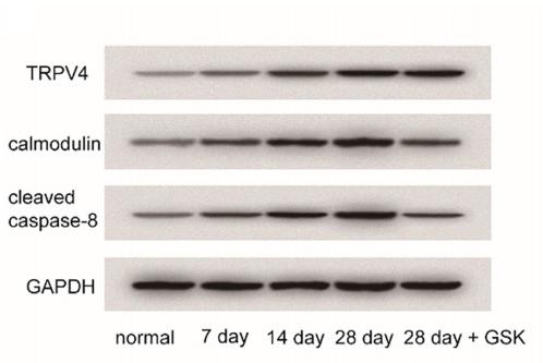
Application: WB    Species: rat    Sample: articular cartilage

Fig. 2. |TRPV4 inhibitor inhibited the high expression of calmodulin and cleaved caspase-8. (A) TRPV4, calmodulin,cleaved caspase-8 protein expressions were detected by western blotting

2). Low-Intensity Extracorporeal Shock Wave Therapy Ameliorates Detrusor Hyperactivity with Impaired Contractility via Transient Potential Vanilloid Channels: A Rat Model for Ovarian Hormone Deficiency. International journal of molecular sciences, 2024 (PubMed: 38732143) [IF=5.6]

Application: WB    Species: Rat    Sample:

Figure 4 LiESWT increased neuronal regeneration, synaptic transmission, and receptor response. The expressions of neuronal endogenous markers (NF, NeuN, and GFAP) and muscarinic receptor (M2 and M3), and purinergic receptor (P2X7) markers were assessed by immunostaining (A–H) and Western blots (I,J). Nuclear DNA was labeled with DAPI (blue). (A–D) The M2 immunostaining was markedly expressed in the UL and SL of the (A) sham group. On the contrary, there was less M2 staining expression in the thinner and defective urothelial mucosa in the UL (yellow arrows) of the (B) OVX group, but the immunostainings in the (C) OVX + SW4 group and the (D) OVX + SW8 group were enhanced. (E–H) Double-labeled analysis of M2 (red, upper panels) and NF (green, lower panels) was distributed in the ML of the (E) sham group. However, the double staining of the (G) OVX + SW4 group and the (H) OVX + SW8 group were widely expressed compared with the (F) OVX group. (I,J) Quantifications of the percentage of neurogenesis-related markers, muscarinic receptors, and purinergic receptors were evaluated by Western blotting. The expressions obviously decreased in the OVX group compared with the sham group. However, the expressions significantly increased in the OVX + SW4 group and OVX + SW8 group compared with the OVX group. Therefore, the LiESWT promoted bladder synaptic transmission, receptor response, and neurogenesis to ameliorate the bladder detrusor contractile response. Nuclear DNA was labeled with DAPI (blue). LiESWT, low-intensity extracorporeal shock wave therapy; OVX + SW4, OHD status for 12 months, followed by once weekly LiESWT for 4 weeks; OVX + SW8, OHD status for 12 months, followed by twice weekly LiESWT for 4 weeks; DAPI, 4′,6-diamidino-2-phenylindole; NF, neurofilament; NeuN, neuronal nuclear antigen and neuron; GFAP, glial fibrillary acidic protein; GAPDH, glyceraldehyde-3-phosphate dehydrogenase; UL, urothelial layer; SL, suburothelial layer. Data are expressed as the means ± SD for n = 8. * p < 0.05 and ** p < 0.01 versus the sham group; ## p < 0.01 versus the OVX group; †† p < 0.01 versus the OVX + SW4 group.

Application: IF/ICC    Species: Rat    Sample:

Figure 4 LiESWT increased neuronal regeneration, synaptic transmission, and receptor response. The expressions of neuronal endogenous markers (NF, NeuN, and GFAP) and muscarinic receptor (M2 and M3), and purinergic receptor (P2X7) markers were assessed by immunostaining (A–H) and Western blots (I,J). Nuclear DNA was labeled with DAPI (blue). (A–D) The M2 immunostaining was markedly expressed in the UL and SL of the (A) sham group. On the contrary, there was less M2 staining expression in the thinner and defective urothelial mucosa in the UL (yellow arrows) of the (B) OVX group, but the immunostainings in the (C) OVX + SW4 group and the (D) OVX + SW8 group were enhanced. (E–H) Double-labeled analysis of M2 (red, upper panels) and NF (green, lower panels) was distributed in the ML of the (E) sham group. However, the double staining of the (G) OVX + SW4 group and the (H) OVX + SW8 group were widely expressed compared with the (F) OVX group. (I,J) Quantifications of the percentage of neurogenesis-related markers, muscarinic receptors, and purinergic receptors were evaluated by Western blotting. The expressions obviously decreased in the OVX group compared with the sham group. However, the expressions significantly increased in the OVX + SW4 group and OVX + SW8 group compared with the OVX group. Therefore, the LiESWT promoted bladder synaptic transmission, receptor response, and neurogenesis to ameliorate the bladder detrusor contractile response. Nuclear DNA was labeled with DAPI (blue). LiESWT, low-intensity extracorporeal shock wave therapy; OVX + SW4, OHD status for 12 months, followed by once weekly LiESWT for 4 weeks; OVX + SW8, OHD status for 12 months, followed by twice weekly LiESWT for 4 weeks; DAPI, 4′,6-diamidino-2-phenylindole; NF, neurofilament; NeuN, neuronal nuclear antigen and neuron; GFAP, glial fibrillary acidic protein; GAPDH, glyceraldehyde-3-phosphate dehydrogenase; UL, urothelial layer; SL, suburothelial layer. Data are expressed as the means ± SD for n = 8. * p < 0.05 and ** p < 0.01 versus the sham group; ## p < 0.01 versus the OVX group; †† p < 0.01 versus the OVX + SW4 group.

3). Treadmill exercise decreases cerebral edema in rats with local cerebral infarction by modulating AQP4 polar expression through the caveolin-1/TRPV4 signaling pathway. BRAIN RESEARCH BULLETIN, 2022 (PubMed: 35961528) [IF=3.8]

4). HDAC1 regulates inflammation and osteogenic differentiation of ankylosing spondylitis fibroblasts through the Wnt-Smad signaling pathway. Journal of Orthopaedic Surgery and Research, 2022 (PubMed: 35794630) [IF=2.6]

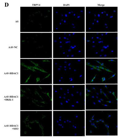
Application: IF/ICC    Species: Human    Sample: AS fibroblasts

Fig. 4 Expression of TRPC1 and TRPV4 in AS fibroblasts after different treatment. A Protein expressions of TRPC1 and TRPV4 in AS fibroblasts were detected by western blot analysis. B Densitometry analysis of protein expression. C Representative images showing immunofluorescence staining of TRPC1. Magnification, × 400. D Representative images showing immunofluorescence staining of TRPV4. Magnification, × 400. E Mean fluorescence intensity of TRPC1. F Mean fluorescence intensity of TRPV4. Data were shown as mean ± SD. *P < 0.05, **P < 0.01 and ***P < 0.001, compared with the AAV-NC group. #P < 0.05, ##P < 0.01 and ###P < 0.001, compared with the AAV-HDAC1 group. &P < 0.05, &&P < 0.01 and &&&P < 0.001, compared with the AAV-HDAC1 + DKK-1 group

Restrictive clause


Affinity Biosciences tests all products strictly. Citations are provided as a resource for additional applications that have not been validated by Affinity Biosciences. Please choose the appropriate format for each application and consult Materials and Methods sections for additional details about the use of any product in these publications.

For Research Use Only.
Not for use in diagnostic or therapeutic procedures. Not for resale. Not for distribution without written consent. Affinity Biosciences will not be held responsible for patent infringement or other violations that may occur with the use of our products. Affinity Biosciences, Affinity Biosciences Logo and all other trademarks are the property of Affinity Biosciences LTD.