OPA1 Antibody - #DF8587
| Product: | OPA1 Antibody |
| Catalog: | DF8587 |
| Description: | Rabbit polyclonal antibody to OPA1 |
| Application: | WB IF/ICC |
| Reactivity: | Human, Mouse, Rat |
| Prediction: | Pig, Zebrafish, Bovine, Horse, Sheep, Rabbit, Chicken, Xenopus |
| Mol.Wt.: | 111kDa, 80-95kDa; 112kD(Calculated). |
| Uniprot: | O60313 |
| RRID: | AB_2841791 |
Related Downloads
Protocols
Product Info
*The optimal dilutions should be determined by the end user.
*Tips:
WB: For western blot detection of denatured protein samples. IHC: For immunohistochemical detection of paraffin sections (IHC-p) or frozen sections (IHC-f) of tissue samples. IF/ICC: For immunofluorescence detection of cell samples. ELISA(peptide): For ELISA detection of antigenic peptide.
Cite Format: Affinity Biosciences Cat# DF8587, RRID:AB_2841791.
Fold/Unfold
Dynamin like 120 kDa protein; Dynamin like 120 kDa protein, mitochondrial; Dynamin-like 120 kDa protein, form S1; FLJ12460; Juvenile kjer type optic atrophy; KIAA0567; KJER type; Large GTP binding protein; largeG; MGM1; Mitochondrial dynamin like 120 kDa protein; Mitochondrial dynamin like GTPase; NPG; NTG; OAK; OPA 1; opa1; OPA1 gene; OPA1_HUMAN; Optic atrophy 1 (autosomal dominant); OPTIC ATROPHY 1; Optic atrophy 1 gene protein; Optic atrophy 1 homolog (human); Optic atrophy protein 1; Optic atrophy protein 1 homolog;
Immunogens
Highly expressed in retina. Also expressed in brain, testis, heart and skeletal muscle. Isoform 1 expressed in retina, skeletal muscle, heart, lung, ovary, colon, thyroid gland, leukocytes and fetal brain. Isoform 2 expressed in colon, liver, kidney, thyroid gland and leukocytes. Low levels of all isoforms expressed in a variety of tissues.
- O60313 OPA1_HUMAN:
- Protein BLAST With
- NCBI/
- ExPASy/
- Uniprot
MWRLRRAAVACEVCQSLVKHSSGIKGSLPLQKLHLVSRSIYHSHHPTLKLQRPQLRTSFQQFSSLTNLPLRKLKFSPIKYGYQPRRNFWPARLATRLLKLRYLILGSAVGGGYTAKKTFDQWKDMIPDLSEYKWIVPDIVWEIDEYIDFEKIRKALPSSEDLVKLAPDFDKIVESLSLLKDFFTSGSPEETAFRATDRGSESDKHFRKVSDKEKIDQLQEELLHTQLKYQRILERLEKENKELRKLVLQKDDKGIHHRKLKKSLIDMYSEVLDVLSDYDASYNTQDHLPRVVVVGDQSAGKTSVLEMIAQARIFPRGSGEMMTRSPVKVTLSEGPHHVALFKDSSREFDLTKEEDLAALRHEIELRMRKNVKEGCTVSPETISLNVKGPGLQRMVLVDLPGVINTVTSGMAPDTKETIFSISKAYMQNPNAIILCIQDGSVDAERSIVTDLVSQMDPHGRRTIFVLTKVDLAEKNVASPSRIQQIIEGKLFPMKALGYFAVVTGKGNSSESIEAIREYEEEFFQNSKLLKTSMLKAHQVTTRNLSLAVSDCFWKMVRESVEQQADSFKATRFNLETEWKNNYPRLRELDRNELFEKAKNEILDEVISLSQVTPKHWEEILQQSLWERVSTHVIENIYLPAAQTMNSGTFNTTVDIKLKQWTDKQLPNKAVEVAWETLQEEFSRFMTEPKGKEHDDIFDKLKEAVKEESIKRHKWNDFAEDSLRVIQHNALEDRSISDKQQWDAAIYFMEEALQARLKDTENAIENMVGPDWKKRWLYWKNRTQEQCVHNETKNELEKMLKCNEEHPAYLASDEITTVRKNLESRGVEVDPSLIKDTWHQVYRRHFLKTALNHCNLCRRGFYYYQRHFVDSELECNDVVLFWRIQRMLAITANTLRQQLTNTEVRRLEKNVKEVLEDFAEDGEKKIKLLTGKRVQLAEDLKKVREIQEKLDAFIEALHQEK
Predictions
Score>80(red) has high confidence and is suggested to be used for WB detection. *The prediction model is mainly based on the alignment of immunogen sequences, the results are for reference only, not as the basis of quality assurance.
High(score>80) Medium(80>score>50) Low(score<50) No confidence
PTMs - O60313 As Substrate
| Site | PTM Type | Enzyme | Source |
|---|---|---|---|
| S21 | Phosphorylation | Uniprot | |
| S22 | Phosphorylation | Uniprot | |
| S107 | Phosphorylation | Uniprot | |
| T114 | Phosphorylation | Uniprot | |
| K117 | Ubiquitination | Uniprot | |
| K123 | Ubiquitination | Uniprot | |
| Y132 | Phosphorylation | Uniprot | |
| K154 | Ubiquitination | Uniprot | |
| K164 | Ubiquitination | Uniprot | |
| K214 | Ubiquitination | Uniprot | |
| K228 | Acetylation | Uniprot | |
| K228 | Ubiquitination | Uniprot | |
| Y268 | Phosphorylation | Uniprot | |
| S298 | Phosphorylation | Uniprot | |
| K301 | Ubiquitination | Uniprot | |
| T302 | Phosphorylation | Uniprot | |
| S344 | Phosphorylation | Uniprot | |
| S345 | Phosphorylation | Uniprot | |
| T351 | Phosphorylation | Uniprot | |
| K352 | Ubiquitination | Uniprot | |
| S383 | Phosphorylation | Uniprot | |
| K387 | Ubiquitination | Uniprot | |
| T405 | Phosphorylation | Uniprot | |
| S440 | Phosphorylation | Uniprot | |
| K474 | Ubiquitination | Uniprot | |
| S478 | Phosphorylation | Uniprot | |
| K489 | Ubiquitination | Uniprot | |
| K527 | Ubiquitination | Uniprot | |
| K530 | Ubiquitination | Uniprot | |
| K535 | Ubiquitination | Uniprot | |
| K568 | Ubiquitination | Uniprot | |
| Y582 | Phosphorylation | Uniprot | |
| K598 | Ubiquitination | Uniprot | |
| S607 | Phosphorylation | Uniprot | |
| T612 | Phosphorylation | Uniprot | |
| K663 | Ubiquitination | Uniprot | |
| S682 | Phosphorylation | Uniprot | |
| K699 | Ubiquitination | Uniprot | |
| S708 | Phosphorylation | Uniprot | |
| K713 | Ubiquitination | Uniprot | |
| R733 | Methylation | Uniprot | |
| K772 | Acetylation | Uniprot | |
| Y808 | Phosphorylation | Uniprot | |
| T815 | Phosphorylation | Uniprot | |
| T816 | Phosphorylation | Uniprot | |
| R824 | Methylation | Uniprot | |
| S831 | Phosphorylation | Uniprot | |
| K834 | Acetylation | Uniprot | |
| K834 | Ubiquitination | Uniprot | |
| K911 | Ubiquitination | Uniprot | |
| K923 | Ubiquitination | Uniprot | |
| K926 | Acetylation | Uniprot | |
| T929 | Phosphorylation | Uniprot | |
| K931 | Acetylation | Uniprot | |
| K940 | Ubiquitination | Uniprot | |
| K948 | Ubiquitination | Uniprot |
Research Backgrounds
Dynamin-related GTPase that is essential for normal mitochondrial morphology by regulating the equilibrium between mitochondrial fusion and mitochondrial fission. Coexpression of isoform 1 with shorter alternative products is required for optimal activity in promoting mitochondrial fusion. Binds lipid membranes enriched in negatively charged phospholipids, such as cardiolipin, and promotes membrane tubulation. The intrinsic GTPase activity is low, and is strongly increased by interaction with lipid membranes. Plays a role in remodeling cristae and the release of cytochrome c during apoptosis (By similarity). Proteolytic processing in response to intrinsic apoptotic signals may lead to disassembly of OPA1 oligomers and release of the caspase activator cytochrome C (CYCS) into the mitochondrial intermembrane space (By similarity). Plays a role in mitochondrial genome maintenance.
Inactive form produced by cleavage at S1 position by OMA1 following stress conditions that induce loss of mitochondrial membrane potential, leading to negative regulation of mitochondrial fusion.
Isoforms that contain the alternative exon 4b (present in isoform 4 and isoform 5) are required for mitochondrial genome maintenance, possibly by anchoring the mitochondrial nucleoids to the inner mitochondrial membrane.
PARL-dependent proteolytic processing releases an antiapoptotic soluble form not required for mitochondrial fusion. Cleaved by OMA1 at position S1 following stress conditions.
Cleavage at position S2 is mediated by YME1L. Cleavage may occur in the sequence motif Leu-Gln-Gln-Gln-Ile-Gln (LQQQIQ). This motif is present in isoform 2, isoform 3, isoform 4 and isoform 7, but is absent in the displayed isoform 1 and in isoform 5.
Mitochondrion inner membrane>Single-pass membrane protein. Mitochondrion intermembrane space. Mitochondrion membrane.
Note: Detected at contact sites between endoplamic reticulum and mitochondrion membranes.
Highly expressed in retina. Also expressed in brain, testis, heart and skeletal muscle. Isoform 1 expressed in retina, skeletal muscle, heart, lung, ovary, colon, thyroid gland, leukocytes and fetal brain. Isoform 2 expressed in colon, liver, kidney, thyroid gland and leukocytes. Low levels of all isoforms expressed in a variety of tissues.
Oligomeric complex consisting of membrane-bound and soluble forms of OPA1. Interacts with RCC1L; this interaction is direct. Interacts with CHCHD3 and IMMT; these interactions occur preferentially with soluble OPA1 forms. Binds PARL (By similarity). Interacts with PRELID1.
Belongs to the TRAFAC class dynamin-like GTPase superfamily. Dynamin/Fzo/YdjA family.
References
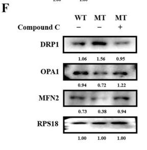
Application: WB Species: human Sample: hepatocytes
Application: WB Species: Human Sample:
Application: WB Species: Mouse Sample:
Application: WB Species: Mice Sample: heart
Restrictive clause
Affinity Biosciences tests all products strictly. Citations are provided as a resource for additional applications that have not been validated by Affinity Biosciences. Please choose the appropriate format for each application and consult Materials and Methods sections for additional details about the use of any product in these publications.
For Research Use Only.
Not for use in diagnostic or therapeutic procedures. Not for resale. Not for distribution without written consent. Affinity Biosciences will not be held responsible for patent infringement or other violations that may occur with the use of our products. Affinity Biosciences, Affinity Biosciences Logo and all other trademarks are the property of Affinity Biosciences LTD.


