Product: TSG101 Antibody
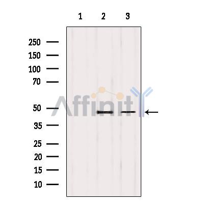
Catalog: DF8427
Description: Rabbit polyclonal antibody to TSG101
Application: WB IHC IF/ICC
Reactivity: Human, Mouse, Rat, Monkey
Prediction: Bovine, Horse, Sheep, Rabbit, Dog
Mol.Wt.: 45 kDa; 44kD(Calculated).
Uniprot: Q99816
RRID: AB_2841675

View similar products>>

   Size Price Inventory
 100ul $280 In stock
 200ul $350 In stock

Lead Time: Same day delivery

For pricing and ordering contact:
Local distributors

Product Info

Source:
Rabbit
Application:
WB 1:1000-3000, IF/ICC 1:100-1:500, IHC 1:50-1:200
*The optimal dilutions should be determined by the end user.
*Tips:

WB: For western blot detection of denatured protein samples. IHC: For immunohistochemical detection of paraffin sections (IHC-p) or frozen sections (IHC-f) of tissue samples. IF/ICC: For immunofluorescence detection of cell samples. ELISA(peptide): For ELISA detection of antigenic peptide.

Reactivity:
Human,Mouse,Rat,Monkey
Prediction:
Bovine(91%), Horse(100%), Sheep(91%), Rabbit(100%), Dog(100%)
Clonality:
Polyclonal
Specificity:
TSG101 Antibody detects endogenous levels of total TSG101.
RRID:
AB_2841675
Cite Format: Affinity Biosciences Cat# DF8427, RRID:AB_2841675.
Conjugate:
Unconjugated.
Purification:
The antiserum was purified by peptide affinity chromatography using SulfoLink™ Coupling Resin (Thermo Fisher Scientific).
Storage:
Rabbit IgG in phosphate buffered saline , pH 7.4, 150mM NaCl, 0.02% sodium azide and 50% glycerol. Store at -20 °C. Stable for 12 months from date of receipt.
Alias:

Fold/Unfold

ESCRT I complex subunit TSG101; ESCRT-I complex subunit TSG101; TS101_HUMAN; TSG 10; TSG 101; TSG10; Tsg101; Tumor susceptibility gene 10; Tumor susceptibility gene 101; Tumor susceptibility gene 101 protein; Tumor susceptibility protein; Tumor susceptibility protein isoform 3; VPS 23; VPS23;

Immunogens

Immunogen:
Uniprot:
Gene(ID):
Expression:
Q99816 TS101_HUMAN:

Heart, brain, placenta, lung, liver, skeletal, kidney and pancreas.

Sequence:
MAVSESQLKKMVSKYKYRDLTVRETVNVITLYKDLKPVLDSYVFNDGSSRELMNLTGTIPVPYRGNTYNIPICLWLLDTYPYNPPICFVKPTSSMTIKTGKHVDANGKIYLPYLHEWKHPQSDLLGLIQVMIVVFGDEPPVFSRPISASYPPYQATGPPNTSYMPGMPGGISPYPSGYPPNPSGYPGCPYPPGGPYPATTSSQYPSQPPVTTVGPSRDGTISEDTIRASLISAVSDKLRWRMKEEMDRAQAELNALKRTEEDLKKGHQKLEEMVTRLDQEVAEVDKNIELLKKKDEELSSALEKMENQSENNDIDEVIIPTAPLYKQILNLYAEENAIEDTIFYLGEALRRGVIDLDVFLKHVRLLSRKQFQLRALMQKARKTAGLSDLY

Predictions

Predictions:

Score>80(red) has high confidence and is suggested to be used for WB detection. *The prediction model is mainly based on the alignment of immunogen sequences, the results are for reference only, not as the basis of quality assurance.

Species
Results
Score
Horse
100
Dog
100
Rabbit
100
Bovine
91
Sheep
91
Pig
0
Xenopus
0
Zebrafish
0
Chicken
0
Model Confidence:
High(score>80) Medium(80>score>50) Low(score<50) No confidence

PTMs - Q99816 As Substrate

Site PTM Type Enzyme
A2 Acetylation
S6 Phosphorylation
K14 Acetylation
Y15 Phosphorylation
K16 Acetylation
Y32 Phosphorylation
S48 Phosphorylation
T56 Phosphorylation
Y63 Phosphorylation
K90 Acetylation
K101 Ubiquitination
T220 Phosphorylation
S222 Phosphorylation
T225 Phosphorylation
K237 Acetylation
K237 Ubiquitination
K257 Ubiquitination
K269 Ubiquitination
K286 Ubiquitination
K292 Ubiquitination
K293 Ubiquitination
S299 Phosphorylation
S300 Phosphorylation
K304 Ubiquitination
S309 Phosphorylation
K361 Ubiquitination
K379 Ubiquitination
Y390 Phosphorylation

Research Backgrounds

Function:

Component of the ESCRT-I complex, a regulator of vesicular trafficking process. Binds to ubiquitinated cargo proteins and is required for the sorting of endocytic ubiquitinated cargos into multivesicular bodies (MVBs). Mediates the association between the ESCRT-0 and ESCRT-I complex. Required for completion of cytokinesis; the function requires CEP55. May be involved in cell growth and differentiation. Acts as a negative growth regulator. Involved in the budding of many viruses through an interaction with viral proteins that contain a late-budding motif P-[ST]-A-P. This interaction is essential for viral particle budding of numerous retroviruses. Required for the exosomal release of SDCBP, CD63 and syndecan. It may also play a role in the extracellular release of microvesicles that differ from the exosomes.

PTMs:

Monoubiquitinated at multiple sites by LRSAM1 and by MGRN1. Ubiquitination inactivates it, possibly by regulating its shuttling between an active membrane-bound protein and an inactive soluble form. Ubiquitination by MGRN1 requires the presence of UBE2D1.

Subcellular Location:

Cytoplasm. Early endosome membrane>Peripheral membrane protein>Cytoplasmic side. Late endosome membrane>Peripheral membrane protein. Cytoplasm>Cytoskeleton>Microtubule organizing center>Centrosome. Midbody>Midbody ring. Nucleus.
Note: Mainly cytoplasmic. Membrane-associated when active and soluble when inactive. Nuclear localization is cell cycle-dependent. Interaction with CEP55 is required for localization to the midbody during cytokinesis.

Extracellular region or secreted Cytosol Plasma membrane Cytoskeleton Lysosome Endosome Peroxisome ER Golgi apparatus Nucleus Mitochondrion Manual annotation Automatic computational assertionSubcellular location
Tissue Specificity:

Heart, brain, placenta, lung, liver, skeletal, kidney and pancreas.

Subunit Structure:

Component of the ESCRT-I complex (endosomal sorting complex required for transport I) which consists of TSG101, VPS28, a VPS37 protein (VPS37A to -D) and MVB12A or MVB12B in a 1:1:1:1 stoichiometry. Interacts with VPS37A, VPS37B and VPS37C. Interacts with DMAP1. Interacts with ubiquitin. Interacts with stathmin, GMCL and AATF (By similarity). Component of an ESCRT-I complex (endosomal sorting complex required for transport I) which consists of TSG101, VPS28, VPS37A and UBAP1 in a 1:1:1:1 stoichiometry. Interacts with HGS; the interaction mediates the association with the ESCRT-0 complex. Interacts with GGA1 and GGA3. Interacts (via UEV domain) with PDCD6IP/AIP1. Interacts with VPS28, SNF8 and VPS36. Self-associates. Interacts with MVB12A; the association appears to be mediated by the TSG101-VPS37 binary subcomplex. Interacts with VPS37D. Interacts with LRSAM1. Interacts with CEP55; the interaction is required for cytokinesis but not for viral budding. Interacts with PDCD6. Interacts with LITAF. Interacts with MGRN1. Interacts with ARRDC1; recruits TSG101 to the plasma membrane.

(Microbial infection) Interacts with HIV-1 p6.

(Microbial infection) Interacts with human spumavirus Gag.

(Microbial infection) Interacts with HTLV-1 Gag.

(Microbial infection) Interacts with Ebola virus VP40.

(Microbial infection) Interacts with EIAV p9; the interaction has been shown in vitro.

(Microbial infection) Interacts with Lassa virus protein Z.

(Microbial infection) Interacts with hepatitis E virus protein ORF3.

Family&Domains:

The UEV domain is required for the interaction of the complex with ubiquitin. It also mediates the interaction with PTAP/PSAP motifs of HIV-1 P6 protein and human spumaretrovirus Gag protein.

The coiled coil domain may interact with stathmin.

The UEV domain binds ubiquitin and P-[ST]-A-P peptide motif independently.

Belongs to the ubiquitin-conjugating enzyme family. UEV subfamily.

Research Fields

· Cellular Processes > Transport and catabolism > Endocytosis.   (View pathway)

References

1). Circulating tumor cells shielded with extracellular vesicle-derived CD45 evade T cell attack to enable metastasis. Signal transduction and targeted therapy, 2024 (PubMed: 38575583) [IF=39.3]

Application: WB    Species: Human    Sample:

Fig. 3 CD45 shed from WBCs could be transferred via EVs to tumor cells. a GFP+ Caco2 cells co-cultured with Jurkat cells at a ratio of 1:4 for different time (0, 1, 2, 4, 8, 16 h), then GFP+ Caco2 cells were collected and stained with anti-CD45-APC to dynamically analyze the MFI shifting of CD45 among GFP+ Caco2 cells using flow cytometry. b Flow cytometric analysis of CD45 transfer from Jurkat cells to Caco2 cells, or from Jurkat cells/ THP1 cells/ WBCs to DLD1 cells following 16 h of indirect co-culture. c, d Bar graph showing the MFI of CD45-PE of Caco2 cells or DLD1 cells after 16 h of indirect co-culture with Jurkat cells/ THP1 cells/ WBCs. e, f Immunoblot analysis showing the makers of EVs isolated from supernatant of THP1 and Jurkat cells. e Density gradient fractions of EVs from Jurkat cells. f Positive (TSG101 and CD63) and negative (Calnexin) markers of EVs. WCL: whole cell lysis. Each ladder was loaded with protein 30 μg. g Nanoparticle tracking analysis of Jurkat cell-derived EVs. h Transmission electron microscope imaging of Jurkat cell-derived EVs. Scale bar = 50 nm. i Plasma EVs charactering by immunoblotting. Jurkat-EVs was used as positive control. P1- and P2-EVs were examples of two CRC cancer patients, HD1 and HD2 were examples of two healthy donors. j Plasma EVs charactering by nano-flow cytometry analysis. k, l CD45 ELISA assay for the measurement of EVs-derived CD45 from healthy donors (HD) and CRC patients (PD) with or without metastasis. m Time series of live cell imaging of DLD1-RFP cells after the addition of EVs (20 μg/mL) derived from CD45-GFP-expressing HEK293T cells for up to 8 h. Scale bar = 5 μm. n Immunofluorescence images of GFP+ Caco2 cells after incubation with PBS or EVs isolated from Jurkat cell supernatant for 12 h. Left scale bar = 20 μm, right scale bar = 5 μm. o Flow cytometric analysis for the measurement of Jurkat cell-derived EVs uptake by Caco2 cells. Caco2 cells were incubated with different concentration of PKH26-stained EVs (0, 1, 2, or 4 μg/mL) for 12 h before analysis. p Caco2 cells were incubated with PKH26-stained EVs (2 μg/mL) and chlorpromazine (CPZ, 0, 5, 10, or 20 μg/mL) for 12 h before collection for flow cytometric analysis of PKH26+ Caco2 cells. All bar graph data are presented as means ± SEM. *P 

2). Isothiazolinone dysregulates the pattern of miRNA secretion: Endocrine implications for neurogenesis. Environment international, 2023 (PubMed: 37939439) [IF=11.8]

3). Mesenchymal Stem Cell Derived Exosomes as Nanodrug Carrier of Doxorubicin for Targeted Osteosarcoma Therapy via SDF1-CXCR4 Axis. International Journal of Nanomedicine, 2022 (PubMed: 35959282) [IF=8.0]

Application: WB    Species: Mouse    Sample: BM-MSCs cells

Figure 1 Characterization of exosomes: the size distributions of blank exosome (A) and exosome-doxorubicin (B) measured by NTA. The mean particle diameters were 141.6 nm for free exosome and 178.1 nm for exosome-doxorubicin. The morphology of blank exosome (C) and exosome-doxorubicin (D) as observed by TEM. (E) Western blotting analysis of the exosomal proteins CD81 and TSG101.

4). Selective CDK9 knockdown sensitizes TRAIL response by suppression of antiapoptotic factors and NF-kappaB pathway. Apoptosis, 2023 (PubMed: 37060507) [IF=7.2]

5). Discovery and validation of extracellular vesicle‐associated miRNAs as noninvasive detection biomarkers for early‐stage non‐small‐cell lung cancer. Molecular Oncology, 2021 (PubMed: 33340250) [IF=6.6]

Application: WB    Species: Human    Sample: lung tissues

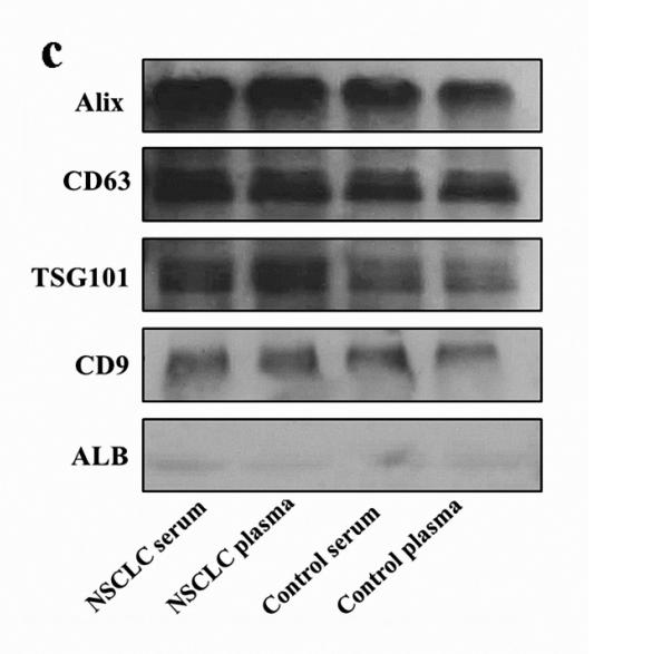
Figure 1. Characterisation of EVs derived from the serum and plasma of NSCLC patients and controls. (a) The shape and structure of serum and plasma EVs isolated by EXOquick kit under TEM. The red arrow represents EVs with typical characteristics (scale bars are 200 nm). (b) The size of EVs derived from control groups and NSCLC groups was analysed by NTA. (c) Western blots of EVs membrane markers, including Alix, CD63, TSG101, CD9, and one negative marker ALB.

6). Mammary Epithelial Cell-Derived Exosomal miR-221-3p Regulates Macrophage Polarization by Targeting Igf2bp2 during Mastitis. Journal of Agricultural and Food Chemistry, 2023 (PubMed: 37757458) [IF=6.1]

7). Exosomes derived from smooth muscle cells ameliorate diabetes‐induced erectile dysfunction by inhibiting fibrosis and modulating the NO/cGMP pathway. Journal of Cellular and Molecular Medicine, 2020 (PubMed: 33009701) [IF=5.3]

Application: WB    Species: Rat    Sample: CCSMCs

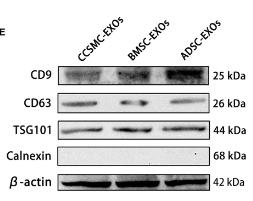
FIGURE 1Cell identification and exosome characterization. (A) Representative flow cytometry histograms of BMSCs and ADSCs show positive staining for CD29 and CD90 but not for CD34 and CD45. (B) BMSCs and ADSCs were successfully induced into osteoblasts (positively stained with Alizarin Red S) and adipocytes (positively stained with Oil Red O). The magnification is 100×. (C) Representative immunofluorescence results of CCSMCs show positive expression for α-SMA and desmin. Scale bars = 50 μm. (D) Exosomes derived from CCSMCs, BMSCs and ADSCs were observed using transmission electron microscopy, and the particle size distributions of the exosomes were measured by nanoparticle tracking analysis. Scale bars = 100 nm. (E) Representative results of Western blot analysis of exosomes derived from CCSMCs, BMSCs and ADSCs show positive expression for CD9, CD63 and TSG101 but not for calnexin. CCSMC: corpus cavernosum smooth muscle cell; BMSC: bone marrow stem cell; ADSC: adipose-derived stem cell; CCSMC-EXOs: exosomes derived from corpus cavernosum smooth muscle cells; BMSC-EXOs: exosomes derived from bone marrow stem cells; ADSC-EXOs: exosomes derived from adipose-derived stem cells; α-SMA: α-smooth muscle actin; DAPI: 4’,6-diamidino-2-phenylindole

8). Pulmonary Delivery of Extracellular Vesicle-Encapsulated Dinaciclib as an Effective Lung Cancer Therapy. Cancers, 2022 (PubMed: 35884614) [IF=5.2]

Application: WB    Species: Human    Sample:

Figure 1 Isolation of TRAIL-expressing extracellular vesicles (EV-T) and post-loading of Dinaciclib (Dina) into EV-T to prepare the complexed therapeutics (EV-T-Dina). (a) Isolation of extracellular vesicles (EVs) by ultracentrifugation; (b) Examination of isolated EV-Ts by transmission electron microscopy (TEM); (c) EV-T analysis by high sensitivity flow cytometry; (d) Assessment of TRAIL expression in EV-Ts by a commercial human TRAIL-specific ELISA kit; (e) Expressional detection of TRAIL and the EV biomarker TSG101 in EVs and EV-Ts by immunoblotting. Uncropped blots are available in Figure S2; (f) Detection of Dina by high-performance liquid chromatography (HPLC); (g) Establishment of a linear regression curve and function for the detection of Dina; (h) Determination and comparison of Dina encapsulation rates by three loading methods of mixing, amphotericin B (AmB), and sonication. Values are means ± SD (n = 3), * p < 0.05, ** p < 0.01, by one-way ANOVA/Bonferroni multiple-comparison post hoc test.

9). Expression level and diagnostic value of exosomal NEAT1/miR‐204/MMP‐9 in acute ST‐segment elevation myocardial infarction. IUBMB Life, 2020 (PubMed: 32916037) [IF=4.6]

Application: WB    Species: human    Sample: serum exosomes

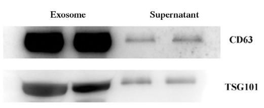
FIGURE 2 |Exosome identification. (a) Structure of exosomes under TEM, magnification 150,000×. (b) Western blot showed the expression of exosome markers CD63 and TSG101

10). Human umbilical cord mesenchymal stem cell-derived exosomes promote neurological function recovery in rat after traumatic brain injury by inhibiting the activation of microglia and astrocyte. Regenerative Therapy, 2022 (PubMed: 36092501) [IF=4.3]

Load more

Restrictive clause


Affinity Biosciences tests all products strictly. Citations are provided as a resource for additional applications that have not been validated by Affinity Biosciences. Please choose the appropriate format for each application and consult Materials and Methods sections for additional details about the use of any product in these publications.

For Research Use Only.
Not for use in diagnostic or therapeutic procedures. Not for resale. Not for distribution without written consent. Affinity Biosciences will not be held responsible for patent infringement or other violations that may occur with the use of our products. Affinity Biosciences, Affinity Biosciences Logo and all other trademarks are the property of Affinity Biosciences LTD.