Product: ABCA1 Antibody
Catalog: DF8233
Description: Rabbit polyclonal antibody to ABCA1
Application: WB
Cited expt.: WB
Reactivity: Human, Mouse
Prediction: Pig, Bovine, Horse, Sheep, Rabbit, Dog, Chicken, Xenopus
Mol.Wt.: 254 kDa; 254kD(Calculated).
Uniprot: O95477
RRID: AB_2841530

View similar products>>

   Size Price Inventory
 100ul $280 In stock
 200ul $350 In stock

Lead Time: Same day delivery

For pricing and ordering contact:
Local distributors

Product Info

Source:
Rabbit
Application:
WB 1:1000-3000
*The optimal dilutions should be determined by the end user. For optimal experimental results, antibody reuse is not recommended.
*Tips:

WB: For western blot detection of denatured protein samples. IHC: For immunohistochemical detection of paraffin sections (IHC-p) or frozen sections (IHC-f) of tissue samples. IF/ICC: For immunofluorescence detection of cell samples. ELISA(peptide): For ELISA detection of antigenic peptide.

Reactivity:
Human,Mouse
Prediction:
Pig(100%), Bovine(100%), Horse(100%), Sheep(100%), Rabbit(100%), Dog(100%), Chicken(92%), Xenopus(100%)
Clonality:
Polyclonal
Specificity:
ABCA1 Antibody detects endogenous levels of total ABCA1.
RRID:
AB_2841530
Cite Format: Affinity Biosciences Cat# DF8233, RRID:AB_2841530.
Conjugate:
Unconjugated.
Purification:
The antiserum was purified by peptide affinity chromatography using SulfoLink™ Coupling Resin (Thermo Fisher Scientific).
Storage:
Rabbit IgG in phosphate buffered saline , pH 7.4, 150mM NaCl, 0.02% sodium azide and 50% glycerol. Store at -20 °C. Stable for 12 months from date of receipt.
Alias:

Fold/Unfold

ABC 1; ABC Transporter 1; ABC-1; ABC1; ABCA 1; ABCA1; ABCA1_HUMAN; ATP binding Cassette 1; ATP binding cassette sub family A ABC1 member 1; ATP binding cassette sub family A member 1; ATP binding Cassette Transporter 1; ATP-binding cassette 1; ATP-binding cassette sub-family A member 1; ATP-binding cassette transporter 1; CERP; Cholesterol efflux regulatory protein; FLJ14958; HDLDT1; Membrane bound; MGC164864; MGC165011; TD; TGD;

Immunogens

Immunogen:

A synthesized peptide derived from human ABCA1, corresponding to a region within C-terminal amino acids.

Uniprot:
Gene(ID):
Expression:
O95477 ABCA1_HUMAN:

Widely expressed, but most abundant in macrophages.

Sequence:
MACWPQLRLLLWKNLTFRRRQTCQLLLEVAWPLFIFLILISVRLSYPPYEQHECHFPNKAMPSAGTLPWVQGIICNANNPCFRYPTPGEAPGVVGNFNKSIVARLFSDARRLLLYSQKDTSMKDMRKVLRTLQQIKKSSSNLKLQDFLVDNETFSGFLYHNLSLPKSTVDKMLRADVILHKVFLQGYQLHLTSLCNGSKSEEMIQLGDQEVSELCGLPREKLAAAERVLRSNMDILKPILRTLNSTSPFPSKELAEATKTLLHSLGTLAQELFSMRSWSDMRQEVMFLTNVNSSSSSTQIYQAVSRIVCGHPEGGGLKIKSLNWYEDNNYKALFGGNGTEEDAETFYDNSTTPYCNDLMKNLESSPLSRIIWKALKPLLVGKILYTPDTPATRQVMAEVNKTFQELAVFHDLEGMWEELSPKIWTFMENSQEMDLVRMLLDSRDNDHFWEQQLDGLDWTAQDIVAFLAKHPEDVQSSNGSVYTWREAFNETNQAIRTISRFMECVNLNKLEPIATEVWLINKSMELLDERKFWAGIVFTGITPGSIELPHHVKYKIRMDIDNVERTNKIKDGYWDPGPRADPFEDMRYVWGGFAYLQDVVEQAIIRVLTGTEKKTGVYMQQMPYPCYVDDIFLRVMSRSMPLFMTLAWIYSVAVIIKGIVYEKEARLKETMRIMGLDNSILWFSWFISSLIPLLVSAGLLVVILKLGNLLPYSDPSVVFVFLSVFAVVTILQCFLISTLFSRANLAAACGGIIYFTLYLPYVLCVAWQDYVGFTLKIFASLLSPVAFGFGCEYFALFEEQGIGVQWDNLFESPVEEDGFNLTTSVSMMLFDTFLYGVMTWYIEAVFPGQYGIPRPWYFPCTKSYWFGEESDEKSHPGSNQKRISEICMEEEPTHLKLGVSIQNLVKVYRDGMKVAVDGLALNFYEGQITSFLGHNGAGKTTTMSILTGLFPPTSGTAYILGKDIRSEMSTIRQNLGVCPQHNVLFDMLTVEEHIWFYARLKGLSEKHVKAEMEQMALDVGLPSSKLKSKTSQLSGGMQRKLSVALAFVGGSKVVILDEPTAGVDPYSRRGIWELLLKYRQGRTIILSTHHMDEADVLGDRIAIISHGKLCCVGSSLFLKNQLGTGYYLTLVKKDVESSLSSCRNSSSTVSYLKKEDSVSQSSSDAGLGSDHESDTLTIDVSAISNLIRKHVSEARLVEDIGHELTYVLPYEAAKEGAFVELFHEIDDRLSDLGISSYGISETTLEEIFLKVAEESGVDAETSDGTLPARRNRRAFGDKQSCLRPFTEDDAADPNDSDIDPESRETDLLSGMDGKGSYQVKGWKLTQQQFVALLWKRLLIARRSRKGFFAQIVLPAVFVCIALVFSLIVPPFGKYPSLELQPWMYNEQYTFVSNDAPEDTGTLELLNALTKDPGFGTRCMEGNPIPDTPCQAGEEEWTTAPVPQTIMDLFQNGNWTMQNPSPACQCSSDKIKKMLPVCPPGAGGLPPPQRKQNTADILQDLTGRNISDYLVKTYVQIIAKSLKNKIWVNEFRYGGFSLGVSNTQALPPSQEVNDAIKQMKKHLKLAKDSSADRFLNSLGRFMTGLDTKNNVKVWFNNKGWHAISSFLNVINNAILRANLQKGENPSHYGITAFNHPLNLTKQQLSEVALMTTSVDVLVSICVIFAMSFVPASFVVFLIQERVSKAKHLQFISGVKPVIYWLSNFVWDMCNYVVPATLVIIIFICFQQKSYVSSTNLPVLALLLLLYGWSITPLMYPASFVFKIPSTAYVVLTSVNLFIGINGSVATFVLELFTDNKLNNINDILKSVFLIFPHFCLGRGLIDMVKNQAMADALERFGENRFVSPLSWDLVGRNLFAMAVEGVVFFLITVLIQYRFFIRPRPVNAKLSPLNDEDEDVRRERQRILDGGGQNDILEIKELTKIYRRKRKPAVDRICVGIPPGECFGLLGVNGAGKSSTFKMLTGDTTVTRGDAFLNKNSILSNIHEVHQNMGYCPQFDAITELLTGREHVEFFALLRGVPEKEVGKVGEWAIRKLGLVKYGEKYAGNYSGGNKRKLSTAMALIGGPPVVFLDEPTTGMDPKARRFLWNCALSVVKEGRSVVLTSHSMEECEALCTRMAIMVNGRFRCLGSVQHLKNRFGDGYTIVVRIAGSNPDLKPVQDFFGLAFPGSVLKEKHRNMLQYQLPSSLSSLARIFSILSQSKKRLHIEDYSVSQTTLDQVFVNFAKDQSDDDHLKDLSLHKNQTVVDVAVLTSFLQDEKVKESYV

Predictions

Predictions:

Score>80(red) has high confidence and is suggested to be used for WB detection. *The prediction model is mainly based on the alignment of immunogen sequences, the results are for reference only, not as the basis of quality assurance.

Species
Results
Score
Pig
100
Horse
100
Bovine
100
Sheep
100
Dog
100
Xenopus
100
Rabbit
100
Chicken
92
Zebrafish
0
Model Confidence:
High(score>80) Medium(80>score>50) Low(score<50) No confidence

Research Backgrounds

Function:

Catalyzes the translocation of specific phospholipids from the cytoplasmic to the extracellular/lumenal leaflet of membrane coupled to the hydrolysis of ATP. Thereby, participates to phospholipids transfer to apoliproteins to form nascent high density lipoproteins/HDLs. Transports preferentially phosphatidylcholine over phosphatidylserine. May play a similar role in the efflux of intracellular cholesterol to apoliproteins and the formation of nascent high density lipoproteins/HDLs.

PTMs:

Phosphorylation on Ser-2054 regulates phospholipid efflux.

Palmitoylation by DHHC8 is essential for membrane localization.

Subcellular Location:

Membrane>Multi-pass membrane protein. Cell membrane. Endosome.

Extracellular region or secreted Cytosol Plasma membrane Cytoskeleton Lysosome Endosome Peroxisome ER Golgi apparatus Nucleus Mitochondrion Manual annotation Automatic computational assertionSubcellular location
Tissue Specificity:

Widely expressed, but most abundant in macrophages.

Family&Domains:

Multifunctional polypeptide with two homologous halves, each containing a hydrophobic membrane-anchoring domain and an ATP binding cassette (ABC) domain.

Belongs to the ABC transporter superfamily. ABCA family.

Research Fields

· Environmental Information Processing > Membrane transport > ABC transporters.

· Organismal Systems > Digestive system > Fat digestion and absorption.

· Organismal Systems > Digestive system > Cholesterol metabolism.

References

1). Multifunctional Nanomedicine for Targeted Atherosclerosis Therapy: Activating Plaque Clearance Cascade and Suppressing Inflammation. ACS Nano, 2025 [IF=15.8]

2). Antisense oligonucleotide targeting TARDBP-EGFR splicing axis inhibits progression of oral squamous cell carcinoma through ABCA1-regulated cholesterol efflux. International journal of oral science, 2026 (PubMed: 41540015) [IF=10.8]

3). Yin-xing-tong-mai decoction attenuates atherosclerosis via activating PPARγ-LXRα-ABCA1/ABCG1 pathway. Pharmacological research, 2021 (PubMed: 33932607) [IF=9.1]

4). The plant extract PNS mitigates atherosclerosis via promoting Nrf2-mediated inhibition of ferroptosis through reducing USP2-mediated Keap1 deubiquitination. British journal of pharmacology, 2024 (PubMed: 39228119) [IF=6.8]

Application: IF/ICC    Species: Mouse    Sample:

FIGURE 2 Panax notoginseng saponins (PNS) reduces foam cell formation in atherosclerotic plaque and peritoneal macrophages. (a) Immunofluorescence analysis of macrophage/foam cells (CD68) in aortic root sections from apoE−/− mice, and mean fluorescence intensity was quantified, n = 10. (b) Analysis of foam cell content in peritoneal macrophages from apoE−/− mice via oil red staining. The number of foam cells was quantified, n = 10. (c) Immunofluorescence analysis of ABCA1 levels in peritoneal macrophages from apoE−/− mice. (d) Quantification of ABCA1/G1 expression in peritoneal macrophages from apoE−/− mice via western blotting, and protein levels were quantified, n = 6. (e) Immunofluorescence analysis of ABCA1/G1 in aortic root frozen sections from apoE−/− mice, and mean fluorescence intensity was quantified; n = 10. Data shown are means ± SEM, *P < 0.05, significantly different from control (Ctrl).

Application: WB    Species: Mouse    Sample:

FIGURE 2 Panax notoginseng saponins (PNS) reduces foam cell formation in atherosclerotic plaque and peritoneal macrophages. (a) Immunofluorescence analysis of macrophage/foam cells (CD68) in aortic root sections from apoE−/− mice, and mean fluorescence intensity was quantified, n = 10. (b) Analysis of foam cell content in peritoneal macrophages from apoE−/− mice via oil red staining. The number of foam cells was quantified, n = 10. (c) Immunofluorescence analysis of ABCA1 levels in peritoneal macrophages from apoE−/− mice. (d) Quantification of ABCA1/G1 expression in peritoneal macrophages from apoE−/− mice via western blotting, and protein levels were quantified, n = 6. (e) Immunofluorescence analysis of ABCA1/G1 in aortic root frozen sections from apoE−/− mice, and mean fluorescence intensity was quantified; n = 10. Data shown are means ± SEM, *P < 0.05, significantly different from control (Ctrl).

5). MaiJiTong granule attenuates atherosclerosis by reducing ferroptosis via activating STAT6-mediated inhibition of DMT1 and SOCS1/p53 pathways in LDLR-/- mice. Phytomedicine : international journal of phytotherapy and phytopharmacology, 2024 (PubMed: 38569295) [IF=6.7]

6). Proteus mirabilis Vesicles Induce Mitochondrial Apoptosis by Regulating miR96-5p/Abca1 to Inhibit Osteoclastogenesis and Bone Loss. Frontiers in Immunology, 2022 (PubMed: 35242136) [IF=5.7]

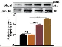
Application: WB    Species: mouse    Sample:

FIGURE 6| Effect of P.M OMVs on miR96/Abca1 mediated mitochondria-dependent apoptosis.(J, K) Expression of Abca1 after 0.15 μg/ml of P.M OMV treatment by RT-qPCR and WB.

7). Fatty acid binding protein 3 deficiency limits atherosclerosis development via macrophage foam cell formation inhibition. EXPERIMENTAL CELL RESEARCH, 2021 (PubMed: 34370993) [IF=3.3]

Application: WB    Species: Mice    Sample: macrophages

Fig. 3. FABP3 knockdown reduced macrophage foam cell formation. Murine peritoneal macrophages were maintained in the special culture medium at 37 ◦ C in a 5 % CO 2 atmosphere. The cells were infected with lentivirus carrying shFABP3 or shRNA NC for 24 h and subsequently exposed with ox-LDL (50 μ g/ml) for 24 h. (A) Representative Oil Red O-stained images of macrophages and quantification of the staining. (B) Immunoblots of SRA1, CD36, SRB1, ABCA1, and ABCG1 in macrophages. (C) Quantification of relative mRNA expression of SRA1, CD36, SRB1, ABCA1, and ABCG1. **p < 0.01 denote significant differences between the Control and the ox-LDL groups, && p < 0.01 denote significant differences between the ox-LDL shRNA NC and ox-LDL + shFABP3 groups.

8). Stigmasterol attenuates atherosclerosis by inhibiting inflammatory signaling and foam cell formation. iMetaOmics, 2025 (PubMed: 41676448)

Application: WB    Species: Mouse    Sample:

Figure 2 Effects of stigmasterol on cholesterol metabolism and phenotypic transition in ox-LDL-treated macrophages. (A) Cells were incubated with stigmasterol (60, 90, and 120 μM) for 6 h, then co-incubated with ox-LDL for 24 h. Morphologic change of foam cells stained by Oil Red O (ORO) and quantitation of lipid drops by extracting ORO dyes from the stained macrophages (magnification ×200). The scale bar in the figure is 50 μm. n = 3. (B) Cells were pretreated with stigmasterol for 6 h, then incubated with 10 μg/mL Dil-ox-LDL and stigmasterol for an additional 4 h. Dil-LDL (red) fluorescence represents DiI-LDL particles while blue fluorescence indicates nucleus. (magnification ×400). The scale bar in the figure is 25 μm. (C) The mean Dil-ox-LDL burden (mFI) was analyzed by flow cytometry. n = 3. (D) Cells were loaded with NBD-cholesterol (5 μg/mL) (green) and stigmasterol (90 μM) for 12 h. Then incubated for 4 h with 0.2% BSA or HDL (50 μg/mL). Green fluorescence represents NBD-cholesterol particles while blue fluorescence indicates the nucleus. (magnification ×200). The scale bar in the figure is 50 μm. (E) HDL-mediated cholesterol efflux (%) was measured in macrophages treated with stigmasterol or control by using fluorescence plate reader. n = 6. (F) Expressions of LDLR, SCARB1, CD36, ABCG1, ABCG5, and ABCA1 after ox-LDL or stigmasterol treatment (60 and 90 μM) were evaluated by western blot. (G) Qualification of LDLR, SCARB1, CD36, ABCG1, ABCG5 and ABCA1. n = 3. (H) Cells were incubated with stigmasterol for 6 h, then co-incubated with ox-LDL for 24 h. The percentage of CD86+ cells was analyzed by flow cytometry. n = 3. (I) Cd86 and Cd206 mRNA detection by q-RT-PCR treated with stigmasterol or ox-LDL. n = 3. (J) Release of TNF-α and IL-10 treated with stigmasterol or ox-LDL. n = 4. Multi-group comparison was measured by one-way ANOVA. The data are presented as the mean ± SD. *p 

Restrictive clause


Affinity Biosciences tests all products strictly. Citations are provided as a resource for additional applications that have not been validated by Affinity Biosciences. Please choose the appropriate format for each application and consult Materials and Methods sections for additional details about the use of any product in these publications.

For Research Use Only.
Not for use in diagnostic or therapeutic procedures. Not for resale. Not for distribution without written consent. Affinity Biosciences will not be held responsible for patent infringement or other violations that may occur with the use of our products. Affinity Biosciences, Affinity Biosciences Logo and all other trademarks are the property of Affinity Biosciences LTD.