| Product: | APOA2 Antibody |
| Catalog: | DF7905 |
| Description: | Rabbit polyclonal antibody to APOA2 |
| Application: | WB IF/ICC |
| Cited expt.: | WB |
| Reactivity: | Human, Mouse, Rat |
| Prediction: | Bovine, Sheep, Dog |
| Mol.Wt.: | 11 kDa(Observed); 11kD(Calculated). |
| Uniprot: | P02652 |
| RRID: | AB_2841322 |
Control Products

Related Downloads
Protocols
Product Info
*The optimal dilutions should be determined by the end user. For optimal experimental results, antibody reuse is not recommended.
*Tips:
WB: For western blot detection of denatured protein samples. IHC: For immunohistochemical detection of paraffin sections (IHC-p) or frozen sections (IHC-f) of tissue samples. IF/ICC: For immunofluorescence detection of cell samples. ELISA(peptide): For ELISA detection of antigenic peptide.
Cite Format: Affinity Biosciences Cat# DF7905, RRID:AB_2841322.
Fold/Unfold
APO A2; Apo AII; Apo-AII; APOA 2; ApoA II; ApoA-II; APOA2; APOA2_HUMAN; APOAII; Apolipoprotein A II; Apolipoprotein A-II(1-76); Apolipoprotein A2; Apolipoprotein AII; ApolipoproteinA II; OTTHUMP00000032244;
Immunogens
A synthesized peptide derived from human APOA2, corresponding to a region within the internal amino acids.
- P02652 APOA2_HUMAN:
- Protein BLAST With
- NCBI/
- ExPASy/
- Uniprot
MKLLAATVLLLTICSLEGALVRRQAKEPCVESLVSQYFQTVTDYGKDLMEKVKSPELQAEAKSYFEKSKEQLTPLIKKAGTELVNFLSYFVELGTQPATQ
Predictions
Score>80(red) has high confidence and is suggested to be used for WB detection. *The prediction model is mainly based on the alignment of immunogen sequences, the results are for reference only, not as the basis of quality assurance.
High(score>80) Medium(80>score>50) Low(score<50) No confidence
Research Backgrounds
May stabilize HDL (high density lipoprotein) structure by its association with lipids, and affect the HDL metabolism.
Phosphorylation sites are present in the extracellular medium.
Apolipoprotein A-II is O-glycosylated.
Secreted.
Plasma; synthesized in the liver and intestine.
Belongs to the apolipoprotein A2 family.
Research Fields
· Organismal Systems > Endocrine system > PPAR signaling pathway.
· Organismal Systems > Digestive system > Cholesterol metabolism.
References
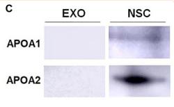
Application: WB Species: Mouse Sample: neural stem cells (NSC)
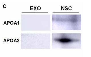
Application: WB Species: mouse Sample: NSCs
Restrictive clause
Affinity Biosciences tests all products strictly. Citations are provided as a resource for additional applications that have not been validated by Affinity Biosciences. Please choose the appropriate format for each application and consult Materials and Methods sections for additional details about the use of any product in these publications.
For Research Use Only.
Not for use in diagnostic or therapeutic procedures. Not for resale. Not for distribution without written consent. Affinity Biosciences will not be held responsible for patent infringement or other violations that may occur with the use of our products. Affinity Biosciences, Affinity Biosciences Logo and all other trademarks are the property of Affinity Biosciences LTD.