| Product: | CCL22 Antibody |
| Catalog: | DF7781 |
| Description: | Rabbit polyclonal antibody to CCL22 |
| Application: | WB |
| Cited expt.: | WB |
| Reactivity: | Human, Mouse, Rat |
| Prediction: | Pig, Bovine, Horse, Sheep, Rabbit, Dog |
| Mol.Wt.: | 10 KD(Observed); 11kD(Calculated). |
| Uniprot: | O00626 |
| RRID: | AB_2841243 |
Control Products

Related Downloads
Protocols
Product Info
*The optimal dilutions should be determined by the end user. For optimal experimental results, antibody reuse is not recommended.
*Tips:
WB: For western blot detection of denatured protein samples. IHC: For immunohistochemical detection of paraffin sections (IHC-p) or frozen sections (IHC-f) of tissue samples. IF/ICC: For immunofluorescence detection of cell samples. ELISA(peptide): For ELISA detection of antigenic peptide.
Cite Format: Affinity Biosciences Cat# DF7781, RRID:AB_2841243.
Fold/Unfold
A 152E5.1; ABCD 1; ABCD1; C C motif chemokine 22; CC chemokine STCP 1; CC chemokine STCP-1; ccl 22; Ccl22; CCL22_HUMAN; Chemokine (C C motif) ligand 22; DC/B CK; DCBCK; Macrophage-derived chemokine; MDC; MDC(1-69); MDC(7-69); MGC34554; SCYA22; Small inducible cytokine subfamily A (Cys Cys) member 22; Small inducible cytokine subfamily A, member 22; Small-inducible cytokine A22; STCP 1; STCP1; Stimulated T cell chemotactic protein 1; Stimulated T-cell chemotactic protein 1;
Immunogens
A synthesized peptide derived from human CCL22, corresponding to a region within the internal amino acids.
Highly expressed in macrophage and in monocyte-derived dendritic cells, and thymus. Also found in lymph node, appendix, activated monocytes, resting and activated macrophages. Lower expression in lung and spleen. Very weak expression in small intestine. In lymph node expressed in a mature subset of Langerhans' cells (CD1a+ and CD83+). Expressed in Langerhans' cell histiocytosis but not in dermatopathic lymphadenopathy. Expressed in atopic dermatitis, allergic contact dermatitis skin, and psoriasis, in both the epidermis and dermis.
- O00626 CCL22_HUMAN:
- Protein BLAST With
- NCBI/
- ExPASy/
- Uniprot
MDRLQTALLVVLVLLAVALQATEAGPYGANMEDSVCCRDYVRYRLPLRVVKHFYWTSDSCPRPGVVLLTFRDKEICADPRVPWVKMILNKLSQ
Predictions
Score>80(red) has high confidence and is suggested to be used for WB detection. *The prediction model is mainly based on the alignment of immunogen sequences, the results are for reference only, not as the basis of quality assurance.
High(score>80) Medium(80>score>50) Low(score<50) No confidence
Research Backgrounds
May play a role in the trafficking of activated/effector T-lymphocytes to inflammatory sites and other aspects of activated T-lymphocyte physiology. Chemotactic for monocytes, dendritic cells and natural killer cells. Mild chemoattractant for primary activated T-lymphocytes and a potent chemoattractant for chronically activated T-lymphocytes but has no chemoattractant activity for neutrophils, eosinophils, and resting T-lymphocytes. Binds to CCR4. Processed forms MDC(3-69), MDC(5-69) and MDC(7-69) seem not be active.
The N-terminal processed forms MDC(3-69), MDC(5-69) and MDC(7-69) are produced by proteolytic cleavage after secretion from monocyte derived dendrocytes.
Secreted.
Highly expressed in macrophage and in monocyte-derived dendritic cells, and thymus. Also found in lymph node, appendix, activated monocytes, resting and activated macrophages. Lower expression in lung and spleen. Very weak expression in small intestine. In lymph node expressed in a mature subset of Langerhans' cells (CD1a+ and CD83+). Expressed in Langerhans' cell histiocytosis but not in dermatopathic lymphadenopathy. Expressed in atopic dermatitis, allergic contact dermatitis skin, and psoriasis, in both the epidermis and dermis.
Belongs to the intercrine beta (chemokine CC) family.
Research Fields
· Environmental Information Processing > Signaling molecules and interaction > Cytokine-cytokine receptor interaction. (View pathway)
· Organismal Systems > Immune system > Chemokine signaling pathway. (View pathway)
References

Application: WB Species: mouse Sample: kidney

Application: IHC Species: mouse Sample: renal
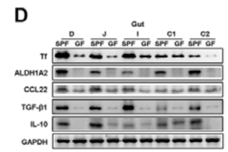
Application: WB Species: Mouse Sample: gut tissue
Restrictive clause
Affinity Biosciences tests all products strictly. Citations are provided as a resource for additional applications that have not been validated by Affinity Biosciences. Please choose the appropriate format for each application and consult Materials and Methods sections for additional details about the use of any product in these publications.
For Research Use Only.
Not for use in diagnostic or therapeutic procedures. Not for resale. Not for distribution without written consent. Affinity Biosciences will not be held responsible for patent infringement or other violations that may occur with the use of our products. Affinity Biosciences, Affinity Biosciences Logo and all other trademarks are the property of Affinity Biosciences LTD.