ZKSCAN3 Antibody - #DF7752
 and mouse anti-beta tubulin Ab(T0023) for 1 hour at 37°C. An AlexaFluor594 conjugated goat anti-rabbit IgG(H+L) Ab(Red) and an AlexaFluor488 conjugated goat anti-mouse IgG(H+L) Ab(Green) were used as the secondary antibody.
The nuclear counter stain is DAPI (blue).")
| Product: | ZKSCAN3 Antibody |
| Catalog: | DF7752 |
| Description: | Rabbit polyclonal antibody to ZKSCAN3 |
| Application: | WB IF/ICC |
| Reactivity: | Human, Mouse |
| Mol.Wt.: | 61 kDa; 61kD(Calculated). |
| Uniprot: | Q9BRR0 |
| RRID: | AB_2841218 |
Related Downloads
Protocols
Product Info
*The optimal dilutions should be determined by the end user.
*Tips:
WB: For western blot detection of denatured protein samples. IHC: For immunohistochemical detection of paraffin sections (IHC-p) or frozen sections (IHC-f) of tissue samples. IF/ICC: For immunofluorescence detection of cell samples. ELISA(peptide): For ELISA detection of antigenic peptide.
Cite Format: Affinity Biosciences Cat# DF7752, RRID:AB_2841218.
Fold/Unfold
dJ874C20.1; mszf35; SCAN-KRAB-zinc finger protein; Skz1; Zf47; Zfp-47; ZFP306; Zfp307; Zfp47; Zinc finger and SCAN domain containing protein 13; Zinc finger and SCAN domain-containing protein 13; Zinc finger protein 306; Zinc finger protein 306, isoform CRA_a; Zinc finger protein 307; Zinc finger protein 309; Zinc finger protein 47 homolog; Zinc finger protein with KRAB and SCAN domains 3; Zinc finger protein with KRAB and SCAN domains 3; Zinc finger protein zfp47; Zinc finger with KRAB and SCAN domains 3; ZKSC3_HUMAN; ZKSCAN3; ZNF309; ZSCAN13; ZSCAN35;
Immunogens
- Q9BRR0 ZKSC3_HUMAN:
- Protein BLAST With
- NCBI/
- ExPASy/
- Uniprot
MARELSESTALDAQSTEDQMELLVIKVEEEEAGFPSSPDLGSEGSRERFRGFRYPEAAGPREALSRLRELCRQWLQPEMHSKEQILELLVLEQFLTILPGNLQSWVREQHPESGEEVVVLLEYLERQLDEPAPQVSGVDQGQELLCCKMALLTPAPGSQSSQFQLMKALLKHESVGSQPLQDRVLQVPVLAHGGCCREDKVVASRLTPESQGLLKVEDVALTLTPEWTQQDSSQGNLCRDEKQENHGSLVSLGDEKQTKSRDLPPAEELPEKEHGKISCHLREDIAQIPTCAEAGEQEGRLQRKQKNATGGRRHICHECGKSFAQSSGLSKHRRIHTGEKPYECEECGKAFIGSSALVIHQRVHTGEKPYECEECGKAFSHSSDLIKHQRTHTGEKPYECDDCGKTFSQSCSLLEHHRIHTGEKPYQCSMCGKAFRRSSHLLRHQRIHTGDKNVQEPEQGEAWKSRMESQLENVETPMSYKCNECERSFTQNTGLIEHQKIHTGEKPYQCNACGKGFTRISYLVQHQRSHVGKNILSQ
PTMs - Q9BRR0 As Substrate
| Site | PTM Type | Enzyme | Source |
|---|---|---|---|
| Y54 | Phosphorylation | Uniprot | |
| S65 | Phosphorylation | Uniprot | |
| K82 | Methylation | Uniprot | |
| K171 | Ubiquitination | Uniprot | |
| T207 | Phosphorylation | Uniprot | |
| T222 | Phosphorylation | Uniprot | |
| T224 | Phosphorylation | Uniprot | |
| T228 | Phosphorylation | Uniprot | |
| T337 | Phosphorylation | Uniprot | |
| T365 | Phosphorylation | Uniprot | |
| T391 | Phosphorylation | Uniprot | |
| T393 | Phosphorylation | Uniprot | |
| T421 | Phosphorylation | Uniprot | |
| T449 | Phosphorylation | Uniprot | |
| Y480 | Phosphorylation | Uniprot | |
| T503 | Phosphorylation | Uniprot | |
| K515 | Acetylation | Uniprot |
Research Backgrounds
Transcriptional factor that binds to the consensus sequence 5'-[GT][AG][AGT]GGGG-3' and acts as a repressor of autophagy. Specifically represses expression of genes involved in autophagy and lysosome biogenesis/function such as MAP1LC3B, ULK1 or WIPI2. Associates with chromatin at the ITGB4 and VEGF promoters. Also acts as a transcription activator and promotes cancer cell progression and/or migration in various tumors and myelomas.
Nucleus. Cytoplasm.
Note: Mainly localizes in the nucleus. Under starvation conditions translocates to the cytoplasm, allowing expression of target genes involved in autophagy and lysosome biogenesis/function.
Belongs to the krueppel C2H2-type zinc-finger protein family.
References
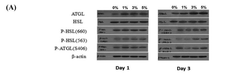
Application: WB Species: Pig Sample: adipose
Restrictive clause
Affinity Biosciences tests all products strictly. Citations are provided as a resource for additional applications that have not been validated by Affinity Biosciences. Please choose the appropriate format for each application and consult Materials and Methods sections for additional details about the use of any product in these publications.
For Research Use Only.
Not for use in diagnostic or therapeutic procedures. Not for resale. Not for distribution without written consent. Affinity Biosciences will not be held responsible for patent infringement or other violations that may occur with the use of our products. Affinity Biosciences, Affinity Biosciences Logo and all other trademarks are the property of Affinity Biosciences LTD.