Product: Perilipin-1 Antibody
Catalog: DF7602
Description: Rabbit polyclonal antibody to Perilipin-1
Application: WB IHC IF/ICC
Reactivity: Human, Mouse, Rat
Prediction: Pig, Bovine, Horse, Sheep, Dog
Mol.Wt.: 50~70kD; 56kD(Calculated).
Uniprot: O60240
RRID: AB_2841093

View similar products>>

   Size Price Inventory
 100ul $280 In stock
 200ul $350 In stock

Lead Time: Same day delivery

For pricing and ordering contact:
Local distributors

Product Info

Source:
Rabbit
Application:
WB 1:1000-3000, IHC 1:50-1:200, IF/ICC 1:100-500
*The optimal dilutions should be determined by the end user.
*Tips:

WB: For western blot detection of denatured protein samples. IHC: For immunohistochemical detection of paraffin sections (IHC-p) or frozen sections (IHC-f) of tissue samples. IF/ICC: For immunofluorescence detection of cell samples. ELISA(peptide): For ELISA detection of antigenic peptide.

Reactivity:
Human,Mouse,Rat
Prediction:
Pig(92%), Bovine(100%), Horse(100%), Sheep(100%), Dog(100%)
Clonality:
Polyclonal
Specificity:
Perilipin-1 Antibody detects endogenous levels of total Perilipin-1.
RRID:
AB_2841093
Cite Format: Affinity Biosciences Cat# DF7602, RRID:AB_2841093.
Conjugate:
Unconjugated.
Purification:
The antiserum was purified by peptide affinity chromatography using SulfoLink™ Coupling Resin (Thermo Fisher Scientific).
Storage:
Rabbit IgG in phosphate buffered saline , pH 7.4, 150mM NaCl, 0.02% sodium azide and 50% glycerol. Store at -20 °C. Stable for 12 months from date of receipt.
Alias:

Fold/Unfold

Lipid droplet associated protein; Lipid droplet-associated protein; PERI; Perilipin; Perilipin-1; PerilipinA; PLIN; PLIN1; PLIN1_HUMAN;

Immunogens

Immunogen:

A synthesized peptide derived from human Perilipin-1, corresponding to a region within C-terminal amino acids.

Uniprot:
Gene(ID):
Expression:
O60240 PLIN1_HUMAN:

Detected in adipocytes from white adipose tissue (at protein level) (PubMed:27832861). Detected in visceral adipose tissue and mammary gland (PubMed:9521880).

Sequence:
MAVNKGLTLLDGDLPEQENVLQRVLQLPVVSGTCECFQKTYTSTKEAHPLVASVCNAYEKGVQSASSLAAWSMEPVVRRLSTQFTAANELACRGLDHLEEKIPALQYPPEKIASELKDTISTRLRSARNSISVPIASTSDKVLGAALAGCELAWGVARDTAEFAANTRAGRLASGGADLALGSIEKVVEYLLPPDKEESAPAPGHQQAQKSPKAKPSLLSRVGALTNTLSRYTVQTMARALEQGHTVAMWIPGVVPLSSLAQWGASVAMQAVSRRRSEVRVPWLHSLAAAQEEDHEDQTDTEGEDTEEEEELETEENKFSEVAALPGPRGLLGGVAHTLQKTLQTTISAVTWAPAAVLGMAGRVLHLTPAPAVSSTKGRAMSLSDALKGVTDNVVDTVVHYVPLPRLSLMEPESEFRDIDNPPAEVERREAERRASGAPSAGPEPAPRLAQPRRSLRSAQSPGAPPGPGLEDEVATPAAPRPGFPAVPREKPKRRVSDSFFRPSVMEPILGRTHYSQLRKKS

Predictions

Predictions:

Score>80(red) has high confidence and is suggested to be used for WB detection. *The prediction model is mainly based on the alignment of immunogen sequences, the results are for reference only, not as the basis of quality assurance.

Species
Results
Score
Horse
100
Bovine
100
Sheep
100
Dog
100
Pig
92
Zebrafish
31
Xenopus
0
Chicken
0
Rabbit
0
Model Confidence:
High(score>80) Medium(80>score>50) Low(score<50) No confidence

PTMs - O60240 As Substrate

Site PTM Type Enzyme
T8 Phosphorylation
T40 Phosphorylation
S53 Phosphorylation
Y58 Phosphorylation
S81 Phosphorylation P17612 (PRKACA)
T85 Phosphorylation
S126 Phosphorylation
S130 Phosphorylation
S174 Phosphorylation
S183 Phosphorylation
S211 Phosphorylation
S277 Phosphorylation
T299 Phosphorylation
T301 Phosphorylation
T306 Phosphorylation
T314 Phosphorylation
S374 Phosphorylation
S382 Phosphorylation
S384 Phosphorylation
Y401 Phosphorylation
S408 Phosphorylation
S436 Phosphorylation
S440 Phosphorylation
S461 Phosphorylation
T476 Phosphorylation
S497 Phosphorylation
S499 Phosphorylation
S504 Phosphorylation
Y515 Phosphorylation
S516 Phosphorylation
S522 Phosphorylation

Research Backgrounds

Function:

Modulator of adipocyte lipid metabolism. Coats lipid storage droplets to protect them from breakdown by hormone-sensitive lipase (HSL). Its absence may result in leanness. Plays a role in unilocular lipid droplet formation by activating CIDEC. Their interaction promotes lipid droplet enlargement and directional net neutral lipid transfer. May modulate lipolysis and triglyceride levels.

PTMs:

Major cAMP-dependent protein kinase-substrate in adipocytes, also dephosphorylated by PP1. When phosphorylated, may be maximally sensitive to HSL and when unphosphorylated, may play a role in the inhibition of lipolysis, by acting as a barrier in lipid droplet (By similarity).

Subcellular Location:

Endoplasmic reticulum. Lipid droplet.
Note: Lipid droplet surface-associated.

Extracellular region or secreted Cytosol Plasma membrane Cytoskeleton Lysosome Endosome Peroxisome ER Golgi apparatus Nucleus Mitochondrion Manual annotation Automatic computational assertionSubcellular location
Tissue Specificity:

Detected in adipocytes from white adipose tissue (at protein level). Detected in visceral adipose tissue and mammary gland.

Subunit Structure:

Interacts with ABHD5 (By similarity). Interacts with CIDEC. Interacts with AQP7.

Family&Domains:

Belongs to the perilipin family.

Research Fields

· Environmental Information Processing > Signal transduction > Apelin signaling pathway.   (View pathway)

· Organismal Systems > Endocrine system > PPAR signaling pathway.

· Organismal Systems > Endocrine system > Regulation of lipolysis in adipocytes.

References

1). Pu-erh tea increases the metabolite Cinnabarinic acid to improve circadian rhythm disorder-induced obesity. Food Chemistry, 2022 (PubMed: 35749873) [IF=8.8]

2). miR-133a silencing rescues glucocorticoid-induced bone loss by regulating the MAPK/ERK signaling pathway. Stem Cell Research & Therapy, 2021 (PubMed: 33781345) [IF=7.5]

Application: IF/ICC    Species: rat    Sample: distal femur

Fig. 7 |Silencing miR-133a attenuates marrow fat accumulation and trabecula damage in distal femurs of GIO rats.a Representative images of H&E staining showing trabecula and marrow adipocytes in distal femur of each group. b Quantification of number of adipocytes in distal femur of each group. c, d Representative images of immunofluorescence staining (c) and quantification of perilipin area /total area (T.Ar) in distal femuro the MP group)

3). Melatonin reduces intramuscular fat deposition by promoting lipolysis and increasing mitochondrial function. JOURNAL OF LIPID RESEARCH, 2019 (PubMed: 30552289) [IF=6.5]

Application: WB    Species: pig    Sample: porcine intramuscular preadipocytes

Fig.?5.|Melatonin-activated PKA and ERK1/2 mediate lipolysis in porcine intramuscular preadipocytes. A–J: Fully differentiated adipocytes were treated with control, 1 mM of melatonin, and 1 mM of melatonin plus 10 M of 4-P-PDOT for 24 h. The expression levels of PKA (A, B), ERK1/2 (C, D), HSL (E, F), PLIN1 (G, H), and ATGL (I, J) and the phosphorylation levels of PKA (p-PKA Thr197) (A, B), ERK1/2 (p-ERK1/2 Thr202/Tyr204) (C, D), HSL (pHSL Ser660) (E, F), PLIN1 (p-PLIN1 Ser522) (G, H), and -tubulin (I, J) were evaluated by Western blotting. The results are represented as the mean ± SEM (*P < 0.05; **P < 0.01; ***P < 0.001; n = 3).

4). Crocetin Alleviates Ovariectomy-Induced Metabolic Dysfunction through Regulating Estrogen Receptor β. Journal of agricultural and food chemistry, 2021 (PubMed: 34851635) [IF=6.1]

5). Sex hormone-binding globulin improves lipid metabolism and reduces inflammation in subcutaneous adipose tissue of metabolic syndrome-affected horses. Frontiers in molecular biosciences, 2023 (PubMed: 38146533) [IF=5.0]

Application: WB    Species: Human    Sample: adipose tissue

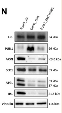
FIGURE 8 Influence of SHBG treatment of lipid metabolism related modulators interplay in SAT (A–N). The relative expression levels of LPL (A), SCD (B), PNPLA2 (C), PLIN1 (D), PPARA (E), FASN (F) in SAT were analyzed by RT-PCR. Protein levels of LPL (G), PLIN1 (H), FASN (I), SCD1 (J), ATGL (K, L) and HSL (M) were analyzed by Western blot ((N), representative membranes). Representative data are shown as mean ± SD.

6). Equisetin inhibits adiposity through AMPK-dependent regulation of brown adipocyte differentiation. Heliyon, 2024 (PubMed: 38327434) [IF=4.0]

Restrictive clause


Affinity Biosciences tests all products strictly. Citations are provided as a resource for additional applications that have not been validated by Affinity Biosciences. Please choose the appropriate format for each application and consult Materials and Methods sections for additional details about the use of any product in these publications.

For Research Use Only.
Not for use in diagnostic or therapeutic procedures. Not for resale. Not for distribution without written consent. Affinity Biosciences will not be held responsible for patent infringement or other violations that may occur with the use of our products. Affinity Biosciences, Affinity Biosciences Logo and all other trademarks are the property of Affinity Biosciences LTD.