Product: Raptor Antibody
Catalog: DF7527
Description: Rabbit polyclonal antibody to Raptor
Application: WB IHC IF/ICC
Reactivity: Human, Mouse, Rat
Prediction: Pig, Zebrafish, Bovine, Horse, Sheep, Dog, Chicken
Mol.Wt.: 149kDa; 149kD(Calculated).
Uniprot: Q8N122
RRID: AB_2841026

View similar products>>

   Size Price Inventory
 100ul $280 In stock
 200ul $350 In stock

Lead Time: Same day delivery

For pricing and ordering contact:
Local distributors

Product Info

Source:
Rabbit
Application:
WB 1:1000-3000, IHC 1:50-1:200, IF/ICC 1:100-1:500
*The optimal dilutions should be determined by the end user.
*Tips:

WB: For western blot detection of denatured protein samples. IHC: For immunohistochemical detection of paraffin sections (IHC-p) or frozen sections (IHC-f) of tissue samples. IF/ICC: For immunofluorescence detection of cell samples. ELISA(peptide): For ELISA detection of antigenic peptide.

Reactivity:
Human,Mouse,Rat
Prediction:
Pig(100%), Zebrafish(94%), Bovine(100%), Horse(100%), Sheep(100%), Dog(100%), Chicken(100%)
Clonality:
Polyclonal
Specificity:
Raptor Antibody detects endogenous levels of total Raptor.
RRID:
AB_2841026
Cite Format: Affinity Biosciences Cat# DF7527, RRID:AB_2841026.
Conjugate:
Unconjugated.
Purification:
The antiserum was purified by peptide affinity chromatography using SulfoLink™ Coupling Resin (Thermo Fisher Scientific).
Storage:
Rabbit IgG in phosphate buffered saline , pH 7.4, 150mM NaCl, 0.02% sodium azide and 50% glycerol. Store at -20 °C. Stable for 12 months from date of receipt.
Alias:

Fold/Unfold

KIAA1303; KOG1; Mip1; P150 target of rapamycin (TOR) scaffold protein; p150 target of rapamycin (TOR) scaffold protein containing WD repeats; P150 target of rapamycin (TOR)-scaffold protein; Raptor; Regulatory associated protein of mTOR; Regulatory associated protein of MTOR complex 1; Regulatory-associated protein of mTOR; RPTOR; RPTOR_HUMAN;

Immunogens

Immunogen:
Uniprot:
Gene(ID):
Expression:
Q8N122 RPTOR_HUMAN:

Highly expressed in skeletal muscle, and in a lesser extent in brain, lung, small intestine, kidney and placenta. Isoform 3 is widely expressed, with highest levels in nasal mucosa and pituitary and lowest in spleen.

Sequence:
MESEMLQSPLLGLGEEDEADLTDWNLPLAFMKKRHCEKIEGSKSLAQSWRMKDRMKTVSVALVLCLNVGVDPPDVVKTTPCARLECWIDPLSMGPQKALETIGANLQKQYENWQPRARYKQSLDPTVDEVKKLCTSLRRNAKEERVLFHYNGHGVPRPTVNGEVWVFNKNYTQYIPLSIYDLQTWMGSPSIFVYDCSNAGLIVKSFKQFALQREQELEVAAINPNHPLAQMPLPPSMKNCIQLAACEATELLPMIPDLPADLFTSCLTTPIKIALRWFCMQKCVSLVPGVTLDLIEKIPGRLNDRRTPLGELNWIFTAITDTIAWNVLPRDLFQKLFRQDLLVASLFRNFLLAERIMRSYNCTPVSSPRLPPTYMHAMWQAWDLAVDICLSQLPTIIEEGTAFRHSPFFAEQLTAFQVWLTMGVENRNPPEQLPIVLQVLLSQVHRLRALDLLGRFLDLGPWAVSLALSVGIFPYVLKLLQSSARELRPLLVFIWAKILAVDSSCQADLVKDNGHKYFLSVLADPYMPAEHRTMTAFILAVIVNSYHTGQEACLQGNLIAICLEQLNDPHPLLRQWVAICLGRIWQNFDSARWCGVRDSAHEKLYSLLSDPIPEVRCAAVFALGTFVGNSAERTDHSTTIDHNVAMMLAQLVSDGSPMVRKELVVALSHLVVQYESNFCTVALQFIEEEKNYALPSPATTEGGSLTPVRDSPCTPRLRSVSSYGNIRAVATARSLNKSLQNLSLTEESGGAVAFSPGNLSTSSSASSTLGSPENEEHILSFETIDKMRRASSYSSLNSLIGVSFNSVYTQIWRVLLHLAADPYPEVSDVAMKVLNSIAYKATVNARPQRVLDTSSLTQSAPASPTNKGVHIHQAGGSPPASSTSSSSLTNDVAKQPVSRDLPSGRPGTTGPAGAQYTPHSHQFPRTRKMFDKGPEQTADDADDAAGHKSFISATVQTGFCDWSARYFAQPVMKIPEEHDLESQIRKEREWRFLRNSRVRRQAQQVIQKGITRLDDQIFLNRNPGVPSVVKFHPFTPCIAVADKDSICFWDWEKGEKLDYFHNGNPRYTRVTAMEYLNGQDCSLLLTATDDGAIRVWKNFADLEKNPEMVTAWQGLSDMLPTTRGAGMVVDWEQETGLLMSSGDVRIVRIWDTDREMKVQDIPTGADSCVTSLSCDSHRSLIVAGLGDGSIRVYDRRMALSECRVMTYREHTAWVVKASLQKRPDGHIVSVSVNGDVRIFDPRMPESVNVLQIVKGLTALDIHPQADLIACGSVNQFTAIYNSSGELINNIKYYDGFMGQRVGAISCLAFHPHWPHLAVGSNDYYISVYSVEKRVR

Predictions

Predictions:

Score>80(red) has high confidence and is suggested to be used for WB detection. *The prediction model is mainly based on the alignment of immunogen sequences, the results are for reference only, not as the basis of quality assurance.

Species
Results
Score
Pig
100
Horse
100
Bovine
100
Sheep
100
Dog
100
Chicken
100
Zebrafish
94
Xenopus
0
Rabbit
0
Model Confidence:
High(score>80) Medium(80>score>50) Low(score<50) No confidence

PTMs - Q8N122 As Substrate

Site PTM Type Enzyme
S8 Phosphorylation P28482 (MAPK1) , P27361 (MAPK3)
K43 Ubiquitination
S48 Phosphorylation
K97 Ubiquitination
K108 Ubiquitination
K120 Ubiquitination
K131 Ubiquitination
K132 Ubiquitination
T159 Phosphorylation
K207 Ubiquitination
K297 Ubiquitination
K335 Ubiquitination
K603 Ubiquitination
Y692 Phosphorylation
S696 Phosphorylation P45983 (MAPK8) , P27361 (MAPK3) , P28482 (MAPK1) , P06493 (CDK1)
T699 Phosphorylation
T700 Phosphorylation
S704 Phosphorylation
T706 Phosphorylation P06493 (CDK1) , P45983 (MAPK8)
S711 Phosphorylation
T714 Phosphorylation
R718 Methylation
S719 Phosphorylation P51812 (RPS6KA3) , Q15418 (RPS6KA1)
S721 Phosphorylation P51812 (RPS6KA3) , Q15418 (RPS6KA1) , Q9UIK4 (DAPK2)
S722 Phosphorylation P51812 (RPS6KA3) , Q15418 (RPS6KA1) , P49841 (GSK3B) , Q13131 (PRKAA1)
Y723 Phosphorylation
S738 Phosphorylation
S771 Phosphorylation Q15759 (MAPK11)
S792 Phosphorylation O75385 (ULK1) , P49841 (GSK3B) , Q13131 (PRKAA1)
S795 Phosphorylation
S836 Phosphorylation
K840 Ubiquitination
T853 Phosphorylation
S854 Phosphorylation
S855 Phosphorylation O75385 (ULK1) , P42345 (MTOR)
T857 Phosphorylation
S859 Phosphorylation O75385 (ULK1) , P42345 (MTOR) , P49841 (GSK3B)
S863 Phosphorylation P54646 (PRKAA2) , P42345 (MTOR) , Q15759 (MAPK11) , P27361 (MAPK3) , P49841 (GSK3B) , P28482 (MAPK1) , P45983 (MAPK8) , Q9UBE8 (NLK)
T865 Phosphorylation
K867 Ubiquitination
S877 Phosphorylation P49841 (GSK3B)
S881 Phosphorylation
S882 Phosphorylation
T883 Phosphorylation
S884 Phosphorylation
S886 Phosphorylation
S887 Phosphorylation
T889 Phosphorylation
K894 Ubiquitination
T908 Phosphorylation Q9UPZ9 (ICK)
Y916 Phosphorylation
T917 Phosphorylation
K973 Ubiquitination
S982 Phosphorylation
K1008 Ubiquitination
K1030 Ubiquitination
K1053 Ubiquitination
K1056 Ubiquitination
K1097 Ubiquitination
K1157 Ubiquitination
S1189 Phosphorylation
Y1193 Phosphorylation
K1221 Ubiquitination

Research Backgrounds

Function:

Involved in the control of the mammalian target of rapamycin complex 1 (mTORC1) activity which regulates cell growth and survival, and autophagy in response to nutrient and hormonal signals; functions as a scaffold for recruiting mTORC1 substrates. mTORC1 is activated in response to growth factors or amino acids. Growth factor-stimulated mTORC1 activation involves a AKT1-mediated phosphorylation of TSC1-TSC2, which leads to the activation of the RHEB GTPase that potently activates the protein kinase activity of mTORC1. Amino acid-signaling to mTORC1 requires its relocalization to the lysosomes mediated by the Ragulator complex and the Rag GTPases. Activated mTORC1 up-regulates protein synthesis by phosphorylating key regulators of mRNA translation and ribosome synthesis. mTORC1 phosphorylates EIF4EBP1 and releases it from inhibiting the elongation initiation factor 4E (eiF4E). mTORC1 phosphorylates and activates S6K1 at 'Thr-389', which then promotes protein synthesis by phosphorylating PDCD4 and targeting it for degradation. Involved in ciliogenesis.

PTMs:

Insulin-stimulated phosphorylation at Ser-863 by MTOR and MAPK8 up-regulates mTORC1 activity. Osmotic stress also induces phosphorylation at Ser-696, Thr-706 and Ser-863 by MAPK8. Ser-863 phosphorylation is required for phosphorylation at Ser-855 and Ser-859. In response to nutrient limitation, phosphorylated by AMPK; phosphorylation promotes interaction with 14-3-3 proteins, leading to negative regulation of the mTORC1 complex. In response to growth factors, phosphorylated at Ser-719, Ser-721 and Ser-722 by RPS6KA1, which stimulates mTORC1 activity.

Subcellular Location:

Cytoplasm. Lysosome. Cytoplasmic granule.
Note: Targeting to lysosomes depends on amino acid availability. In arsenite-stressed cells, accumulates in stress granules when associated with SPAG5 and association with lysosomes is drastically decreased.

Extracellular region or secreted Cytosol Plasma membrane Cytoskeleton Lysosome Endosome Peroxisome ER Golgi apparatus Nucleus Mitochondrion Manual annotation Automatic computational assertionSubcellular location
Tissue Specificity:

Highly expressed in skeletal muscle, and in a lesser extent in brain, lung, small intestine, kidney and placenta. Isoform 3 is widely expressed, with highest levels in nasal mucosa and pituitary and lowest in spleen.

Subunit Structure:

Part of the mammalian target of rapamycin complex 1 (mTORC1) which contains MTOR, MLST8, RPTOR, AKT1S1/PRAS40 and DEPTOR. mTORC1 binds to and is inhibited by FKBP12-rapamycin. Binds directly to 4EBP1 and RPS6KB1 independently of its association with MTOR. Binds preferentially to poorly or non-phosphorylated forms of EIF4EBP1, and this binding is critical to the ability of MTOR to catalyze phosphorylation. Forms a complex with MTOR under both leucine-rich and -poor conditions. Interacts with ULK1 in a nutrient-dependent manner; the interaction is reduced during starvation. Interacts (when phosphorylated by AMPK) with 14-3-3 protein, leading to inhibition of its activity. Interacts with SPAG5; SPAG5 competes with MTOR for RPTOR-binding, resulting in decreased mTORC1 formation. Interacts with WAC; WAC positively regulates MTOR activity by promoting the assembly of the TTT complex composed of TELO2, TTI1 and TTI2 and the RUVBL complex composed of RUVBL1 and RUVBL2 into the TTT-RUVBL complex which leads to the dimerization of the mTORC1 complex and its subsequent activation. Interacts with G3BP1. The complex formed with G3BP1 AND SPAG5 is increased by oxidative stress. Interacts with HTR6. Interacts with PIH1D1. Interacts with LARP1. Interacts with BRAT1. Interacts with SIK3.

(Microbial infection) Interacts with vaccinia virus protein F17; this interaction dysregulates mTOR.

Family&Domains:

Belongs to the WD repeat RAPTOR family.

Research Fields

· Cellular Processes > Transport and catabolism > Autophagy - other.   (View pathway)

· Cellular Processes > Transport and catabolism > Autophagy - animal.   (View pathway)

· Environmental Information Processing > Signal transduction > mTOR signaling pathway.   (View pathway)

· Environmental Information Processing > Signal transduction > PI3K-Akt signaling pathway.   (View pathway)

· Environmental Information Processing > Signal transduction > AMPK signaling pathway.   (View pathway)

· Human Diseases > Cancers: Overview > MicroRNAs in cancer.

· Organismal Systems > Aging > Longevity regulating pathway.   (View pathway)

· Organismal Systems > Aging > Longevity regulating pathway - multiple species.   (View pathway)

· Organismal Systems > Endocrine system > Insulin signaling pathway.   (View pathway)

References

1). Dioscin ameliorates murine ulcerative colitis by regulating macrophage polarization. Pharmacological research, 2021 (PubMed: 34343656) [IF=9.3]

2). Potential role of mTORC1 and the PI3K-Akt pathway in anti-acne properties of licorice flavonoids. Journal of Functional Foods, 2020 [IF=5.6]

Application: WB    Species: Rat    Sample:

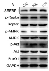
Fig. 6. Validation of the proteins in the regulation of the PI38-Akt signaling pathway and mitochondrial activity. A) Representative Western blotting images of SREBP-1, p-Raptor, Raptor, p-AMPK, AMPK, p-Akt, Akt, FoxO1. B) Relative expression of srebp-1 **P < 0.01. C) Relative expression of mTORC1. D) PI3K-Akt signaling pathway. E) Grey value analyses of SREBP-1, p-Raptor, p-AMPK, p-Akt, and FoxO1 *P < 0.05, n = 3 rats per group. F) Directionality of the changes in mTORC1 and PI3K-Akt signaling pathway, frame with red background: proteins up-regulated in MDL, frame with claybank background: proteins down-regulated in MDL, green arrows: proteins regulated by LCF. (For interpretation of the references to color in this figure legend, the reader is referred to the web version of this article.)

3). Inhibition of mammalian target of rapamycin complex 1 in the brain microvascular endothelium ameliorates diabetic Aβ brain deposition and cognitive impairment via the sterol-regulatory element-binding protein 1/lipoprotein receptor-associated protein 1 signaling pathway. CNS Neuroscience & Therapeutics, 2023 (PubMed: 36890627) [IF=5.5]

Application: WB    Species: Mouse    Sample:

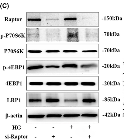
FIGURE 2 mTORC1 impairs Aβ efflux in HBMECs under high-glucose conditions by downregulating LRP1 expression. Cultured HBMECs pretreated with or without rapamycin (20 nmol/L) were stimulated with 50 mM glucose. After 72 h, (A) the efflux rate of Aβ was evaluated in a Transwell in vitro blood–brain barrier model. (B) The protein levels of p-P70S6K, P70S6K, p-4EBP1, 4EBP1, and LRP1 in whole cell lysates were detected by Western blotting (left). The bar graph shows the quantification of all target proteins (right). HBMECs were transfected with Raptor siRNA (si-Raptor) and the corresponding negative control (Ctrl.), followed by 50 mM glucose stimulation for 72 h, and then lysis. (C) Western blotting with Raptor, p-P70S6K, P70S6K, p-4EBP1, 4EBP1, and LRP1 antibodies was performed, and (D) relative mRNA expressions were detected by quantitative reverse transcription–polymerase chain reaction (qRT-PCR). mRNA expression was normalized to β-actin. All data were obtained from three independent experiments.

4). Rapamycin Supplementation May Ameliorate Erectile Function in Rats With Streptozotocin-Induced Type 1 Diabetes by Inducing Autophagy and Inhibiting Apoptosis, Endothelial Dysfunction, and Corporal Fibrosis. Journal of Sexual Medicine, 2018 (PubMed: 30224017) [IF=3.5]

Application: WB    Species: rat    Sample: corpus cavernosum

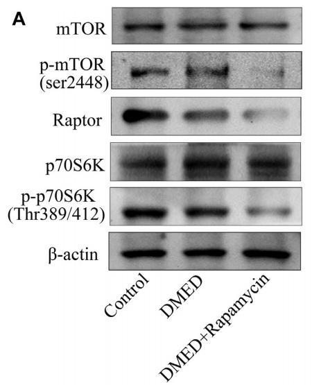
Figure 6. |Effect of rapamycin on the corpus cavernosum (control, n ¼ 6; DMED, n ¼ 6; DMED þ rapamycin, n ¼ 6). A, B, Representative Western blot for mTOR, p-mTOR, p70S6K, p-p70S6K, raptor, rictor, AMPKa, p- AMPKa, Akt, and p-Akt.

Restrictive clause


Affinity Biosciences tests all products strictly. Citations are provided as a resource for additional applications that have not been validated by Affinity Biosciences. Please choose the appropriate format for each application and consult Materials and Methods sections for additional details about the use of any product in these publications.

For Research Use Only.
Not for use in diagnostic or therapeutic procedures. Not for resale. Not for distribution without written consent. Affinity Biosciences will not be held responsible for patent infringement or other violations that may occur with the use of our products. Affinity Biosciences, Affinity Biosciences Logo and all other trademarks are the property of Affinity Biosciences LTD.