, using CD68 Antibody at 1/1000 dilution.
5ug/NC membrane strip.
Exposure for 10s with Affinity™ ECL Kit(#KF8003).
Bands result from membrane strip incubation.")


 and mouse anti-beta tubulin Ab(T0023 1:200) for 1 hour at 37°C. An AlexaFluor594 conjugated goat anti-rabbit IgG(H+L) Ab(Red) and an AlexaFluor488 conjugated goat anti-mouse IgG(H+L) Ab(Green) were used as the secondary antibody.
The nuclear counter stain is DAPI(blue).")
. A, Representative images of IL-33+ cell, CD206+ cell and CD68+ cell in non-tumour and tumour tissue (scale bar = 50 μm).")
 were injected using 25 μl of 200 μM BBR, followed by 25 μl rhSP-D (40 μg/mL) 1 day later. Sham-operated and OA-induction groups received an injection of 50 μl PBS into the right knee joint. For rats receiving rAAV-GFP or rAAV-SP-D shRNA, injections were performed for 10 consecutive days beginning 1 day after the initial BBR injection. At 10 weeks post-surgery, animals were sacrificed and the knee joints were collected for TUNEL and IHC analysis. (A) TUNEL staining (400 ×) was used to assess apoptosis. The p-p65, TLR4, F4/80, CD68, and CD34 expression was assessed via IHC (400 × with higher 800 × magnification inset). (B) Quantification of the indicated proteins in IHC images. Each column represents the mean ± SEM (n = 5). ***P < 0.001 versus the sham-operated group; ##P < 0.01 and ###P < 0.001 versus the OA-induction group; &P < 0.05 and &&P < 0.01 versus the OA + BBR group.")
 HE staining followed by quantitative analysis of proportion of plaque versus media. (B)
Masson staining and quantification of collagen fibrils relative expression in plaque and media. Immunohistochemistry staining for α-SMA (C) and CD68 (D) followed
by quantification in plaque and media. n = 5, Bars = 200 μm or 50 μm. Data are presented as mean ± SD. *P < 0.05, **P < 0.01, ***P < 0.001.")
 Analysis of the carotid arteries by H&E staining (the bar is 100 μm). (B) ELISA assay of plasma AGEs
expression. (C) ELISA for plasma TNF-α in vivo. (D) ELISA for plasma IL-6 levels in vivo. (E) ELISA for plasma IL-10 levels in vivo. (F) qPCR analysis of iNOS expression
in carotid atherosclerotic tissues. (G) qPCR analysis of Arg1 expression in carotid atherosclerotic tissues. (H) Western blot analysis of iNOS and Arg1 expression in
carotid atherosclerotic tissues. β-actin was detected as control. (I) Representative photographs of immunofluorescence staining about the composition of macrophages in carotid artery sections. Scale bars: 100 μm. DAPI, a nuclear marker, in blue; CD68+, a pan-macrophage marker, in green; iNOS, an M1 macrophage marker,
in red and Arg1, an M2 macrophage marker, in red. Sham: right carotid artery of atherosclerotic apoE−/- mice, Injury: left carotid artery of atherosclerotic apoE−/-
mice, STZ: right carotid artery of atherosclerotic apoE−/- mice with diabetes mellitus, Injury + STZ: left carotid artery of atherosclerotic apoE−/- mice with diabetes
mellitus. Data are presented as the mean ± SD. p < 0.01.")
 Mesenteric lymph nodes were harvested from the Sham-operated, SAE, and FMT-challenged SAE rats. Protein expression of CD80, CD68, CD206, and CD163 as determined by WB in the mesenteric lymph nodes from different rats.")
 Immunofluorescence staining confirmation for the appearance of PD-1-positive TAMs. (B) mRNA expression of CD68 showed a correlation with PD-1 expression in the TCGA cohort. (C) mRNA expression of CD68 showed a correlation with PD-L1 expression in the TCGA cohort.")
 Both THP-1 cells and THP-1 derived macrophages expressed PD-1 and CD68. T24 cells expressed PD-L1. (B) A co-IP assay showing the binding between CD68 and PD-1, hinting at the possibility of a CD68 and PD-1 interaction. (C) The binding of CD68 and PD-1 promoted THP-1 derived macrophages to M2 polarization whereas the blockage can reverse the process. (D) Molecular docking showing the interactions of CD68 and PD-1 through possible LAMP-like and IgV domains. SP indicates signal peptide and TM indicates transmembrane domain. (E) The cell viability assays of T24, THP-1 derived macrophages and PBMC co-culture experiments. T24 cell group and THP-1 derived macrophage group were the control groups. ***p < 0.001 (Student’s t-test).")
 by targeting ATP2A2 and CACNA1H. The pregnant mice were injected with 20 μg LPS at P16, and the premature labor rate was calculated as PTL mice number/total mice number in each group (10 valid mice in each group) (a). The expression of miR-25-3p in the myometrium was examined by real-time PCR (b). The expression of CACNA1H and ATP2A2 in the myometrium was examined by real-time PCR (c). The expression of Cav3.2 and SERCA2a in the myometrium was examined by western blot (d). The expression of miR-25-3p in LPS-stimulated HMSMs or HTR-8/SVneo was determined by real-time PCR (e). HMSMs: human myometrial smooth muscle cells.")
/CD68+ PLA2G7+ cells(lower). E AUCell analysis of the relative gene set enrichment scores in Inflammatory response signature. F Scatter plot showing the correlation between M1 and M2 marker gene expression. G, H AUCell analyses of the relative gene set enrichment scores in M1 (G) and M2 (H) macrophage signatures. The t-SNE maps showing AUC scores of selected gene signatures (left); the box plots showing AUC scores of selected gene signatures in each cell type (middle) or sample group (right). I Trajectory analysis of FN1high macrophages, PLA2G7high macrophages, MS4A6Ahigh macrophages and other macrophages using Monocle 2. J STAT1 motif showed greater enrichment of regulon activity for PLA2G7high and MS4A6Ahigh macrophages using SCENIC analysis. K t-SNE visualization of AUC values of STAT1 motif in macrophages. L Schematic developmental trajectories of macrophage subpopulations in FHP lungs. *P < 0.05, **P < 0.01, ***P < 0.001, ****P < 0.0001.")
 The relative mRNA expression of MCP-1, CD86, and CD206 in left ventricular tissues was detected by qRT-PCR. (D and F) The contents of M1 (CD68+, iNOS+) and M2 (CD68+, Arg-1+) macrophages were examined by double-immunofluorescence staining. Scale bars, 50 μm. Arrows point to double-immunofluorescence staining cells. (E and G) The percentage of iNOS-positive CD68 macrophages and the percentage of Arg-1-positive CD68 macrophages (six mice/group, three sections/mouse). The data are expressed as means ± SD (n = 6/group). ***, P < 0.001; **, P < 0.01; *, P < 0.05 versus control mice. †††, P < 0.001; ††, P < 0.01; †, P < 0.05 versus DM mice. ###, P < 0.001 versus GVDM + shNC mice. Arg-1, arginase 1; DAPI, 4',6-diamidino-2-phenylindole; iNOS, inducible nitric-oxide synthase.")
 Representative sections of AT from rats intragastrically administered with 0.5% sodium carboxymethylcellulose (n = 5) or UA (n = 6). Tendon tissues showed remarkable differences in the morphology by hematoxylin and eosin staining ((a), left) and in the collagen expression by Masson's trichrome (MT) staining ((a), middle) and Sirius red staining ((a), right). Bars = 50 μm. (b, c) Representative sections were stained for CD3 (b) and CD68 (c) in red, and nuclei were stained with Hoechst 33258 (blue). Bars = 100 μm. (d–g) Comparison of mechanical testing results of the rat AT tissue postinduction of UA (n = 6) or the vehicle control (n = 7) in the failure load (d), the ultimate stress level (e), the stiffness (f), and the levels of Young's modulus (g). Values are the mean ± SEM of average data from 2 independent experiments. ∗P < 0.05; ∗∗P < 0.01 versus vehicle alone.")
, consuming microplastics (P) with acute DSS-induced colitis (D) and with acute colitis against the background of microplastic consumption (PD), stained with alcian blue at pH 1.0 (highly sulfated mucins), the PAS reaction (neutral mucins), immunofluorescent staining with antibodies to CD68 (macrophages) and chromogranin A (endocrine cells). Photographs of sections with immunofluorescent staining are discolored and inverted for better contrast.")
 immunofluorescence staining (original magnification, × 40, bar = 50 μm) and quantitative analysis of macrophage accumulation and macrophage polarization by detection of (a) the macrophage marker cluster of differentiation (CD)68 and M1 marker inducible nitric oxide synthase (iNOS), and (b) the M2 markers CD206 and arginase (Arg)-1, in aortic sections from MiR-22-3p agomir or normal control (NC) mice; and (c) western blot and quantitative analysis of relative protein levels of the M1 markers (iNOS, interleukin [IL]-6, and tumour necrosis factor [TNF]-α) and M2 markers (Arg1 and CD206) in aortic sections from MiR-22-3p agomir or NC mice.")
 Nissl staining of mice’ prefrontal cortex (Thin arrows, normal neurons; Arrowheads, damaged neurons). Scale bar, 100 µm; Original magnification, 200 ×. (B) Immunofluorescence staining of Iba-1 and CD68 in mice’ prefrontal cortex (Thin arrows, the colocalization of Iba-1 and CD68 in microglia; Hollow arrowheads, CD68-positive macrophages). Cell nucleus was stained with DAPI. Scale bar, 50 µm; Original magnification, 400×. (C) Western blot analysis of Iba-1 expression in mice’ prefrontal cortex. ns, no significance;")
 Oil Red O staining of HVSMCs in vitro. (B) Immunofluorescence assay to determine the levels of α-SMA and CD68 in vitro. (C) Relative protein expression of KLF4 in vitro. (D) Immunofluorescence for α-SMA and CD68 expression in the aorta. (E) Immunohistochemistry for KLF4 expression in the aorta. ***P")
 reduced macrophage infiltration in liver cancer, and inhibited lung metastasis. The rats were treated with JDF 3 days after resection for 2 weeks. JDF-L: 12.74 g/kg/day, JDF-M: 38.22 g/kg/day, and JDF-H: 63.7 g/kg/day. A. The results of H&E staining of lung tissue; scale bar: 200 μm; 400 μm. B. Immunofluorescence double-labeling of CD206 and CD68 in liver cancer tissue, scale bar: 130 μm; **P < 0.01 vs. Saline.")
. (a) HE staining of the Achilles tendon tissues. (b) Masson staining of the Achilles tendon tissues. Representative images of immunofluorescence staining of (c) CD68, (d) p-mTOR, (e) p-p70S6K, and (f) Collagen I in Achilles tendon tissues in three groups of animals were shown.")
 Western blotting was utilized to assess the protein expression levels of CD68, α-SMA, and MMP-9 in VSMCs following AIF-1 knockdown. (C, D) Western blotting was used to evaluate the protein expression levels of CD68, α-SMA, and MMP-9 in VSMCs that overexpress AIF-1. (E, G) Fluorescence photomicrographs display CD68 (red), α-SMA (green), and DAPI (blue) staining in VSMCs with AIF-1 knockdown. (F, H) Fluorescence photomicrographs illustrate the staining of CD68 (red), α-SMA (green), and DAPI (blue) in VSMCs that overexpress AIF-1. (I, J) Fluorescence photomicrographs show the staining of CD68 (red), α-SMA (green), and DAPI (blue) in atherosclerotic plaques of ApoE−/− mice fed a high-fat diet (HFD). Each experiment was conducted with three independent replicates. ∗P < 0.05, ∗∗P < 0.01, ∗∗∗P < 0.001. (For interpretation of the references to color in this figure legend, the reader is referred to the Web version of this article.)")
 Body weight statistics of mice at 0–12 weeks. (B,C) Mice thoracic and abdominal aortic tissue stained with oil red O. (D-G) Mouse Lipid Panel (T-CHO, TG, HDL-C, LDL-C). (H,I) Immunofluorescence staining of partial aortic arch in mice (LY86 and HMCGR). (J) The immunofluorescence co-localization of LY86 and CD68 in partial aortic arch of mice. The findings were presented as mean ± SD (n = 3); *P")
 CD68 expression (n Comparison of lung tissue HE staining (n = 3). (B) Immunohistochemical detection of the = 3). (C) Comparison of i—NOS and CD68 protein expression in the lung tissues by dual immunofluorescence staining (Control: normal control group; LPS group; LPS+EXO group: LPS + alveolar epithelial cell exosomes).")
 CD68 expression (n Comparison of lung tissue HE staining (n = 3). (B) Immunohistochemical detection of the = 3). (C) Comparison of i—NOS and CD68 protein expression in the lung tissues by dual immunofluorescence staining (Control: normal control group; LPS group; LPS+EXO group: LPS + alveolar epithelial cell exosomes).")
 exacerbated atherosclerotic plaque progression and pyroptosis in ApoE−/− mice. ApoE−/− mice treated with adeno-associated virus serotype 9 (AAV9)–mediated overexpression of HCC-1 (AAV9-HCC-1 mice) and the controls (AAV9-control mice) were fed with high-fat diet for 12 weeks. A, Oil red O staining of aorta en face in different groups (control, n=5 vs AAV9-HCC-1, n=4; Mann-Whitney U test, P=0.0159, Shapiro-Wilk test [SW] showed no normal distribution). H&E (hematoxylin-eosin staining; B), oil red O (C), Masson trichrome (D), and sirius red (E) staining and quantification of aortic root cross sections (control, n=8 vs AAV9-HCC-1, n=6; unpaired t test, SW showed normal distribution). Anti-LyC6 antibody–stained (F) and anti-CD68 antibody–stained (G) aortic root cross sections and quantification (control, n=8 vs AAV9-HCC-1, n=6; unpaired t test, SW showed normal distribution). Anti-caspase-1 antibody–stained (H) and TUNEL (terminal deoxynucleotidyl transferase [TdT]-mediated dUTP nick-end labeling)-stained (I) aortic root cross (Continued )sections and quantification (control, n=8 vs AAV9-HCC-1, n=6; unpaired t test, SW showed normal distribution). Anti-NF-κB (nuclear factor-κB) antibody–stained (J) and anti-NLRP3 (NLR family pyrin domain containing 3) antibody–stained (K) aortic root cross sections and quantification (control, n=8 vs AAV9-HCC-1, n=6; unpaired t test, SW showed normal distribution). Serum levels of TNF-α (tumor necrosis factor-α; L) and IL (interleukin)-1β (M) in mice (control, n=8 vs AAV9-HCC-1, n=6; unpaired t test, SW showed normal distribution). Data were exhibited as mean±SD. Scale bar=100 µm")
 Rats were injected intra-articularly with rAAV encoding SP-D-specific shRNA for 10 consecutive days and sacrificed at the fourth week of the first injection. H&E staining and immunohistochemical staining of SP-D, CD68, F4/80, and TLR4 protein levels in cartilage were assessed following rAAV-GFP or rAAV-SP-D shRNA injection into the rat knee joints. The ratios of immunoreactive cells of SP-D (B), CD68 (C), F4/80 (D), and TLR4 (E) were analyzed. Data were expressed as mean ± SEM (n = 5). **P < 0.01 and ***P < 0.001 vs. rAAV-GFP group.")
, n = 6 for (A–C, E).")
 Representative immunoblot and relative quantification of TLR4 and MyD88 in RAW264.7 cells. (D,E) Representative immunofluorescence images of TLR4-positive and MyD88-positive in RAW264.7 cells. Scale bars: 25 μm. (F–H) Representative immunoblot and relative quantification of p-IκBα (S32/S36), IκBα, p-p65 (S536), and p65 in RAW264.7 cells. (I) Representative immunofluorescence images of NF-κB p65 in RAW264.7 cells. Scale bars: 100 μm. (J–N) Representative immunoblot and relative quantification of CD68, MCP-1, ICAM1, and VCAM1 in RAW264.7 cells. (O–R) mRNA levels of Ccl2, Ccl3, Ccl4, and Cxcl10 in RAW264.7 cells. Data are presented as mean ± SEM (n = 3). * p < 0.05, ** p < 0.01, *** p < 0.001, and **** p < 0.0001 vs. the Con group. # p < 0.05, ## p < 0.01, ### p < 0.001, and #### p < 0.0001 vs. the LPS group.")
 The protein expression of α7nAchR, CD68, iNOS, p-JAK2, and p-STAT in M1/M2 cells of each group was detected by Western blot. (A-G) M1 macrophages. (H-N) M2 macrophages. * indicates P < 0.05 compared with M1/M2 group; # indicates P < 0.05 compared with M1/M2+si-NC+FMN group.")
 Representative images of immunofluorescence staining of CD68 in rat intestinal tissues. Scale bar: 100 μm. (B) Determination of CD68-positive cells (%) based on staining results in (A). (C–E) ELISA measurements of IL-1β, IL-6, and TNF-α levels in intestinal tissue homogenates of rats. (F–L) RT-qPCR analysis of IL-1β and IL-6 mRNA levels in rat intestinal tissues. N = 8. ***p")
 Schematic timeline of the experimental protocol. Days 0, 14, 28, and 42 represent intraperitoneal (i.p.) injections of ovalbumin (OVA). Days 21–54 represent exposure to OVA aerosol. Aerobic exercise adaptation occurred from days 21–23, and days 25 and 53 represent the initial and final physical test. AE was initiated on day 28 and ended on day 52. Euthanasia was performed on day 56. The C and E groups were replaced with normal saline. (B) Representative immunofluorescence images of CitH3, MPO, and CD68 staining of lung tissues (n = 6). (C) The protein expression of CitH3 and MPO was detected by Western blot analysis (n = 6). C: control group; E: exercise group; A: asthma group; AE: asthma + exercise. Glyceraldehyde-3-phosphate dehydrogenase (GAPDH) was used as a loading control for all Western blot assays. Data are expressed as means ± standard deviation (SD). ∗P < 0.05.")
 The schematic timeline for experimental design. (B) The immunofluorescence staining of Iba1 in the hippocampus and the statitical analysis of number of Iba1+ cells/105 μm2. n = 5 mice for each group. (C) Example trajectories in NORT and the statistical analysis of DI, n = 10 mice for each group. (D) Statistical analysis of the rate of alteration in YMT in different groups, n = 10 mice for each group. (E) Expressions of PSD95, SYN, and CD68 in different groups, n = 3 mice for each group. Unpaired t-test was applied for all comparisons. *p < 0.05, **p < 0.01, ***p < 0.001.")
 The mRNA level of LILRB2 was upregulated in the atherosclerotic artery tissues (n = 7) with enhanced LILRB2 expression. (B) The protein level of LILRB2 was upregulated in the atherosclerotic artery tissues with enhanced LILRB2 expression. (C) The levels of METs were determined using CD68, CitH3, and MPO immunostaining. MET formation was upregulated in the atherosclerotic artery tissues (n = 3) with enhanced LILRB2 expression. (D) The PI3K/AKT signaling pathway was activated in the atherosclerotic artery tissues with enhanced LILRB2 expression. **p < 0.01, compared with the LILRB2-low group. LILRB2, leukocyte immunoglobulin-like receptor B2; MET, macrophage extracellular trap; MPO, myeloperoxidase.")
 but applied to ATC. C Differences of number and strength overall information flow within the inferred networks between PTC and ATC. D The potential outgoing and incoming interaction strength of each cellular cluster in PTC and ATC. Heatmap showing the relative signaling contribution of each cell group based on the number and strength compared PTC with ATC. E Heatmap showing the relative signaling contribution of each cell group based on the number and strength compared PTC with ATC. F Differential strength of network centrality based on APOE+ macrophages (PTC vs. ATC). G Bubble heatmap showing the cell–cell communication of selected ligand–receptor pairs between APOE+ macrophage and CD8+ PDCD1+ T cells. Dot size indicates P value, colored by communication probability. H Multiplex IHC shows the cross-talk between APOE+ macrophages and T cells via the ligand–receptor of THBS1-CD47.")
 Cells were incubated with stigmasterol (50 and 100 μM) for 6 h, then co-incubated with ox-LDL for 24 h. Morphologic change of foam cells stained by ORO and quantitation of lipid drops by extracting ORO dyes from the stained SMCs (magnification ×200). The scale bar in the figure is 50 μm. n = 5. (B) Cells were loaded with NBD-cholesterol (5 μg/mL) (green) and stigmasterol (50 or 100 μM) for 12 h. Then incubated for 4 h with 0.2% BSA or HDL (50 μg/mL). HDL-mediated cholesterol efflux (%) was measured by using fluorescence microplate reader. n = 5. (C) Representative fluorescence image of SMCs cultured with NBD-cholesterol (green) and stigmasterol after 4 h of HDL incubation (magnification ×400). The scale bar in the figure is 25 μm. (D) Cells were pretreated with stigmasterol for 6 h, then incubated with 10 μg/mL Dil-ox-LDL and stigmasterol for an additional 4 h. Dil-LDL (red) fluorescence represents Dil-ox-LDL particles while blue fluorescence indicates nucleus (magnification ×200). The scale bar in the figure is 50 μm. (E) Expression of SRA, CD36, ABCA1, and ABCG1 after ox-LDL or stigmasterol treatment was evaluated by western blot. (F) Quantification of SRA, CD36, ABCA1, and ABCG1. n = 3. (G) Immunofluorescent staining with CD68 (red) and ACTA2 (green) antibodies in SMCs after stigmasterol treatment (magnification ×200). The scale bar in the figure is 50 μm. n = 3. (H) CD68 and α-SMA in SMCs were evaluated by Western blot after stigmasterol treatment. (I) Quantification of CD68 and α-SMA. n = 3. Multi-group comparison was measured by one-way ANOVA. The data are presented as the mean ± SD. *p")
 Cells were incubated with stigmasterol (50 and 100 μM) for 6 h, then co-incubated with ox-LDL for 24 h. Morphologic change of foam cells stained by ORO and quantitation of lipid drops by extracting ORO dyes from the stained SMCs (magnification ×200). The scale bar in the figure is 50 μm. n = 5. (B) Cells were loaded with NBD-cholesterol (5 μg/mL) (green) and stigmasterol (50 or 100 μM) for 12 h. Then incubated for 4 h with 0.2% BSA or HDL (50 μg/mL). HDL-mediated cholesterol efflux (%) was measured by using fluorescence microplate reader. n = 5. (C) Representative fluorescence image of SMCs cultured with NBD-cholesterol (green) and stigmasterol after 4 h of HDL incubation (magnification ×400). The scale bar in the figure is 25 μm. (D) Cells were pretreated with stigmasterol for 6 h, then incubated with 10 μg/mL Dil-ox-LDL and stigmasterol for an additional 4 h. Dil-LDL (red) fluorescence represents Dil-ox-LDL particles while blue fluorescence indicates nucleus (magnification ×200). The scale bar in the figure is 50 μm. (E) Expression of SRA, CD36, ABCA1, and ABCG1 after ox-LDL or stigmasterol treatment was evaluated by western blot. (F) Quantification of SRA, CD36, ABCA1, and ABCG1. n = 3. (G) Immunofluorescent staining with CD68 (red) and ACTA2 (green) antibodies in SMCs after stigmasterol treatment (magnification ×200). The scale bar in the figure is 50 μm. n = 3. (H) CD68 and α-SMA in SMCs were evaluated by Western blot after stigmasterol treatment. (I) Quantification of CD68 and α-SMA. n = 3. Multi-group comparison was measured by one-way ANOVA. The data are presented as the mean ± SD. *p")
| Product: | CD68 Antibody |
| Catalog: | DF7518 |
| Description: | Rabbit polyclonal antibody to CD68 |
| Application: | WB IHC IF/ICC |
| Cited expt.: | WB, IHC, IF/ICC |
| Reactivity: | Human, Mouse, Rat |
| Prediction: | Bovine, Horse, Sheep, Rabbit, Dog |
| Mol.Wt.: | 37kD,70-140kD(glycosylated)(Observed); 37kD(Calculated). |
| Uniprot: | P34810 |
| RRID: | AB_2841017 |
Control Products

Related Downloads
Protocols
Product Info
*The optimal dilutions should be determined by the end user. For optimal experimental results, antibody reuse is not recommended.
*Tips:
WB: For western blot detection of denatured protein samples. IHC: For immunohistochemical detection of paraffin sections (IHC-p) or frozen sections (IHC-f) of tissue samples. IF/ICC: For immunofluorescence detection of cell samples. ELISA(peptide): For ELISA detection of antigenic peptide.
Cite Format: Affinity Biosciences Cat# DF7518, RRID:AB_2841017.
Fold/Unfold
CD 68; CD68; CD68 antigen; CD68 molecule; CD68_HUMAN; DKFZp686M18236; gp11; Gp110; LAMP4; Macrophage antigen CD68 (microsialin); MACROPHAGE ANTIGEN CD68; Macrosialin; SCARD1; Scavenger receptor class D member 1;
Immunogens
A synthesized peptide derived from human CD68, corresponding to a region within the internal amino acids.
Highly expressed by blood monocytes and tissue macrophages. Also expressed in lymphocytes, fibroblasts and endothelial cells. Expressed in many tumor cell lines which could allow them to attach to selectins on vascular endothelium, facilitating their dissemination to secondary sites.
- P34810 CD68_HUMAN:
- Protein BLAST With
- NCBI/
- ExPASy/
- Uniprot
MRLAVLFSGALLGLLAAQGTGNDCPHKKSATLLPSFTVTPTVTESTGTTSHRTTKSHKTTTHRTTTTGTTSHGPTTATHNPTTTSHGNVTVHPTSNSTATSQGPSTATHSPATTSHGNATVHPTSNSTATSPGFTSSAHPEPPPPSPSPSPTSKETIGDYTWTNGSQPCVHLQAQIQIRVMYTTQGGGEAWGISVLNPNKTKVQGSCEGAHPHLLLSFPYGHLSFGFMQDLQQKVVYLSYMAVEYNVSFPHAAQWTFSAQNASLRDLQAPLGQSFSCSNSSIILSPAVHLDLLSLRLQAAQLPHTGVFGQSFSCPSDRSILLPLIIGLILLGLLALVLIAFCIIRRRPSAYQAL
Predictions
Score>80(red) has high confidence and is suggested to be used for WB detection. *The prediction model is mainly based on the alignment of immunogen sequences, the results are for reference only, not as the basis of quality assurance.
High(score>80) Medium(80>score>50) Low(score<50) No confidence
Research Backgrounds
Could play a role in phagocytic activities of tissue macrophages, both in intracellular lysosomal metabolism and extracellular cell-cell and cell-pathogen interactions. Binds to tissue- and organ-specific lectins or selectins, allowing homing of macrophage subsets to particular sites. Rapid recirculation of CD68 from endosomes and lysosomes to the plasma membrane may allow macrophages to crawl over selectin-bearing substrates or other cells.
N- and O-glycosylated.
Cell membrane>Single-pass type I membrane protein.
Endosome membrane>Single-pass type I membrane protein. Lysosome membrane>Single-pass type I membrane protein.
Highly expressed by blood monocytes and tissue macrophages. Also expressed in lymphocytes, fibroblasts and endothelial cells. Expressed in many tumor cell lines which could allow them to attach to selectins on vascular endothelium, facilitating their dissemination to secondary sites.
Belongs to the LAMP family.
Research Fields
· Cellular Processes > Transport and catabolism > Lysosome. (View pathway)
References
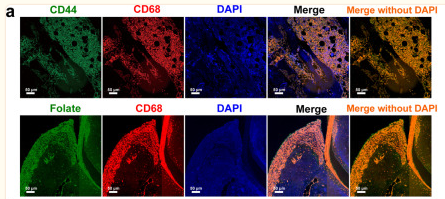
Application: IF/ICC Species: Rat Sample:
Restrictive clause
Affinity Biosciences tests all products strictly. Citations are provided as a resource for additional applications that have not been validated by Affinity Biosciences. Please choose the appropriate format for each application and consult Materials and Methods sections for additional details about the use of any product in these publications.
For Research Use Only.
Not for use in diagnostic or therapeutic procedures. Not for resale. Not for distribution without written consent. Affinity Biosciences will not be held responsible for patent infringement or other violations that may occur with the use of our products. Affinity Biosciences, Affinity Biosciences Logo and all other trademarks are the property of Affinity Biosciences LTD.