


.
Bands result from membrane strip incubation.")
.
Bands result from membrane strip incubation.")
, using Occludin Antibody at 1/1000 dilution.
5ug/NC membrane strip.
Exposure for 10s with Affinity™ ECL Kit(#KF8003).
Bands result from membrane strip incubation.")


 Ab, diluted at 1/600, was used as the secondary antibody.")
 ZO-1 mRNA level, (b) occludin mRNA level, (c) ZO-1 protein level, and (d) occludin protein level. Expression of the selected genes was quantified by quantitative reverse transcription-PCR. Equal loading was assessed by")
 Representative photographs of treated and untreated cells. PTL treatment reduced p-ERK 2, NF-κB, and Snail staining compared with sections obtained from control mice. (B and D) Representative photographs of treated and untreated cells. PTL treatment increased E-cadherin and Occludin staining and reduced Vimentin and N-cadherin staining compared with sections obtained from control mice. Each experiment was performed in triplicate. Results show the means of the three experiments, and the error bars represent standard deviation (*P < 0.05 and **P < 0.01).")
 Representative TEM images in colon tissues (white arrows indicate TJs, black arrows indicate intestinal villi loss).(B) The mRNA level of TJP1 in colon tissues. (C) The mRNA level of OCLN in colon tissues. (D) The fluorescence intensity of ZO-1. (E) The fluorescence intensity of occludin.(F) Representative fluorescent images of ZO-1 and occludin in colon tissues. All data were shown as mean ± SEM. (n = 6 for each group). ⁎P < 0.05. ⁎⁎P < 0.01 compared with control group; #P < 0.05. ##P < 0.01 compared with DSS group.")
 and mucin-2 (B). (C) Representative
Western blotting images of TJs protein, and the relative protein expressions were
normalized to β-actin. (D-H) Changes in the relative protein expression levels of
ZO-1, ZO-2, occludin, JAM-A, and claudin-1 were measured respectively. Data are
shown as mean ± SEM (n = 3). # P < 0.05, ## P < 0.01 vs. Control group, * P < 0.05, **
P < 0.01 vs. DSS group, & P < 0.05, && P < 0.01 vs. BBR group.")
 Representative photographs of treated and untreated cells. PTL treatment reduced p-ERK 2, NF-κB, and Snail staining compared with sections obtained from control mice. (B and D) Representative photographs of treated and untreated cells. PTL treatment increased E-cadherin and Occludin staining and reduced Vimentin and N-cadherin staining compared with sections obtained from control mice. Each experiment was performed in triplicate. Results show the means of the three experiments, and the error bars represent standard deviation (*P < 0.05 and **P < 0.01).")
 and Western blot (C). Representative immunostaining for ZO-1 in the jejunum (original magnification ×200) (D). #P < 0.05 compared to control mice, *P < 0.05 compared to EHEC-treated mice.")
. Knockdown of SUMO1 inhibited the secretion of IL-6,
IL-8 and TNF-α in HTEC cells treated under hypoxic condition for 24 h. ***p < 0.001 vs control, !!p < 0.01 vs siNC. (B). SUMO1 silencing promoted the TEER value in
HTEC cells treated under hypoxic condition for 24 h. ***p < 0.001 vs control, !!!p < 0.001 vs siNC. (C). SUMO-1 silencing reduced the permeability of HTEC cells
treated under hypoxic condition for 24 h. *p < 0.05 vs control, ***p < 0.001 vs siNC; !!!p < 0.001 vs siNC. (D). Western blotting was used to examine the protein
levels of SUMO1, HIF-1α, VEGF, Occluding, Claudin-1 and ZO-1 in cells as indicated. (E). Immunohistochemistry staining assay was performed to determine the
levels of ZO-1, Occluding and Claudin-1 in cells as indicated above.")
 Relative mRNA expression of MLCK, ZO-1, occludin and claudin-1 (n = 4); (e–i) Expressions of MLC,p-MLC, ZO-1, occludin and claudin-1proteins (n = 3). Values were represented the mean ± SEM. **P < 0.01, *P < 0.05 versus 5-FU group and ##P < 0.01 versus normal group.")
 Protein expression levels of claudin‑1, claudin‑2, occludin and ZO‑1 in each group were measured using western blot analysis.")
 VE-cad, p-VE-cad, and Occludin protein levels in HUVECs were determined (n=5).")
. a
p < 0.05 group HI vs. group S. b
p < 0.05 group HI vs. group HL. (n = 5). The data represent the mean ± SEM.")
 urea (TPPU) recovered expression of tight junction proteins down-regulated by permanent cerebral middle artery occlusion (pMCAO). (A) Brains were removed from animals at 1 or 3 days after surgery, and cortical tissue was homogenized and assayed by Western blot against Occludin, ZO-1, and Claudin-5. (B) Quantitation of relative levels of ZO-1 (n = 5). (C) Quantitation of Occludin levels (n = 5). (D) Quantitation of relative levels of Claudin-5 (n = 5). Data are the mean ± SEM. **p < 0.01, ***p < 0.001, ****p < 0.0001.")
. Effect of miR-429 on the permeability of the BSCB via HRP flux. Data represent the mean ± S.D. (n ¼ 3, each). *P < 0.05 vs. agomiR-429 NC group, #P < 0.05 vs.
antagomiR-429 NC group. (B). RT-qPCR analysis of TJ-related protein expression in the BSCB following changes in the expression of miR-429. Data represent the mean ± S.D. (n ¼ 3,
each). *P < 0.05 vs. agomiR-429 NC group, #P < 0.05 vs. antagomiR-429 NC group. (CeD). Western blotting analysis of TJ-related protein expression in the BSCB following changes in
the expression of miR-429. GAPDH was used as an endogenous control. Representative protein expression and their integrated light density values (IDVs) are shown. Data represent
the mean ± S.D. (n ¼ 3, each). *P < 0.05, vs. agomiR-429 NC group, #P < 0.05, vs. antagomiR-429 NC group. (E). Immunofluorescence staining of TJ-related proteins in the different
groups. ZO-1 (red), occludin (red), and claudin-5 (red) are labeled with their respective antibodies. Nuclei (blue) are labeled with DAPI (n ¼ 3). Scale bars represent 20 mm. (For
interpretation of the references to colour in this figure legend, the reader is referred to the Web version of this article.)")
 HE staining of mouse intestinal tissues (scale bar = 100 μm). (B) Western blot analysis of ZO-1 and occludin in the intestine. (C) Protein quantification by western blotting. All data are shown as the mean ± SEM (n = 3 for each group). ## p < 0.01 versus the R1-Ct group and *p < 0.05 versus the P8-Ct group.")
 The relative protein levels of ZO-1 and occludin
in colon tissue. (C and D) The relative protein levels of ZO-1 and occludin in cecum tissue. (E) The levels of serum glucose in mice. (F) The levels of
glucose in the content of colon and cecum in mice. (G and H) The relative protein levels and distribution of ZO-1 and occludin in Caco-2 cells
exposed to 5.56 mmol L−1
, 30 mmol L−1 and 60 mmol L−1 glucose for 72 h. (I and J) The relative protein levels and distribution of ZO-1 and occludin
in Caco-2 cells exposed to 60 mmol L−1 glucose for 0 h, 12 h, 24 h and 72 h. Each bar represents the mean ± SEM for groups of three or six. *P <
0.05, **P < 0.01, compared to db/m mice, or NG as indicated.")
 induced colitis. (a) IL-22 and occludin expression in various groups assessed using western blotting. (b) Relative protein expression of IL-22 and occludin. (n = 3 per group). ∗P < 0.05, DSS group compared with control group; #P < 0.05, CM+DSS group compared with DSS group; &P < 0.05, CM+DSS+aIL-22 group compared with CM+DSS group.")
 Representative western blots and quantification data of TJ and AJ proteins in M, IM groups.
Columns represent mean ± SD, n = 5. (C) Claudin-5, Occludin, p120-Catenin, β-Catenin/CD31/Hoechst staining of sections from the spinal cord in M, IM groups. Red:
Claudin-5/Occludin/p120-Catenin/β-Catenin; green: CD31; blue: Hoechst. Scale bar, 20 μm. (
$
p < 0.05; $$p < 0.01; $$$p < 0.001).")
 and qRT-PCR (B); Expression of Occludin in the colon was measured by immunohistochemistry (C) and qRT-PCR (D); The mRNA expression of inflammatory factors in the intestinal tissues (E); The mRNA expression of ABCG2 in the intestine (F). “ns” represents not significant; “*” represents p < 0.05, “**” represents p < 0.01 and “***” represents p < 0.001. N = 7/group.")
 Immunopositivity (arrows) of HO-1, MPO, occludin, and ZO-1 proteins (A) and the percentage of positive cells (B–E). The results showed that compared with those in the other groups, the expression levels of HO-1 and MPO in the TBI + DM + L-NIO group increased significantly, whereas the expression levels of ZO-1 and occludin decreased significantly. Scale bars: 400 µm and 40 µm (as labeled). Data are expressed as the mean ± SD (n = 6). *P < 0.05 (one-way analysis of variance followed by Bonferroni post hoc test). DM: Diabetes mellitus; HO-1: heme oxygenase-1; L-NIO: N(5)-(1-iminoethyl)-L-ornithine; MPO: myeloperoxidase; TBI: traumatic brain injury; ZO-1: zonula occludens-1.")
 proteins. (A) Expression of claudin-1, Occludin, and ZO-1 mRNA are altered in Caco-2 cells 48 h after treatment with Ts-ML-ES. mRNAs expression of stimulated cells (normalized with GAPDH) relative to unstimulated cells as mean ± SD from three independent experiments. ∗∗∗∗p < 0.0001 by one-way ANOVA with Tukey’s posttest. (B) The effect of Ts-ML-ES on the expression of Claudin-1, Occludin, and ZO-1 in Caco-2 cells. Fully differentiated Caco-2 cells were incubated with 25 μg/ml Ts-ML-ES (48 h). Cells without any treatments are designated as the negative control (NC) and those treated with TNF-α as the positive control (PC). β-actin was used as the internal control. Representative blots of three independent experiments are shown. (C) Effect of apical membrane exposure in control, TNF-α, or Ts-ML-ES treatment of Ts on the location of Claudin-1, Occludin, or ZO-1 in the TJs of Caco-2 cell monolayers. The merged images demonstrate the three TJ proteins in green. DNA was stained with DAPI (blue) to reveal the positions of the nuclei. (D) Transmission electron microscopy (TME) of Caco-2 cells incubated for 48 h with 20 ng/ml TNF-α, and 25 μg/ml Ts-ML-ES. The cells were then fixed with glutaraldehyde in preparation for TEM imaging. Arrowheads: Structure of TJ protein.")
 proteins. (A) Expression of claudin-1, Occludin, and ZO-1 mRNA are altered in Caco-2 cells 48 h after treatment with Ts-ML-ES. mRNAs expression of stimulated cells (normalized with GAPDH) relative to unstimulated cells as mean ± SD from three independent experiments. ∗∗∗∗p < 0.0001 by one-way ANOVA with Tukey’s posttest. (B) The effect of Ts-ML-ES on the expression of Claudin-1, Occludin, and ZO-1 in Caco-2 cells. Fully differentiated Caco-2 cells were incubated with 25 μg/ml Ts-ML-ES (48 h). Cells without any treatments are designated as the negative control (NC) and those treated with TNF-α as the positive control (PC). β-actin was used as the internal control. Representative blots of three independent experiments are shown. (C) Effect of apical membrane exposure in control, TNF-α, or Ts-ML-ES treatment of Ts on the location of Claudin-1, Occludin, or ZO-1 in the TJs of Caco-2 cell monolayers. The merged images demonstrate the three TJ proteins in green. DNA was stained with DAPI (blue) to reveal the positions of the nuclei. (D) Transmission electron microscopy (TME) of Caco-2 cells incubated for 48 h with 20 ng/ml TNF-α, and 25 μg/ml Ts-ML-ES. The cells were then fixed with glutaraldehyde in preparation for TEM imaging. Arrowheads: Structure of TJ protein.")
 The protein expressions of occludin and (B) ZO-1 in the colon were detected by immunoblotting. The results were represented as mean±SEM (n= 6 each group). *P <0.05, **P <0.01, ***P<0.001, compared to HFD + Sed group; #P<0.05,compared to SD + Sed group")
 H&E stain of colon tissues; red arrow was muscular layer, black arrow was inflammation cells, green arrow was serosal layer, blue arrow was mucous layer, yellow arrow was gland hyperplasia; (B) the histology scoring of colon histology; (C) effects of OPA on claudin-1, ZO-1, occludin, and NLRP3 of colon tissue in colitis mice; The integrated optical density (IOD) of claudin-1 (D), ZO-1 (E), occluding (F), and NLRP3 (G) positive cells. The red arrows represent nucleus; the green arrows represent positive cells. Con was control group, M was model group, H was the high dose OPA group, L was low dose OPA group, P was positive group. Data were expressed as the mean ± SD (n = 3), the statistical analyses were done with t-test; ** p < 0.01, * p < 0.05 compared with the model group; ## p < 0.01, # p < 0.05 compared with the control group.")
 Western blot analysis of Hpa-1, Sdc-1, occludin-1 and ZO-1 expression. Densitometric analysis of (B) Hpa-1 and (C) Sdc-1 expression. (D) Expression levels of Sdc-1 in the cell culture supernatant. (E) Densitometric analysis of occludin-1 and (F) ZO-1 expression. Data are presented as the mean ± standard deviation of three repeats. *P<0.05, **P<0.01, ***P<0.001 vs. the control group; #P<0.05, ##P<0.01 vs. HG. AMPK, adenosine monophosphate-activated protein kinase; Hpa-1, heparanase-1; TJ, tight junction; Sdc-1, syndecan-1; ZO-1, Zonula occludens-1; HG, high glucose.")
 AB/
PAS and immunofluorescence imaging
detected the mucin-producing cells and the
expression of mucin-2, respectively. (B)
Analysis of the expression levels of the
mucin-producing cells and mucin-2. (C) The
expressions of occludin, claudin-1,2, and ZO1 protein were verified by western blot. (D)
Analysis of the expression levels of occludin,
claudin-1,2, and ZO-1. Data are expressed as
means ± SEM; n = 5 for each group. * P <
0.05, ** P < 0.01, *** P < 0.001, and ****P
< 0.0001.")
 Comparison of occluding expression in the CLP and FMT groups at 12, 24 add 48 h, respectively, and observed under a digital microscope (20 × 10). Significant results are shown in the box plot on the right, ****P < 0.0001; (B) Relative expression of occludin and ZO-1 compared with β-actin expression in the Sham, CLP, and FMT groups at 24 or 48 h. SB, CB, TB, SD, CD, and TD represent the 24 h Sham, CLP, and FMT groups and 48 h Sham, CLP, and FMT groups, respectively.")
 The expression of
Claudin-1 in sham-operated group, NS+CLP and ZJPs+CLP group by immunohistochemistry (IHC). (d) Relative expression of Claudin-1 in
sham-operated group, NS+CLP and ZJPs+CLP group. (e-g) The expression of Occludin in sham-operated group, NS+CLP and ZJPs+CLP
group by immunohistochemistry (IHC). (h) Relative expression of Occludin in sham-operated group, NS+CLP and ZJPs+CLP group. (i-k)
The expression of ZO-1 in sham-operated group, NS+CLP and ZJPs+CLP group by immunohistochemistry (IHC). (l) Relative expression of
ZO-1 in sham-operated group, NS+CLP and ZJPs+CLP group. (m) Western blot assay for tight junction proteins in sham-operated group,
NS+CLP and ZJPs+CLP group. (n-p) The relative density of tight junction proteins for Claudin-1(N), Occludin(O), ZO-1(P). *
p < .05, **p < .01, ****p < .0001, nsp > .05 versus sham-operated group. #
p < .05, ####p < .0001 versus NS+CLP group")
 LPS in serum and (B) the protein expression of tight junctions in the colon of high-fat diet-induced hyperglycaemia. ND: normal diet; HFD: high-fat diet; HCE: high dosage of crude extract; LPS: lipopolysaccharide. Values represent the mean ± SEM (n = 6). The statistical methods used one-way ANOVA, and the values with different letters are significantly different at p < 0.05.")
 The expressions of occludin, claudin-1,2, and ZO1 protein were verified by western blot.")
, occludin-1 (H), ZO-1 (I) and mucin-2 (J)determined by immunohistochemistry (original magnification 200×) (n = 4 images/group).")
 Western blotting results of ZO1, occludin, and claudin-5 in each group and quantification (n = 6/group, mean ± SD). (b) Brain water content in each group after TBI (n = 6/group, mean ± SD). (c, d) Fluorescence of Evans blue and Evans blue extravasation in each group around the injury site (n = 12/group, mean ± SD) (scale bar = 50 μm, 200x) (∗P < 0.05 vs. the overexpression vector group, &P < 0.05 vs. the knockdown vector group by one-way ANOVA).")
 The protein expressions of occludin and (B) ZO-1 in the colon were detected by immunoblotting. The results were represented as mean±SEM (n= 6 each group). *P <0.05, **P <0.01, ***P<0.001, compared to HFD + Sed group; #P<0.05, compared to SD + Sed group.")
 Effect of PSG on MPO level in the colon of IBD mice. (b) Effect of PSG on DAO level in the colon of IBD mice. The expressions of tight junction protein ZO1 and occludin in the colon tissues of IBD mice were evaluated by Western blot assay (c) and quantitative analysis for Western blot results (n = 3) (d). (e) The expression of MUC2 in the colon tissues of IBD mice was detected by IHC. Results were expressed as the mean ± SEM. ∗p < 0.05 and ∗∗p < 0.01 vs. the control group; #p < 0.05 and ##p < 0.01 vs. the model group (DSS only).")
 The expression of Ki-67, LPS 100 μg/mL, 24 h. (B) The expression of ZO-1, LPS 100 μg/mL, 24 h. (C) The expression of occludins, LPS 100 μg/mL, 24 h. (D) The expression of claudin-1, LPS 100 μg/mL, 24 h. (E) Quantitative analysis of Ki-67 with different concentrations of LPS stimulated for 8 h or 24 h. (F) Quantitative analysis of ZO-1 with different concentrations of LPS stimulated for 8 h or 24 h. (G) Quantitative analysis of occludins with different concentrations of LPS stimulated for 8 h or 24 h. (H) Quantitative analysis of claudin-1 with different concentrations of LPS stimulated for 8 h or 24 h. Data are presented as means ± SD, compared with LPS 0 μg/mL 8 h or 24 h group accordingly using Student's t-tests. ∗ is used for 24 h. SD: Standard deviation; LPS: Lipopolysaccharide; ZO-1: Zonula occludens-1.")
 Representative the occludin, claudin-1 and ZO-1 protein repressive at day 8. CK group administrate with 0.9% saline; PE group administrate with L. paracasei; NC, CIP and TE groups administrate with E. coli. (B,D,F) Representative the occludin, claudin-1 and ZO-1 protein repressive at day 15. CK and NC group administrate with 0.9% saline; CIP group administrate with ciprofloxacin; PE group administrate with E. coli, and TE group administrate with L. paracasei. *Representative compared with CK group. *P < 0.05; **P < 0.01. ▲Representative compared with NC group. ▲▲P < 0.01, ▲▲▲P < 0.001, ▲▲▲▲P < 0.0001 as determined by Student’s t-test.")
 and 15 (B). “M” representative Protein Marker; *Compared with CK group. *P < 0.05; **P < 0.01; ▲Compared with NC group. ▲P < 0.05; ▲▲P < 0.01 as determined by Student’s t-test.")
 Serum E2 and P4 levels and the ratios of E2 to P4. (B) The protein expression of claudin-1, claudin-3, occludin, and ZO-1 in goat mammary glands. Data are presented as the means ± SD, different letters indicate the hormones are different (p < 0.05).")
 Serum DAO activity; (B) serum D-lactate concentration; (C,D) the mRNA abundance of jejunal and ileal OCLN, ZO-1, and CLDN-1; (E) the protein levels of jejunal OCLN, ZO-1, and CLDN-1; (F) the protein levels of ileal OCLN, ZO-1, and CLDN-1. Values were presented as mean with standard error represented by vertical bars. a,bMeans with different letters differ significantly among the groups (P < 0.05). OCLN, occludin; ZO-1, zonula occludens-1; CLDN-1, claudin-1.")
 PAECs were co-cultured with wild-type or resistin knockout PAMs, and the PAMs were infected by H. parasuis (100 MOI) for 2 h. mRNA levels of claudin-5, occludin, JAM-1, JAM-2, and VE-cadherin in PAECs were determined using qRT-PCR (a, b), and relative protein levels were detected using western blot analysis (c). (d, e, f) PAECs were stimulated or unstimulated with recombined resistin protein (2.5, 5, or 0 nM) for 2 h. Cells were lysed, mRNA levels of claudin-5 and occludin were analysed using qRT-PCR (d, e), and protein levels were analysed using western blot analysis (f). The mRNA level of each gene was standardized to GAPDH. The antibodies against claudin-5, occludin, JAM-1, JAM-2, and VE-cadherin were diluted at 1/1000, and the antibody against β-actin was diluted at 1/50000. β-actin served as a loading control, and the relative protein level was calculated with ImageJ software and normalized to β-actin. **p")

 villi and (B) crypts of small intestine segments, measured by comparison of the optical density (OD) of the microscopic images converted to 8-bit, grey-scale images. (C) Representative images of the occludin IR in sections of duodenum. All scale bars represent 100 µm. (D) Representative images of the cadherin IR in sections of duodenum. Red lines show the presence of cadherins. All scale bars represent 40 µm. A, B: Bar plots show lsmeans value and standard deviation (whiskers). A p-value range was attributed above plots when two groups exhibit significant differences: * p value")
 Effect on MPO concentration of the colonic homogenate. (b) Effect on DAO concentration of the colonic homogenate. The expression levels of tight junction proteins ZO1 and occludin in colon tissues were evaluated by Western blot (c) and quantitative analysis for Western blot results by normalizing to β-actin (d). Results were expressed as mean ± SEM. ∗p < 0.05, ∗∗p < 0.01 vs. the control group; #p < 0.05, ##p < 0.01 vs. the model group (DSS only).")
 H&E staining of jejunum; (B) TG content in jejunum; (C) Protein expressions of Zo-1, Claudin-1 and Occludin in jejunum; (D) Densitometry analysis of Zo-1, Claudin-1 and Occludin; (E) mRNA levels of Zo-1, Claudin-1 and Occludin in jejunum; (F) Protein expressions of p-JNK, JNK, p-p38 and p38 in jejunum; (G) Densitometry analysis of p-JNK, JNK, p-p38 and p38; (H) mRNA levels of Inos, Tnf-α and Il-1β in jejunum; (I) mRNA levels of yGcs, Mnsod, Nqo-1, Gpx1 and Ho-1 in jejunum. Data are presented as mean ± SEM, (A-B, n = 3; C-I, n = 6). *p < 0.05, **p < 0.01, vs. control group, #p < 0.05, ##p < 0.01 vs. HFD group.")
 on the expression of TJs in enteritis mice: (A,B) WB of ZO-1 and occludin and statistical analysis of WB in the colonic tissue. (C,D) qRT-PCR was used to quantify the ZO-1 and occludin mRNA levels in colonic tissue. Every dot in the figure represents a mouse, and different colors represent different groups. The data are presented as the mean ± SEM of 3 (ELISA) and 5 (qPCR) mice per group, respectively. ## p < 0.01 vs. the normal group; ns indicates p > 0.05, ** p < 0.01 vs. the model group.")

 Densitometric analysis revealed decreased Cld-1 under the action of TNFα, decreased claudin (Cld)-1, -2 and -4 and increased occludin (Occl) after IL10 action, Tricellulin (Tricell), * p < 0.05, Mann–Whitney U-test, n for each protein = 4. The values were normalized to total protein amount. The number of symbols corresponds to the number of samples. (B) Representative Western blot bands.")
 Immunopositivity (arrows) of HO-1, MPO, occludin, and ZO-1 proteins (A) and the percentage of positive cells (B–E). The results showed that compared with those in the other groups, the expression levels of HO-1 and MPO in the TBI + DM + L-NIO group increased significantly, whereas the expression levels of ZO-1 and occludin decreased significantly. Scale bars: 400 µm and 40 µm (as labeled). Data are expressed as the mean ± SD (n = 6). *P < 0.05 (one-way analysis of variance followed by Bonferroni post hoc test). DM: Diabetes mellitus; HO-1: heme oxygenase-1; L-NIO: N(5)-(1-iminoethyl)-L-ornithine; MPO: myeloperoxidase; TBI: traumatic brain injury; ZO-1: zonula occludens-1.")
 The level of occludin and claudin-1 by Western blotting. (B) Quantification of occludin level. (C) Quantification of claudin-1 level. (D) Macrophage infiltration by IHC for F4/80. (E) mRNA levels of MCP-1, TNF-α, IL-1β, and IL-6 by RT-PCR. n = 4 mice per group. The data are mean ± SEM (error bars). † P < 0.05, ††P < 0.01, †††P < 0.001, BSA vs palmitoleic acid; ‡ P < 0.05, ‡‡P < 0.01, ‡‡‡P < 0.001, oleic acid vs palmitoleic acid; §§§P < 0.001, BSA vs oleic acid.")
 E-cadherin, (B), ZO-1, (C) Occludin, (D) Claudin-1. Data are expressed in mean ± SEM (n = 5–7). Significant difference presented as different symbol. *p")

 The protein expression levels of ACSL4, ACSF2, COX2, FTH1, Nrf2 and GPX4 after the indicated treatments. (B–G) The relative protein expression levels of ACSL4 (B), ACSF2 (C), COX2 (D), FTH1 (E), Nrf2 (F) and GPX4 (G) were quantitated by ImageJ software. (H–I) IHC staining of Claudin-1 and Occludin in colon tissues, magnification: 200 . Data are expressed as the mean SEM (n = 3). **p < 0.01 and *p < 0.05 vs. the control group, ##p < 0.01 and #p < 0.05 vs. the DSS group. ACSL4, acyl-CoA synthetase long-chain family member 4; ACSF2, acyl-CoA synthetase family member 2; COX2, cyclooxygenase 2; FTH1, ferritin heavy chain 1; Nrf2, nuclear factor erythroid-related Factor 2; GPX4, glutathione peroxidase 4.")
 HE staining results of colon tissue; (B) AB staining results; (C) PAS staining results; (D) IHC results for Muc2 protein; (E) mRNA expression of ZO-1, occludin and claudin 1; (F) Western blotting results for ZO-1, occludin and claudin 1; (G) normalized analysis results for the ZO-1, occludin and claudin 1 proteins. Data are presented as mean ± SD (A–D,G) (n = 3–5 per group).")
;B 为大鼠结肠组织 Occludin 和 ZO-1 蛋白的表达 (×300);C 为大鼠 Occludin 蛋白阳性面积;D 为大鼠 ZO-1 蛋白阳性面 积;E 为大鼠血清 D- 乳酸水平;F 为大鼠 血清脂多糖水平,与健康对照组比较,a")
 Serum DAO activity; (B) serum D-lactate concentration; (C and D) the mRNA abundance of jejunal and ileal OCLN, ZO-1, and CLDN-1; (E and F) the relative protein abundance of jejunal and ileal OCLN, ZO-1, and CLDN-1. Values were expressed as mean with standard error represented by vertical bars. a−c Means with different letters differ significantly among the groups (P < 0.05). OCLN, occludin; ZO-1, zonula occludens-1; CLDN-1, claudin-1.")
 Hematoxylin and eosin staining of representative colonic tissues from the three groups. Compared with the WT and MgT groups, the APP/PS1 group showed slight mucosal epithelial cell damage, hyperchromatic nuclear pyknosis, enhanced eosinophilia in the cytoplasm (black arrows), and slight inflammatory cell infiltration in the lamina propria (red arrows). (B) Western blotting and quantification of protein levels of ZO-1, occludin, and claudin-5 in the WT, APP/PS1, and MgT groups. Results presented as means ± SEM (n = 3), analyzed by one-way analysis of variance followed by Tukey’s honestly significant difference test; *P < 0.05, **P < 0.01, and ***P < 0.001 between the APP/PS1 and MgT groups. APP/PS1: Amyloid-β precursor protein and mutant human presenilin 1; MGT: magnesium-L-threonate; WT: wild-type; ZO-1: zona occludens 1.")
. E. Representative immunohistochemical image of the mammary glands by ZO-1, Occludin, and Claudin-3 antibodies staining (scale bars, 20 μm). Data are expressed as mean ± SD (B-D). ns, no significance, *p < 0.05, **p < 0.01, and ***p < 0.001 indicate significant statistical difference by one-way ANOVA (B-D).")
. E. Representative immunohistochemical image of the mammary glands by ZO-1, Occludin, and Claudin-3 antibodies staining (scale bars, 20 μm). Data are expressed as mean ± SD (B-D). ns, no significance, *p < 0.05, **p < 0.01, and ***p < 0.001 indicate significant statistical difference by one-way ANOVA (B-D).")
 for 2 h and S. aureus treatment (105 CFU) for 24 h (n = 5). E. Immunofluorescence staining was performed to detect TJs protein ZO-1, Occludin, and Claudin-3 expressions in MMECs (scale bars, 10 μm). F-H. Protein levels in the NF-κB signaling pathway, including IκB, p65, and phosphorylation levels of IκB and p65, were detected with HEX pretreatment (0, 2.5, and 5 μM) for 2 h and S. aureus treatment (105 CFU) for 24 h (n = 5). One-way ANOVA (B-D and G-H) was used, and values are presented as the mean ± SD (B-D and G-H). *p < 0.05, **p < 0.01, and ***p < 0.001 indicate significantly different from each group. ns, no significance.")
. E: Ultrastructure comparison among the sham group, MCAO/r group, VNS treatment group, VNS + Vehicle group, and VNS + C48/80 group by transmission electron microscopy (TEM). The MCAO/r group exhibited sparse epithelial microvilli, damaged epithelial cells, a reduced amount of electron-dense material, a sparse structure, and widened intercellular gaps. VNS treatment ameliorated tight junction disruption and epithelial cell injury, while the VNS + C48/80 group howed a disrupted epithelial microvilli arrangement and structural damage. F: ZO-1 immunofluorescence staining (green) and WGA cell membrane staining (red) showing that VNS treatment alleviated colonic barrier damage. The data are presented as the mean ± SD. * P < 0.05, ** P < 0.01, *** P < 0.001 using one-way ANOVA. (For interpretation of the references to colour in this figure legend, the reader is referred to the web version of this article.)")
 The protein expression levels of TJP1, CLDN1 and OCLN in different groups. (B) The ratio of TJP1, CLDN1, or OCLN grayscale values to β-actin grayscale values in different groups (n=3). (C) Bacterial load in the colonic contents of different groups (n=5). *P")
 perfusion and also measured the protein levels of tight junction in the peri-hematoma region at day 3 after ICH. A, Immunofluorescence images of mice brain perfused with FITC-dextran. we co-stained CD31 and ZO-1 as markers of blood vessels. The image on the right shows the sites taken by confocal microscopy. Scale bars = 100um, all images captured with 40 × objective. magnification = 400x. B, OLT1177 significantly reduces the extent of FITC-dextran Leakage at day 3 after ICH. n = 5–6 per group. **P < 0.01C, Western blot images show the expression of ZO-1, occludin, and claudin-5 in the brains of indicated groups of mice receiving vehicle or OLT1177. D, Data points show the protein expression levels of ZO-1, occludin, and claudin-5 in the brains of indicated groups of mice receiving vehicle or OLT1177. n = 4 mice per group. Data are presented as mean ± SD.")
 Description of the steps of FMT experiment. (B) The score for Severity of sepsis in Sen and Res. (C) H&E staining in the GA muscle. Representative histological images on day 9 postoperatively (400× magnification). Scale bar = 100 μm. Comparison of muscle fibre diameter and CSA of muscle fibre in mice from each group. (D) Western blot analysis of MuRF‐1 and MAFbx protein levels in TA tissue in Sen and Res groups. The results were normalized to the expression of GAPDH. (E) Western blot analysis of ZO‐1 and occludin protein levels in colon tissue in Sen and Res groups. The results were normalized to the expression of GAPDH. Data are means ± SEM. P values determined through Student's t test and ordinary one‐way ANOVA. Sen versus Res: # P < 0.05, ## P < 0.01, ### P < 0.001. Sen and Res versus Ctrl: **P < 0.01, ***P < 0.001. Sen versus ABX: $ P < 0.05.")
 Representative Western blotting images of the tight junction proteins ZO-1, Occludin and Claudin-1. (B) Relative expression of the ZO-1 protein. (C) Relative expression of the occludin protein. (D) Relative expression of the claudin-1 protein. (E) AB-PAS staining of the colon samples from different groups.")
. The results are expressed as the mean ± SD (n = 6/group). ∗p < 0.05 vs. the control group; #p < 0.05 vs. the DSS-treated group. IPEC-J2 cells pretreated with different concentrations of GPA for 6 h were exposed to LPS (1 μg/ml) for 3 h. ZO-1 and occludin protein levels were analyzed by western blotting in IPEC-J2 cells (c, d). In vitro IPEC-J2 cell permeability was monitored using the transepithelial electrical resistance (TEER) assay (f). The results are expressed as mean ± SD, three independent experiments. ∗p < 0.05, ∗∗p < 0.01, and ∗∗∗p < 0.001 vs. the control group; #p < 0.05, ##p < 0.01, and ###p < 0.001 vs. the LPS-treated group.")
 Experimental design. The mice were randomly divided into three groups: the control group, the Vag group, and the vagus nerve stimulation group (n = 7). After 3 days, the mice were sacrificed for the collection of mammary tissue. (B) Representative H&E-stained images of mammary tissues from three different groups (scale bar, 50 μm). (C) Histopathological score of the mammary gland (n = 7). ns, not significant. (D–F) Inflammatory cytokines, including TNF-α (D) and IL-1β (E) concentrations and MPO activity (F) in the three groups of mammary tissues, were detected by the kit (n = 7). (G–J) The expression levels of the TJ proteins ZO-1, Occludin, and Claudin-3 in representative mammary tissues as detected by western blotting (G) and the relative intensity of the three proteins using β-actin as an internal reference (H–J) (n = 3). (K) The localization and expression of TJ proteins in the mammary gland was determined by immunohistochemistry (scale bar, 20 μm). (L and M) Serum levels of TNF-α (L) and IL-1β (M) (n = 7). Data are presented as means ± SDs (C–F, H–J, L, and M), and one-way ANOVA was performed for statistical analysis (B–F and H–J). ∗p < 0.05; ∗∗p < 0.01; ∗∗∗p < 0.001; and ∗∗∗∗p < 0.0001 indicate significant differences. See also Figures S1 and S2.")
 Experimental design. The mice were randomly divided into three groups: the control group, the Vag group, and the vagus nerve stimulation group (n = 7). After 3 days, the mice were sacrificed for the collection of mammary tissue. (B) Representative H&E-stained images of mammary tissues from three different groups (scale bar, 50 μm). (C) Histopathological score of the mammary gland (n = 7). ns, not significant. (D–F) Inflammatory cytokines, including TNF-α (D) and IL-1β (E) concentrations and MPO activity (F) in the three groups of mammary tissues, were detected by the kit (n = 7). (G–J) The expression levels of the TJ proteins ZO-1, Occludin, and Claudin-3 in representative mammary tissues as detected by western blotting (G) and the relative intensity of the three proteins using β-actin as an internal reference (H–J) (n = 3). (K) The localization and expression of TJ proteins in the mammary gland was determined by immunohistochemistry (scale bar, 20 μm). (L and M) Serum levels of TNF-α (L) and IL-1β (M) (n = 7). Data are presented as means ± SDs (C–F, H–J, L, and M), and one-way ANOVA was performed for statistical analysis (B–F and H–J). ∗p < 0.05; ∗∗p < 0.01; ∗∗∗p < 0.001; and ∗∗∗∗p < 0.0001 indicate significant differences. See also Figures S1 and S2.")
 on intestinal permeability in diabetic nephropathy rats")
; semiquantitative analysis of fluorescence intensity (B). Scale bar = 20 μm (magnification: ×400). The number of samples was 6. One‐way ANOVA and Tukey tests were applied for multigroup comparisons. # p")
 Expression of intestinal barrier markers (claudin-1, occludin, and ZO-l) and TLR4/NF-κB/NLRP3/IL1-β (IHC staining ×200); (B) mRNA expression of IL-6, TNF-α, IL-17A, ICAM-1, MCP-1, and MIP-2 in the colon tissue of rat in each treatment group (nsp > .05 vs. sham group; **p < .01 vs. sham group; ##p < .01 vs. AKI group).")
 Serum diamine oxidase activity and (B) D-lactate contents. (C–F) The relative protein expression of ZO-1, Occludin, and MUC2 in the jejunum. CON, basal diet; EQ, basal diet + 200 g/ton ethoxyquin; AC, basal diet + 200 g/ton antioxidant compound. Abbreviations: DAO, diamine oxidase. ab Different superscript letters indicate significant differences (p < 0.05).")
 Effect on the DAO concentration of the colonic homogenate. (B) Evaluation of TJ permeability by FITC-dextran. (C) The expressions of the tight junction proteins ZO1, Claudin and occludin in colon tissues were evaluated by Western blotting. (D) Quantitative analysis of the Western blot results normalized to that of β-actin. (E) Expression of MUC2 in the colon tissues of UC mice was detected by IHC (original magnification, ×200; scale bar = 100 μm). (F) Quantitative analysis of the IHC results (n = 3). The results are expressed as the mean ± SEM. ###p < 0.001, ##p < 0.01, #p < 0.05 vs. the control group; ***p < 0.001, *p < 0.05 vs. the DSS group.")
 IL-17RA was knocked down in organoids, then the organoids were stimulated with 0.01% DSS and treated with GA for 10 h. (A) The levels of pro-inflammatory cytokines and chemokines (n = 3). (B) The mRNA expressions of TJP1, Occludin and Muc-2 (n = 3). (C-D) Act1 was knocked down in organoids, then the organoids were stimulated with 0.01% DSS and treated with GA for 10 h. (C) The levels of pro-inflammatory cytokines and chemokines (n = 3). (D) The mRNA expressions of TJP1, Occludin and Muc-2 (n = 3). (E-K) C57BL/6 mice with colits induced by 3% DSS for 7 days, and then the mice were orally administered with GA (20 mg/kg, 7 days) or intraperitoneally injected with brodalumab (0.55 mg/kg, 7 days), and the co-treatment of GA and brodalumab in colitis mice was also performed in this study for 7 days. (E) The percentage of body weight change. (F) DAI score. (G) The representative image of colon length. (H-J) The levels of pro-inflammatory cytokines (IL-1β, IL-6 and TNF-α) in colinic tissues. (K) The representative fluorescent images of ZO-1, occludin and Muc-2 in colonic tissues. All data are expressed as mean ± S.E.M (n = 6). *P < 0.05 and **P < 0.01 compared with the control group, #P < 0.05 and ##P < 0.01 compared with the DSS group, &&P < 0.01 compared Sh-NC group, n.s indicates not significant (P > 0.05).")
 Amylase; (b) Lipase; (c) CRP; (d) Pancreas pathological score; (e) Pancreatic HE staining. N = 6, * p < 0.01 vs. control; # p < 0.05 vs. SAP.")
 Representative H&E-stained images of rumen tissues from three different groups (100×/200×) (n = 3). (B) Histopathological score of the rumen tissues. (C–F) The expression levels of the TJs ZO-1, Occludin, and Claudin-3 in representative rumen tissues as detected by western blotting (C), and the relative intensity of the three proteins using β-actin as an internal reference (D–F) (n = 3). * p < 0.05, ** p < 0.01 and **** p < 0.0001 indicate significant differences. One-way ANOVA (B,D–F) was used, and values are presented as means ± SDs (B,D–F).")
 Caco2 cells stably expressing shRNAs against SIRT1 were subjected to Western blotting. (B) Caco2 cell lysates were subjected to immunoprecipitation (IP) with anti-β-TrCP1, using normal mouse or rabbit IgG as a control, followed by Western blot analyses. (C) Caco2 cells stably coexpressing β-TrCP1 and shRNAs against SIRT1 were subjected to Western blotting. (D) Caco2 cells treated with or without DSS were subjected to Western blotting. (E) HCT116 cells were treated with 4% DSS for 8 hours and the whole cell lysates were subjected to Western blotting. (F) Caco2 cells stably overexpressing SIRT1 were treated with 4% DSS for 8 hours. Whole-cell lysates were subjected to Western blotting. (G) Caco2 cells stably overexpressing β-TrCP1 were treated with 4% DSS for 8 hours. Whole-cell lysates were subjected to Western blotting. (H) Caco2 cells pretreated with 4% DSS for 8 hours or stably expressing shRNAs against SIRT1 were subjected to qPCR analyses for β-TrCP1. (I) To examine the effects of DSS on ubiquitination of β-TrCP1, 293FT cells were cotransfected with the relevant expression plasmids for 48 hours, followed by treatment with 4% DSS for 12 hours. Before harvesting, cells underwent treatment with 20 μM MG132 for 6 hours. Ubiquitination of β-TrCP1 was examined by denature-IP-western analyses. (J) The β-TrCP1 protein half-lives were measured by CHX assays in Caco2 cells pretreated with 4% DSS for 8 hours. (K and L) To examine the effects of SIRT1 on ubiquitination of β-TrCP1 and Snail1, 293FT cells were cotransfected with indicated expressing plasmids. Cells were treated with 20 μM MG132 for 6 hours before collection. Ubiquitination of Snail1 was examined by denature-IP-western analyses. (M) The β-TrCP1 and Snail1 protein half-lives were measured by CHX assays in Caco2 cells stably overexpressing SIRT1. (N and O) Caco2 cell lysates were subjected to IP with anti-SIRT1 (H) or anti-β-TrCP1 (I), using normal mouse or rabbit IgG as a control, followed by Western blot analyses. (P) HEK-293T cells cotransfected with His/Myc-β-TrCP1 and Flag-SIRT1 were subjected to IP and Western blotting analyses.")
 group and the high concentrate (HC) group. The LC group was fed a LCD and the HC group was fed a HCD for 8 weeks. A Changes in rumen pH within 12-h in LC and HC groups of goats. B Mammary gland histological examination using H&E-stained sections (scale bar, 50 μm). C Mammary gland histological scores in various groups based on sections stained with H&E. D Number of SCC in milk samples from dairy goats. E–G The inflammatory parameters of mammary glands of various groups focus on IL-6, IL-1β, and TNF-α concentrations. H MPO activity in mammary tissue. I Immunohistochemical analysis of Occludin levels in mammary tissue. Occludin-positive staining is shown as a yellow–brown precipitate. J and K Western blotting and intensity analysis were used to measure the tight junction levels of mammary Claudin-3, Occludin, and ZO-1. Data are expressed as the mean ± SD.")
 The expressions of ZO-1, occludin and Claudin-3 in the mammary tissue were detected by western blotting. (B–D) Quantification of relative protein expression of ZO-1 (B), occludin (C) and Claudin-3 (D) to β-actin. (E) The expressions of p65, p-p65, IκB and p-IκB in the mammary tissue were detected by western blotting. (F,G) Quantification of relative protein expression of p-p65 (F) and p-IκB (G) to β-actin. Data were expressed as means ± SEM (B–D,F,G) and statistical significances were analyzed with ANOVA, followed by the Tukey test (B–D,F,G). *p")
 Representative histological HE sections of the colon (scale bar, 100 or 200 μm). Red arrows indicate major pathologic changes. (B) Representative images of ZO-1 and Occludin in the colon (scale bar, 50μm, 20×) from four groups. (C) Histological colitis score, reflecting colonic inflammation severity, are shown. Black dots represent individual scores from HE-stained colon sections. (n=3 animals per group). (D) Quantitative analysis of immunohistochemical data for Occludin expression, black dots represent individual IOD measurements from colon sections. (n=3 animals per group). (E) Quantitative analysis of immunohistochemical data for ZO-1 expression, black dots represent individual IOD measurements from colon sections. (n=3 animals per group). (F) Western blotting showing protein level of LRRK2 in the colon, with GAPDH serving as the internal reference. (G) Quantification of the relative expression level of LRRK2. (H) The mRNA expression level of LRRK2 measured by RT-qPCR. n=4-7. (I) The mRNA level of TNF-α. n=6-11. (J) The mRNA level of IL-1β. n=5-11. # Intra-group comparisons, *Inter-group comparisons. */#p")
 for Claudin-5 (a) or ZO-1 (b) or Occludin (c) (green). Scale bar = 100 μm. B VEGF protein expression plots on cropped gels and blots, and VEGF mRNA expression plots. C–E Intergroup comparison of mRNA quantification and immunofluorescence densitometric semi-quantification of the three TJ proteins, Claudin-5, ZO-1 and Occludin. The data are presented as the mean ± SD (n=3/group, *p < 0.05, **p < 0.01, ***p < 0.001). (For interpretation of the references to color in this figure legend, the reader is referred to the web version of this article.)")
 The effect of Fer-1 pretreatment followed by LPS stimulation on MMEC activity was assessed by the CCK-8 assay. (B–E) The effects of Fer-1 pretreatment on TNF-α (B) and IL-1β (E) in cell supernatants and TNF-α (C) and IL-1β (D) mRNA levels in cells were investigated. (F) Fer-1 significantly inhibited LPS-induced ROS accumulation in MMEC (scale bars, 25 μm). (G) GSH content. (H) MDA level. (I–L) Fer-1 pretreatment significantly restored TJ protein expression in LPS-induced MMEC (n = 3). (M–Q) Representative images and relative intensities of ferroptosis-associated proteins in MMEC after Fer-1 preprocessing (n = 3). Data are presented as the means ± SD, and one-way analysis of variance (ANOVA) was performed for statistical analysis. ∗p < 0.05, ∗∗p < 0.01, ∗∗∗p < 0.001, and ∗∗∗∗p < 0.0001 indicate significant differences.")
 Polyacrylamide gel electrophoresis and protein expression levels of claudin-5, occludin, E-cadherin, and VE-cadherin in the upper lobe of the right lung. (B) Polyacrylamide gel electrophoresis and protein expression levels of claudin-5, occludin, E-cadherin, and VE-cadherin in the middle lobe of the right lung. (C) Polyacrylamide gel electrophoresis and protein expression levels of claudin-5, occludin, E-cadherin, and VE-cadherin in the lower lobe of the right lung. (D) Protein expression levels of claudin-5, occludin, E-cadherin, and VE-cadherin in the whole right lung. The average WB results of the upper, middle, and lower lobes were taken to compare the expression of protein in the whole lung. *p < 0.05. APRV, airway pressure release ventilation; LTV, low tidal volume ventilation.")

 The content of S100β in hippocampus on the third day following exposure to acute noise at different intensities. (b) Expression levels of blood-brain barrier-related proteins in the hippocampus of male rats on the third day following exposure to acute noise at different intensities. Quantification of ZO-1 (c), Claudin-5 (d), and Occludin (e) protein expression. (f) Expression levels of blood-brain barrier-related proteins in the hippocampus of male rats one month after exposure to acute noise at 135dB. Quantification of ZO-1 (g), Claudin-5 (h), and Occludin (i) protein expression. N = 4 rats per condition.")
 HE staining of ileum. (B) Villus width, villus height and crypt depth were analyzed. (C-E) ZO-1, claudin-1, and occludin were observed in the ileum by immunochemistry. The nuclei were stained blue by hematoxylin, and the positive expression of DAB was brownish-yellow. (F) The expression of ZO-1, claudin-1, and occludin proteins in the ileum. Data are shown as means ± SD (n = 6 mice/group). (G) The expression of ZO-1, claudin-1, and occludin mRNA in the ileum. #P")
α diversity analysis of the microbiome was performed by measuring Chao1, ACE, Shannon, and Simpson indices. (E) β diversity analysis was performed using PCoA. (F,G) Relative abundance percentages of gut microbes at the phylum and genus levels. (H) Clustering heatmap at the genus level. (I,J) Relative abundance of Akkermansia and Muribaculum at the genus level. (K) Phylogenetic tree generated by Linear Discriminant Analysis Effect Size (LEfSe) analysis. (L,M) mRNA expression of ZO-1 and Occludin in mouse colon tissue. (N–P) Immunoblot analysis of occludin and ZO-1 in the colon. (Q,R) Concentrations of acetate and butyrate in mouse brain tissue. *p")
 H-E staining and histological score of colorectal tissues from PNC-ONC, PNC-OHFD, PHFD-OHFD, and PHFD-ONC mice (scale bar: 50 µm). (C and D) PAS staining and quantification of colorectal tissues from PNC-ONC, PNC-OHFD, PHFD-OHFD, and PHFD-ONC mice (scale bar: 25 µm). Positive cells indicate goblet cells. E-F. IHC staining and quantification of occludin and ZO-1 in the colorectal tissues of PNC-ONC, PNC-OHFD, PHFD-OHFD, and PHFD-ONC mice (scale bar: 50 µm). Occludin and ZO-1 are expressed between colorectal epithelial cells. G. ELISA results of serum TNF-a, IL-1β, and IL-6 in PNC-ONC, PNC-OHFD, PHFD-OHFD, and PHFD-ONC mice. n=5 per group. *P")
 Schematic diagram of the co-culture of Caco2 cells and THP-1 cells in a high-lipid environment. (B–D) Cell IF staining and quantification of ZO-1 and occludin in Caco2 cells stimulated by normal or low MARCO-expressing THP-1 cells and/or a high-lipid environment (400 mg/mL sodium palmitate; scale bar: 50 µm). (E) WB of ZO-1 and occludin protein expression in Caco2 cells stimulated by normal or low MARCO-expressing THP-1 cells and/or a high-lipid environment (400 mg/mL sodium palmitate). n=5 per group. *P")
 Schematic diagram of the co-culture of Caco2 cells and THP-1 cells in a high-lipid environment. (B–D) Cell IF staining and quantification of ZO-1 and occludin in Caco2 cells stimulated by normal or low MARCO-expressing THP-1 cells and/or a high-lipid environment (400 mg/mL sodium palmitate; scale bar: 50 µm). (E) WB of ZO-1 and occludin protein expression in Caco2 cells stimulated by normal or low MARCO-expressing THP-1 cells and/or a high-lipid environment (400 mg/mL sodium palmitate). n=5 per group. *P")
 HE staining of ileum (n = 4). (B) Ileal Z O-1 immunohistochemical staining (n = 3). (C) Ileal Occludin immunohistochemic al staining (n = 3). (D–F) ZO-1, Occludin protein expression levels in ileal tissue s (n = 3).")
 Western blots for occludin in the intestinal tissues. Actin was used as the loading control. (B) The relative expression of intestinal occludin analyzed by Image J. Abbreviations: Results are shown as the means ± SD. ** p < 0.01, ns, no significant difference, as determined by Mann-Whitney U test between two groups.")
 H&E and Nissl staining of PFC (×200, scale bar = 100 μm), arrows indicate changes in cell morphology (n = 3). (C-E) The protein expression level of ZO-1, claudin-5 and occludin in PFC (n = 3). (F, G) Serum TNF-α and IL-6 by ELISA (n = 6). (H, I) PFC TNF-α and IL-6 by ELISA (n = 6). (J, K) PFC 5-HT and BDNF by ELISA (n = 6).")
. CN: control group; COR: 100 µM cortisol group; COR + GABA: 1 mg/mL GABA + 100 µM cortisol group; * indicates p")
 improve cecal mucosa barrier function in low-birth-weight piglets. Proteins expression of (a) Wnt family member 8B (WNT8B); (b) interleukin-1β (IL-1β) and signal transduction and activator of transcription-4 (STAT-4); (c) bile acid main receptors G protein-coupled bile acid receptor (GPBAR1) and farnesoid x receptor (FXR); (d) tight junction proteins including Occludin, Claudin-1, Claudin-4, and zonula occludens-1 (ZO-1). (e) Representative immunofluorescence images of mucin 2 (MUC-2, red) and the nuclei stained with DAPI (blue). Full-length blots are presented in the Supporting Information, n = 6 per group.")
 Serum levels of IL-1β (A), IL-6 (B), TNF-α (C), KC/GRO (D), IL-10 (E), and IFN-γ (F) in each group (n = 6 biological replicates). (G–J) Representative immunohistochemical staining (from each group) and corresponding semi-quantitative analysis of Occludin and ZO-1 expressions in colonic tissues (n = 3 biological replicates). Images were acquired at × 400 magnification; scale bar = 50 μm ∗P < 0.05, ∗∗P < 0.01, ∗∗∗P < 0.001, ∗∗∗∗P < 0.0001, nsP>0.05, Compared with the Model. The results were expressed as mean ± SD.")
 Representative images of HE-stained ileal tissues showing histopathological changes. Scale bar: 100 μm. (B) Quantification of villus lengths in ileal tissues from (A). (C) Representative images of immunohistochemical analysis of occludin and ZO-1 expression in rat ileal tissues. Scale bar: 100 μm. (D, E) Quantification of occludin and ZO-1 expression based on (A). (F, G) RT-qPCR measurement of occludin and ZO-1 mRNA levels in rat intestinal tissues. (H–J) Western blotting showing occludin and ZO-1 protein levels in rat intestinal tissues. N = 8. **p")
 Representative images of HE-stained ileal tissues showing histopathological changes. Scale bar: 100 μm. (B) Quantification of villus lengths in ileal tissues from (A). (C) Representative images of immunohistochemical analysis of occludin and ZO-1 expression in rat ileal tissues. Scale bar: 100 μm. (D, E) Quantification of occludin and ZO-1 expression based on (A). (F, G) RT-qPCR measurement of occludin and ZO-1 mRNA levels in rat intestinal tissues. (H–J) Western blotting showing occludin and ZO-1 protein levels in rat intestinal tissues. N = 8. **p")
 Typical images of HUVECs stained with SA-β-gal (blue stain indicates senescent cells). The number of SA-β-gal+ cells decreased in the Cur group compared to the HG/IL-1β + DMSO group, whereas it increased after brusatol treatment. Scale bar = 200 μm. (B) Quantification of SA-β-gal+ cells (n = 5). (C) Typical images of Claudin-5 staining (green, TJ protein). The intensity of Claudin-5 increased in the Cur group compared to the HG/IL-1β + DMSO group, which was decreased by brusatol treatment. Scale bar = 100 μm. (D) Quantification of the relative fluorescence intensity of Claudin-5 (n = 5). (E) Analysis of VE-Cadherin, ZO-1, Occludin and Claudin-5 expression in HUVECs by WB (TJ protein). The level of TJ protein was increased in the Cur group compared to the HG/IL-1β + DMSO group, which was decreased by brusatol treatment. (F–I) Quantification of the level of VE-Cadherin, ZO-1, Claudin-5, Occludin (n = 3). (J) Analysis of NRF2, HO-1, P21 and P16 expression in HUVECs by WB. Note: (1) The levels of NRF2 and HO-1 were upregulated in the Cur group compared to the HG/IL-1β + DMSO group, which was reduced by brusatol treatment. (2) The levels of P21 and P16 were decreased in the Cur group compared to the HG/IL-1β + DMSO group, which were increased by brusatol treatment. (K–N) Quantification of NRF2, HO-1, P21, and P16 levels (n = 3). ∗P < 0.05, ∗∗P < 0.01. (For interpretation of the references to colour in this figure legend, the reader is referred to the Web version of this article.)")
| Product: | Occludin Antibody |
| Catalog: | DF7504 |
| Description: | Rabbit polyclonal antibody to Occludin |
| Application: | WB IHC IF/ICC |
| Cited expt.: | WB, IHC, IF/ICC |
| Reactivity: | Human, Mouse, Rat, Pig |
| Prediction: | Pig, Bovine, Horse, Sheep, Rabbit, Dog, Chicken |
| Mol.Wt.: | 23~30kD, 52~70kD(Observed); 59kD(Calculated). |
| Uniprot: | Q16625 |
| RRID: | AB_2841004 |
Control Products

Related Downloads
Protocols
Product Info
*The optimal dilutions should be determined by the end user. For optimal experimental results, antibody reuse is not recommended.
*Tips:
WB: For western blot detection of denatured protein samples. IHC: For immunohistochemical detection of paraffin sections (IHC-p) or frozen sections (IHC-f) of tissue samples. IF/ICC: For immunofluorescence detection of cell samples. ELISA(peptide): For ELISA detection of antigenic peptide.
Cite Format: Affinity Biosciences Cat# DF7504, RRID:AB_2841004.
Fold/Unfold
BLCPMG; FLJ08163; FLJ18079; FLJ77961; FLJ94056; MGC34277; Occludin; Ocln; OCLN_HUMAN; Tight junction protein occludin;
Immunogens
A synthesized peptide derived from human Occludin, corresponding to a region within the internal amino acids.
Localized at tight junctions of both epithelial and endothelial cells. Highly expressed in kidney. Not detected in testis.
- Q16625 OCLN_HUMAN:
- Protein BLAST With
- NCBI/
- ExPASy/
- Uniprot
MSSRPLESPPPYRPDEFKPNHYAPSNDIYGGEMHVRPMLSQPAYSFYPEDEILHFYKWTSPPGVIRILSMLIIVMCIAIFACVASTLAWDRGYGTSLLGGSVGYPYGGSGFGSYGSGYGYGYGYGYGYGGYTDPRAAKGFMLAMAAFCFIAALVIFVTSVIRSEMSRTRRYYLSVIIVSAILGIMVFIATIVYIMGVNPTAQSSGSLYGSQIYALCNQFYTPAATGLYVDQYLYHYCVVDPQEAIAIVLGFMIIVAFALIIFFAVKTRRKMDRYDKSNILWDKEHIYDEQPPNVEEWVKNVSAGTQDVPSPPSDYVERVDSPMAYSSNGKVNDKRFYPESSYKSTPVPEVVQELPLTSPVDDFRQPRYSSGGNFETPSKRAPAKGRAGRSKRTEQDHYETDYTTGGESCDELEEDWIREYPPITSDQQRQLYKRNFDTGLQEYKSLQSELDEINKELSRLDKELDDYREESEEYMAAADEYNRLKQVKGSADYKSKKNHCKQLKSKLSHIKKMVGDYDRQKT
Predictions
Score>80(red) has high confidence and is suggested to be used for WB detection. *The prediction model is mainly based on the alignment of immunogen sequences, the results are for reference only, not as the basis of quality assurance.
High(score>80) Medium(80>score>50) Low(score<50) No confidence
Research Backgrounds
May play a role in the formation and regulation of the tight junction (TJ) paracellular permeability barrier. It is able to induce adhesion when expressed in cells lacking tight junctions.
Dephosphorylated by PTPRJ. The tyrosine phosphorylation on Tyr-398 and Tyr-402 reduces its ability to interact with TJP1. Phosphorylation at Ser-490 also attenuates the interaction with TJP1.
Cell membrane>Multi-pass membrane protein. Cell junction>Tight junction.
Localized at tight junctions of both epithelial and endothelial cells. Highly expressed in kidney. Not detected in testis.
The C-terminal is cytoplasmic and is important for interaction with ZO-1. Sufficient for the tight junction localization. Involved in the regulation of the permeability barrier function of the tight junction (By similarity). The first extracellular loop participates in an adhesive interaction.
Belongs to the ELL/occludin family.
Research Fields
· Cellular Processes > Cellular community - eukaryotes > Tight junction. (View pathway)
· Environmental Information Processing > Signaling molecules and interaction > Cell adhesion molecules (CAMs). (View pathway)
· Human Diseases > Infectious diseases: Bacterial > Pathogenic Escherichia coli infection.
· Human Diseases > Infectious diseases: Viral > Hepatitis C.
· Organismal Systems > Immune system > Leukocyte transendothelial migration. (View pathway)
References

Application: IF/ICC Species: Mouse Sample:
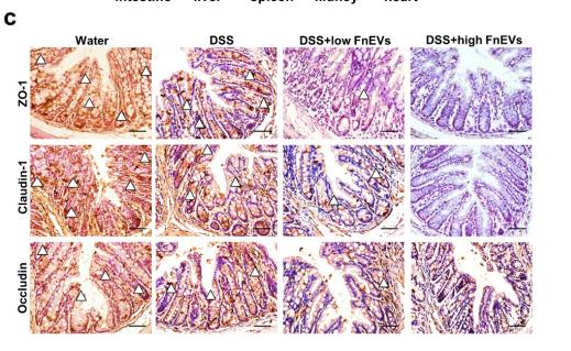
Application: IHC Species: mouse Sample: colon
Restrictive clause
Affinity Biosciences tests all products strictly. Citations are provided as a resource for additional applications that have not been validated by Affinity Biosciences. Please choose the appropriate format for each application and consult Materials and Methods sections for additional details about the use of any product in these publications.
For Research Use Only.
Not for use in diagnostic or therapeutic procedures. Not for resale. Not for distribution without written consent. Affinity Biosciences will not be held responsible for patent infringement or other violations that may occur with the use of our products. Affinity Biosciences, Affinity Biosciences Logo and all other trademarks are the property of Affinity Biosciences LTD.