Nephrin Antibody - #DF7501


 and mouse anti-beta tubulin Ab(T0023 1:200) for 1 hour at 37°C. An AlexaFluor594 conjugated goat anti-rabbit IgG(H+L) Ab(Red) and an AlexaFluor488 conjugated goat anti-mouse IgG(H+L) Ab(Green) were used as the secondary antibody.
The nuclear counter stain is DAPI(blue).")
| Product: | Nephrin Antibody |
| Catalog: | DF7501 |
| Description: | Rabbit polyclonal antibody to Nephrin |
| Application: | WB IHC IF/ICC |
| Cited expt.: | WB, IHC |
| Reactivity: | Human, Mouse, Rat |
| Prediction: | Pig, Zebrafish, Bovine, Horse, Sheep, Rabbit, Dog |
| Mol.Wt.: | 135kD,180kD; 135kD(Calculated). |
| Uniprot: | O60500 |
| RRID: | AB_2841001 |
Related Downloads
Protocols
Product Info
*The optimal dilutions should be determined by the end user. For optimal experimental results, antibody reuse is not recommended.
*Tips:
WB: For western blot detection of denatured protein samples. IHC: For immunohistochemical detection of paraffin sections (IHC-p) or frozen sections (IHC-f) of tissue samples. IF/ICC: For immunofluorescence detection of cell samples. ELISA(peptide): For ELISA detection of antigenic peptide.
Cite Format: Affinity Biosciences Cat# DF7501, RRID:AB_2841001.
Fold/Unfold
CNF; Nephrin; Nephrosis 1 congenital Finnish type; Nephrosis 1, congenital, Finnish type (nephrin); NPHN; NPHN_HUMAN; NPHS 1; Nphs1; Renal glomerulus specific cell adhesion receptor; Renal glomerulus-specific cell adhesion receptor;
Immunogens
A synthesized peptide derived from human Nephrin
- O60500 NPHN_HUMAN:
- Protein BLAST With
- NCBI/
- ExPASy/
- Uniprot
MALGTTLRASLLLLGLLTEGLAQLAIPASVPRGFWALPENLTVVEGASVELRCGVSTPGSAVQWAKDGLLLGPDPRIPGFPRYRLEGDPARGEFHLHIEACDLSDDAEYECQVGRSEMGPELVSPRVILSILVPPKLLLLTPEAGTMVTWVAGQEYVVNCVSGDAKPAPDITILLSGQTISDISANVNEGSQQKLFTVEATARVTPRSSDNRQLLVCEASSPALEAPIKASFTVNVLFPPGPPVIEWPGLDEGHVRAGQSLELPCVARGGNPLATLQWLKNGQPVSTAWGTEHTQAVARSVLVMTVRPEDHGAQLSCEAHNSVSAGTQEHGITLQVTFPPSAIIILGSASQTENKNVTLSCVSKSSRPRVLLRWWLGWRQLLPMEETVMDGLHGGHISMSNLTFLARREDNGLTLTCEAFSEAFTKETFKKSLILNVKYPAQKLWIEGPPEGQKLRAGTRVRLVCLAIGGNPEPSLMWYKDSRTVTESRLPQESRRVHLGSVEKSGSTFSRELVLVTGPSDNQAKFTCKAGQLSASTQLAVQFPPTNVTILANASALRPGDALNLTCVSVSSNPPVNLSWDKEGERLEGVAAPPRRAPFKGSAAARSVLLQVSSRDHGQRVTCRAHSAELRETVSSFYRLNVLYRPEFLGEQVLVVTAVEQGEALLPVSVSANPAPEAFNWTFRGYRLSPAGGPRHRILSSGALHLWNVTRADDGLYQLHCQNSEGTAEARLRLDVHYAPTIRALQDPTEVNVGGSVDIVCTVDANPILPGMFNWERLGEDEEDQSLDDMEKISRGPTGRLRIHHAKLAQAGAYQCIVDNGVAPPARRLLRLVVRFAPQVEHPTPLTKVAAAGDSTSSATLHCRARGVPNIVFTWTKNGVPLDLQDPRYTEHTYHQGGVHSSLLTIANVSAAQDYALFTCTATNALGSDQTNIQLVSISRPDPPSGLKVVSLTPHSVGLEWKPGFDGGLPQRFCIRYEALGTPGFHYVDVVPPQATTFTLTGLQPSTRYRVWLLASNALGDSGLADKGTQLPITTPGLHQPSGEPEDQLPTEPPSGPSGLPLLPVLFALGGLLLLSNASCVGGVLWQRRLRRLAEGISEKTEAGSEEDRVRNEYEESQWTGERDTQSSTVSTTEAEPYYRSLRDFSPQLPPTQEEVSYSRGFTGEDEDMAFPGHLYDEVERTYPPSGAWGPLYDEVQMGPWDLHWPEDTYQDPRGIYDQVAGDLDTLEPDSLPFELRGHLV
Predictions
Score>80(red) has high confidence and is suggested to be used for WB detection. *The prediction model is mainly based on the alignment of immunogen sequences, the results are for reference only, not as the basis of quality assurance.
High(score>80) Medium(80>score>50) Low(score<50) No confidence
Research Backgrounds
Seems to play a role in the development or function of the kidney glomerular filtration barrier. Regulates glomerular vascular permeability. May anchor the podocyte slit diaphragm to the actin cytoskeleton. Plays a role in skeletal muscle formation through regulation of myoblast fusion (By similarity).
Phosphorylated at Tyr-1193 by FYN, leading to the recruitment and activation of phospholipase C-gamma-1/PLCG1.
Cell membrane>Single-pass type I membrane protein.
Note: Predominantly located at podocyte slit diaphragm between podocyte foot processes. Also associated with podocyte apical plasma membrane.
Specifically expressed in podocytes of kidney glomeruli.
Belongs to the immunoglobulin superfamily.
References

Application: IHC Species: Rat Sample: glomeruli
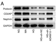
Application: WB Species: Mouse Sample:

Application: WB Species: Rat Sample: renal tissues
Restrictive clause
Affinity Biosciences tests all products strictly. Citations are provided as a resource for additional applications that have not been validated by Affinity Biosciences. Please choose the appropriate format for each application and consult Materials and Methods sections for additional details about the use of any product in these publications.
For Research Use Only.
Not for use in diagnostic or therapeutic procedures. Not for resale. Not for distribution without written consent. Affinity Biosciences will not be held responsible for patent infringement or other violations that may occur with the use of our products. Affinity Biosciences, Affinity Biosciences Logo and all other trademarks are the property of Affinity Biosciences LTD.