
 HepG2 cells were treated with the indicated concentrations of 25-HC for 24 hours before mRNA was extracted and expressions of TLR2, TLR4, TLR5, TLR7 and TLR9 were examined by RT-qPCR. (B) HepG2 cells were treated with the indicated concentrations of 25-HC for 24 hours or treated with 25-HC at 10 μM for the indicated times before protein was collected to detect the TLR2, TLR4, TLR5, TLR7 and TLR9 expressions by Western blotting. HepG2 cells were transfected with siNC or siTLR4 or siTLR9 for 24 hours, (C) then cells were treated with the indicated concentrations of 25-HC for 24 hours before proteins were collected and the expressions of MMP1, MMP2, MMP3 and MMP9 were determined by Western blotting. (D) Or migratory ability of HepG2 cells after treated with 25-HC for 36 hours was determined by Transwell assay. Results were obtained from 3 independent experiments and are expressed as the means ± SEM. Statistical significance was determined by Student’s t-test. *P<0.05, ***P<0.001.")
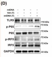
 signaling pathway A-B. qRT-PCR analysis for the mRNA expression of genes encoding TAM recruitment associated cytokines and chemokines including CCL2, CSF1, TGF-β, IL-6, and CCL22 in OVISE (a) and CAOV4 (b) cells 48 h after lentiviral mediated TFB2M overexpression. C-D. qRT-PCR (c) analysis of IL-6 transcription in cells and ELISA (d) assay of IL-6 secretion in the supernatants of cultured OVISE and CAOV4 cells with TFB2M overexpression alone, treated with DNase I alone or with combined TFB2M overexpression and DNase I treatment. E. qPCR was used to determine the copy number of mtDNA in the cytoplasm of OVISE and CAOV4 cells with TFB2M overexpression alone, treated with DNase I alone or with combined TFB2M overexpression and DNase I treatment. F. Immunofluorescent staining of TLR9 in OVISE and CAOV4 cells with or without TFB2M overexpression. The interactions between TLR9 and cytosolic mtDNA were indicated by white arrows. G-J. Western blot analyses were performed to detect the protein levels of TFB2M in whole cells and phospho-NF-κB p65 (p-p65) (Ser536) and p65 in the cytoplasm and nucleus of OVISE and CAOV4 cells with TFB2M overexpression alone or with combined ODN INH-18 (a TLR9 antagonist), DNase I or PDTC (an NF-κB inhibitor) treatment. Total TFB2M, nucleus p-p65, and p65 in each group were quantified using ImageJ software (h-j). Data shown are the mean ± SD. from three independent experiments. EV: empty vector. **p < 0.01, ***p < 0.001, n.s., not significant.")
. (A) Expression of mit-cyto C detected by WB. (B) Expression of cyt-cyto C detected by WB. (C) Comparison of relative expression of cyt-cyto C at different time points in each group. (D) Intergroup comparison of relative expression of cyt-cyto C in rats at different time points. (E) Comparison of relative expression of mit-cyto C at different time points in each group. (F) Intergroup comparison of relative expression of mit-cyto C in rats at different time points. (G) Comparison of relative expression of mtDNA at different time points in each group detected by RT-qPCR. (H) Intergroup comparison of relative expression of mtDNA in rats at different time points detected by RT-qPCR. (I) Comparison of relative expression of TLR9 at different time points in each group. (J) Intergroup comparison of relative expression of TLR9 in rats at different time points. △p<0.05 vs 6h, △△p<0.01 vs 6h, △△△p<0.001 vs 6h; ■p<0.05 vs 24h, ■■p<0.01 vs 24h, ■■■p<0.001 vs 24h; *p<0.05 vs Sham, **p<0.01 vs Sham, ***p<0.001 vs Sham; #p<0.05 vs MIRI, ##p<0.01 vs MIRI, ###p<0.001 vs MIRI. Sham: sham operation group; MIRI: MIRI model group; EMIRI: EA preconditioning plus MIRI model group. 6h: 6h after reperfusion; 24h: 24h after reperfusion; 3d: 3d after reperfusion.")
| Product: | TLR9 Antibody |
| Catalog: | DF2970 |
| Description: | Rabbit polyclonal antibody to TLR9 |
| Application: | WB IHC IF/ICC |
| Cited expt.: | WB, IF/ICC |
| Reactivity: | Human, Mouse, Rat |
| Prediction: | Horse, Rabbit, Dog |
| Mol.Wt.: | 117kD,140kD(full), 45kD,75kD(cleaved)(Observed); 116kD(Calculated). |
| Uniprot: | Q9NR96 |
| RRID: | AB_2840950 |
Control Products

Related Downloads
Protocols
Product Info
*The optimal dilutions should be determined by the end user. For optimal experimental results, antibody reuse is not recommended.
*Tips:
WB: For western blot detection of denatured protein samples. IHC: For immunohistochemical detection of paraffin sections (IHC-p) or frozen sections (IHC-f) of tissue samples. IF/ICC: For immunofluorescence detection of cell samples. ELISA(peptide): For ELISA detection of antigenic peptide.
Cite Format: Affinity Biosciences Cat# DF2970, RRID:AB_2840950.
Fold/Unfold
CD 289; CD289; TLR 9; TLR9; TLR9_HUMAN; Toll like receptor 9; Toll like receptor 9 isoform A precursor; Toll like receptor 9 isoform B; Toll-like receptor 9;
Immunogens
A synthesized peptide derived from human TLR9, corresponding to a region within the internal amino acids.
Highly expressed in spleen, lymph node, tonsil and peripheral blood leukocytes, especially in plasmacytoid pre-dendritic cells. Levels are much lower in monocytes and CD11c+ immature dendritic cells. Also detected in lung and liver.
- Q9NR96 TLR9_HUMAN:
- Protein BLAST With
- NCBI/
- ExPASy/
- Uniprot
MGFCRSALHPLSLLVQAIMLAMTLALGTLPAFLPCELQPHGLVNCNWLFLKSVPHFSMAAPRGNVTSLSLSSNRIHHLHDSDFAHLPSLRHLNLKWNCPPVGLSPMHFPCHMTIEPSTFLAVPTLEELNLSYNNIMTVPALPKSLISLSLSHTNILMLDSASLAGLHALRFLFMDGNCYYKNPCRQALEVAPGALLGLGNLTHLSLKYNNLTVVPRNLPSSLEYLLLSYNRIVKLAPEDLANLTALRVLDVGGNCRRCDHAPNPCMECPRHFPQLHPDTFSHLSRLEGLVLKDSSLSWLNASWFRGLGNLRVLDLSENFLYKCITKTKAFQGLTQLRKLNLSFNYQKRVSFAHLSLAPSFGSLVALKELDMHGIFFRSLDETTLRPLARLPMLQTLRLQMNFINQAQLGIFRAFPGLRYVDLSDNRISGASELTATMGEADGGEKVWLQPGDLAPAPVDTPSSEDFRPNCSTLNFTLDLSRNNLVTVQPEMFAQLSHLQCLRLSHNCISQAVNGSQFLPLTGLQVLDLSHNKLDLYHEHSFTELPRLEALDLSYNSQPFGMQGVGHNFSFVAHLRTLRHLSLAHNNIHSQVSQQLCSTSLRALDFSGNALGHMWAEGDLYLHFFQGLSGLIWLDLSQNRLHTLLPQTLRNLPKSLQVLRLRDNYLAFFKWWSLHFLPKLEVLDLAGNQLKALTNGSLPAGTRLRRLDVSCNSISFVAPGFFSKAKELRELNLSANALKTVDHSWFGPLASALQILDVSANPLHCACGAAFMDFLLEVQAAVPGLPSRVKCGSPGQLQGLSIFAQDLRLCLDEALSWDCFALSLLAVALGLGVPMLHHLCGWDLWYCFHLCLAWLPWRGRQSGRDEDALPYDAFVVFDKTQSAVADWVYNELRGQLEECRGRWALRLCLEERDWLPGKTLFENLWASVYGSRKTLFVLAHTDRVSGLLRASFLLAQQRLLEDRKDVVVLVILSPDGRRSRYVRLRQRLCRQSVLLWPHQPSGQRSFWAQLGMALTRDNHHFYNRNFCQGPTAE
Predictions
Score>80(red) has high confidence and is suggested to be used for WB detection. *The prediction model is mainly based on the alignment of immunogen sequences, the results are for reference only, not as the basis of quality assurance.
High(score>80) Medium(80>score>50) Low(score<50) No confidence
Research Backgrounds
Key component of innate and adaptive immunity. TLRs (Toll-like receptors) control host immune response against pathogens through recognition of molecular patterns specific to microorganisms. TLR9 is a nucleotide-sensing TLR which is activated by unmethylated cytidine-phosphate-guanosine (CpG) dinucleotides. Acts via MYD88 and TRAF6, leading to NF-kappa-B activation, cytokine secretion and the inflammatory response. Controls lymphocyte response to Helicobacter infection (By similarity). Upon CpG stimulation, induces B-cell proliferation, activation, survival and antibody production.
Activated by proteolytic cleavage of the flexible loop between repeats LRR14 and LRR15 within the ectodomain. Cleavage requires UNC93B1. Proteolytically processed by first removing the majority of the ectodomain by either asparagine endopeptidase (AEP) or a cathepsin followed by a trimming event that is solely cathepsin mediated and required for optimal receptor signaling.
Endoplasmic reticulum membrane>Single-pass type I membrane protein. Endosome. Lysosome. Cytoplasmic vesicle>Phagosome.
Note: Relocalizes from endoplasmic reticulum to endosome and lysosome upon stimulation with agonist. Exit from the ER requires UNC93B1. Endolysosomal localization is required for proteolytic cleavage and subsequent activation. Intracellular localization of the active receptor may prevent from responding to self nucleic acid.
Highly expressed in spleen, lymph node, tonsil and peripheral blood leukocytes, especially in plasmacytoid pre-dendritic cells. Levels are much lower in monocytes and CD11c+ immature dendritic cells. Also detected in lung and liver.
Belongs to the Toll-like receptor family.
Research Fields
· Human Diseases > Infectious diseases: Parasitic > Chagas disease (American trypanosomiasis).
· Human Diseases > Infectious diseases: Parasitic > African trypanosomiasis.
· Human Diseases > Infectious diseases: Parasitic > Malaria.
· Human Diseases > Infectious diseases: Bacterial > Tuberculosis.
· Human Diseases > Infectious diseases: Viral > Measles.
· Human Diseases > Infectious diseases: Viral > Herpes simplex infection.
· Organismal Systems > Immune system > Toll-like receptor signaling pathway. (View pathway)
References

Application: WB Species: human Sample: HUVECs
Application: WB Species: Mice Sample: BMDCs

Application: WB Species: mouse Sample: RAW264.7cells
Application: WB Species: mouse Sample: macrophages
Restrictive clause
Affinity Biosciences tests all products strictly. Citations are provided as a resource for additional applications that have not been validated by Affinity Biosciences. Please choose the appropriate format for each application and consult Materials and Methods sections for additional details about the use of any product in these publications.
For Research Use Only.
Not for use in diagnostic or therapeutic procedures. Not for resale. Not for distribution without written consent. Affinity Biosciences will not be held responsible for patent infringement or other violations that may occur with the use of our products. Affinity Biosciences, Affinity Biosciences Logo and all other trademarks are the property of Affinity Biosciences LTD.