
, using Granzyme B/H Antibody at 1/1000 dilution.
5ug/NC membrane strip.
Exposure for 6s with Affinity™ ECL Kit(#KF8003).
Bands result from membrane strip incubation.")
, using Granzyme B/H Antibody at 1/1000 dilution.
5ug/NC membrane strip.
Exposure for 30s with Affinity™ ECL Kit(#KF8001).
Bands result from membrane strip incubation.")
.
Bands result from membrane strip incubation.")


| Product: | Granzyme B/H Antibody |
| Catalog: | AF0175 |
| Description: | Rabbit polyclonal antibody to Granzyme B/H |
| Application: | WB IHC IF/ICC |
| Cited expt.: | IHC, IF/ICC |
| Reactivity: | Human, Mouse, Rat |
| Prediction: | Pig, Bovine, Horse, Dog |
| Mol.Wt.: | 28-40kDa(Glycosylation)(Observed); 28kD,27kD(Calculated). |
| Uniprot: | P10144 | P20718 |
| RRID: | AB_2833368 |
Control Products

Related Downloads
Protocols
Product Info
*The optimal dilutions should be determined by the end user. For optimal experimental results, antibody reuse is not recommended.
*Tips:
WB: For western blot detection of denatured protein samples. IHC: For immunohistochemical detection of paraffin sections (IHC-p) or frozen sections (IHC-f) of tissue samples. IF/ICC: For immunofluorescence detection of cell samples. ELISA(peptide): For ELISA detection of antigenic peptide.
Cite Format: Affinity Biosciences Cat# AF0175, RRID:AB_2833368.
Fold/Unfold
C11; Cathepsin G like 1; Cathepsin G-like 1; CCPI; CGL 1; CGL1; CSP B; CSPB; CTLA-1; CTLA1; CTSGL1; Cytotoxic serine protease B; Cytotoxic T lymphocyte associated serine esterase 1; Cytotoxic T lymphocyte proteinase 2; Cytotoxic T-lymphocyte proteinase 2; Fragmentin 2; Fragmentin-2; GRAB_HUMAN; Granzyme 2; Granzyme B (granzyme 2, cytotoxic T lymphocyte associated serine esterase 1); Granzyme B; Granzyme-2; GranzymeB; GRB; Gzmb; Hlp; Human lymphocyte protein; Lymphocyte protease; Protease, serine, B; SECT; T cell serine protease 1-3E; T-cell serine protease 1-3E; Cathepsin G like 2; Cathepsin G-like 2; CCP X; CCP-X; CGL 2; CGL2; CSP C; CSP-C; CTLA1; CTSGL2; Cytotoxic serine protease C; Cytotoxic T lymphocyte associated serine esterase 1; Cytotoxic T lymphocyte proteinase; Cytotoxic T-lymphocyte proteinase; EC 3.4.21.-; GRAH_HUMAN; Granzyme H (cathepsin G-like 2, protein h-CCPX); Granzyme H; GZMH; Protein h CCPX;
Immunogens
A synthesized peptide derived from human Granzyme B/H, corresponding to a region within N-terminal amino acids.
- P10144 GRAB_HUMAN:
- Protein BLAST With
- NCBI/
- ExPASy/
- Uniprot
MQPILLLLAFLLLPRADAGEIIGGHEAKPHSRPYMAYLMIWDQKSLKRCGGFLIRDDFVLTAAHCWGSSINVTLGAHNIKEQEPTQQFIPVKRPIPHPAYNPKNFSNDIMLLQLERKAKRTRAVQPLRLPSNKAQVKPGQTCSVAGWGQTAPLGKHSHTLQEVKMTVQEDRKCESDLRHYYDSTIELCVGDPEIKKTSFKGDSGGPLVCNKVAQGIVSYGRNNGMPPRACTKVSSFVHWIKKTMKRY
- P20718 GRAH_HUMAN:
- Protein BLAST With
- NCBI/
- ExPASy/
- Uniprot
MQPFLLLLAFLLTPGAGTEEIIGGHEAKPHSRPYMAFVQFLQEKSRKRCGGILVRKDFVLTAAHCQGSSINVTLGAHNIKEQERTQQFIPVKRPIPHPAYNPKNFSNDIMLLQLERKAKWTTAVRPLRLPSSKAQVKPGQLCSVAGWGYVSMSTLATTLQEVLLTVQKDCQCERLFHGNYSRATEICVGDPKKTQTGFKGDSGGPLVCKDVAQGILSYGNKKGTPPGVYIKVSHFLPWIKRTMKRL
Predictions
Score>80(red) has high confidence and is suggested to be used for WB detection. *The prediction model is mainly based on the alignment of immunogen sequences, the results are for reference only, not as the basis of quality assurance.
High(score>80) Medium(80>score>50) Low(score<50) No confidence
Research Backgrounds
This enzyme is necessary for target cell lysis in cell-mediated immune responses. It cleaves after Asp. Seems to be linked to an activation cascade of caspases (aspartate-specific cysteine proteases) responsible for apoptosis execution. Cleaves caspase-3, -7, -9 and 10 to give rise to active enzymes mediating apoptosis.
Cytoplasmic granule.
Note: Cytoplasmic granules of cytolytic T-lymphocytes and natural killer cells.
Belongs to the peptidase S1 family. Granzyme subfamily.
Cytotoxic chymotrypsin-like serine protease with preference for bulky and aromatic residues at the P1 position and acidic residues at the P3' and P4' sites. Probably necessary for target cell lysis in cell-mediated immune responses. Participates in the antiviral response via direct cleavage of several proteins essential for viral replication.
Cytoplasmic granule.
Note: Cytoplasmic granules of cytolytic T-lymphocytes.
Constitutively expressed in NK cells.
Belongs to the peptidase S1 family. Granzyme subfamily.
Research Fields
· Cellular Processes > Cell growth and death > Apoptosis. (View pathway)
· Human Diseases > Endocrine and metabolic diseases > Type I diabetes mellitus.
· Human Diseases > Cancers: Overview > Transcriptional misregulation in cancer.
· Human Diseases > Immune diseases > Autoimmune thyroid disease.
· Human Diseases > Immune diseases > Allograft rejection.
· Human Diseases > Immune diseases > Graft-versus-host disease.
· Organismal Systems > Immune system > Natural killer cell mediated cytotoxicity. (View pathway)
References

Application: IF/ICC Species: Mouse Sample:
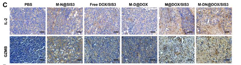
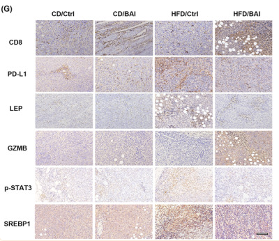
Application: IHC Species: mice Sample: NK cells

Application: IHC Species: Mouse Sample:
Application: IHC Species: Mouse Sample:
Restrictive clause
Affinity Biosciences tests all products strictly. Citations are provided as a resource for additional applications that have not been validated by Affinity Biosciences. Please choose the appropriate format for each application and consult Materials and Methods sections for additional details about the use of any product in these publications.
For Research Use Only.
Not for use in diagnostic or therapeutic procedures. Not for resale. Not for distribution without written consent. Affinity Biosciences will not be held responsible for patent infringement or other violations that may occur with the use of our products. Affinity Biosciences, Affinity Biosciences Logo and all other trademarks are the property of Affinity Biosciences LTD.