MRC2 Antibody - #AF0564

, blocked with antigen-specific peptides,
Lane 2: Hela cells(uv treatment),
Lane 3: RAW264.7 cells(serum starvation treatment).")
 and mouse anti-beta tubulin Ab(T0023) for 1 hour at 37°C. An AlexaFluor594 conjugated goat anti-rabbit IgG(H+L) Ab(Red) and an AlexaFluor488 conjugated goat anti-mouse IgG(H+L) Ab(Green) were used as the secondary antibody.
The nuclear counter stain is DAPI (blue).")
| Product: | MRC2 Antibody |
| Catalog: | AF0564 |
| Description: | Rabbit polyclonal antibody to MRC2 |
| Application: | WB IHC IF/ICC |
| Cited expt.: | IHC |
| Reactivity: | Human, Mouse, Rat |
| Prediction: | Pig, Bovine, Horse, Sheep, Rabbit, Dog |
| Mol.Wt.: | 167kDa(Observed); 167kD(Calculated). |
| Uniprot: | Q9UBG0 |
| RRID: | AB_2834341 |
Related Downloads
Protocols
Product Info
*The optimal dilutions should be determined by the end user. For optimal experimental results, antibody reuse is not recommended.
*Tips:
WB: For western blot detection of denatured protein samples. IHC: For immunohistochemical detection of paraffin sections (IHC-p) or frozen sections (IHC-f) of tissue samples. IF/ICC: For immunofluorescence detection of cell samples. ELISA(peptide): For ELISA detection of antigenic peptide.
Cite Format: Affinity Biosciences Cat# AF0564, RRID:AB_2834341.
Fold/Unfold
C-type lectin domain family 13 member E; C-type mannose receptor 2; CD 280; CD280; CD280 antigen; CLEC 13E; CLEC13E; ENDO 180; ENDO180; Endocytic receptor (macrophage mannose receptor family); Endocytic receptor 180; FLJ35911; KIAA0709; Macrophage mannose receptor 2; Mannose receptor C type 2; MRC 2; MRC2; MRC2_HUMAN; UPAR-associated protein; UPARAP; Urokinase plasminogen activator receptor associated protein; Urokinase receptor associated protein; Urokinase receptor-associated protein; Urokinase-type plasminogen activator receptor-associated protein;
Immunogens
A synthesized peptide derived from human MRC2, corresponding to a region within N-terminal amino acids.
Ubiquitous with low expression in brain, placenta, lung, kidney, pancreas, spleen, thymus and colon. Expressed in endothelial cells, fibroblasts and macrophages. Highly expressed in fetal lung and kidney.
- Q9UBG0 MRC2_HUMAN:
- Protein BLAST With
- NCBI/
- ExPASy/
- Uniprot
MGPGRPAPAPWPRHLLRCVLLLGCLHLGRPGAPGDAALPEPNVFLIFSHGLQGCLEAQGGQVRVTPACNTSLPAQRWKWVSRNRLFNLGTMQCLGTGWPGTNTTASLGMYECDREALNLRWHCRTLGDQLSLLLGARTSNISKPGTLERGDQTRSGQWRIYGSEEDLCALPYHEVYTIQGNSHGKPCTIPFKYDNQWFHGCTSTGREDGHLWCATTQDYGKDERWGFCPIKSNDCETFWDKDQLTDSCYQFNFQSTLSWREAWASCEQQGADLLSITEIHEQTYINGLLTGYSSTLWIGLNDLDTSGGWQWSDNSPLKYLNWESDQPDNPSEENCGVIRTESSGGWQNRDCSIALPYVCKKKPNATAEPTPPDRWANVKVECEPSWQPFQGHCYRLQAEKRSWQESKKACLRGGGDLVSIHSMAELEFITKQIKQEVEELWIGLNDLKLQMNFEWSDGSLVSFTHWHPFEPNNFRDSLEDCVTIWGPEGRWNDSPCNQSLPSICKKAGQLSQGAAEEDHGCRKGWTWHSPSCYWLGEDQVTYSEARRLCTDHGSQLVTITNRFEQAFVSSLIYNWEGEYFWTALQDLNSTGSFFWLSGDEVMYTHWNRDQPGYSRGGCVALATGSAMGLWEVKNCTSFRARYICRQSLGTPVTPELPGPDPTPSLTGSCPQGWASDTKLRYCYKVFSSERLQDKKSWVQAQGACQELGAQLLSLASYEEEHFVANMLNKIFGESEPEIHEQHWFWIGLNRRDPRGGQSWRWSDGVGFSYHNFDRSRHDDDDIRGCAVLDLASLQWVAMQCDTQLDWICKIPRGTDVREPDDSPQGRREWLRFQEAEYKFFEHHSTWAQAQRICTWFQAELTSVHSQAELDFLSHNLQKFSRAQEQHWWIGLHTSESDGRFRWTDGSIINFISWAPGKPRPVGKDKKCVYMTASREDWGDQRCLTALPYICKRSNVTKETQPPDLPTTALGGCPSDWIQFLNKCFQVQGQEPQSRVKWSEAQFSCEQQEAQLVTITNPLEQAFITASLPNVTFDLWIGLHASQRDFQWVEQEPLMYANWAPGEPSGPSPAPSGNKPTSCAVVLHSPSAHFTGRWDDRSCTEETHGFICQKGTDPSLSPSPAALPPAPGTELSYLNGTFRLLQKPLRWHDALLLCESRNASLAYVPDPYTQAFLTQAARGLRTPLWIGLAGEEGSRRYSWVSEEPLNYVGWQDGEPQQPGGCTYVDVDGAWRTTSCDTKLQGAVCGVSSGPPPPRRISYHGSCPQGLADSAWIPFREHCYSFHMELLLGHKEARQRCQRAGGAVLSILDEMENVFVWEHLQSYEGQSRGAWLGMNFNPKGGTLVWQDNTAVNYSNWGPPGLGPSMLSHNSCYWIQSNSGLWRPGACTNITMGVVCKLPRAEQSSFSPSALPENPAALVVVLMAVLLLLALLTAALILYRRRQSIERGAFEGARYSRSSSSPTEATEKNILVSDMEMNEQQE
Predictions
Score>80(red) has high confidence and is suggested to be used for WB detection. *The prediction model is mainly based on the alignment of immunogen sequences, the results are for reference only, not as the basis of quality assurance.
High(score>80) Medium(80>score>50) Low(score<50) No confidence
Research Backgrounds
May play a role as endocytotic lectin receptor displaying calcium-dependent lectin activity. Internalizes glycosylated ligands from the extracellular space for release in an endosomal compartment via clathrin-mediated endocytosis. May be involved in plasminogen activation system controlling the extracellular level of PLAUR/PLAU, and thus may regulate protease activity at the cell surface. May contribute to cellular uptake, remodeling and degradation of extracellular collagen matrices. May play a role during cancer progression as well as in other chronic tissue destructive diseases acting on collagen turnover. May participate in remodeling of extracellular matrix cooperating with the matrix metalloproteinases (MMPs).
N-glycosylated.
Membrane>Single-pass type I membrane protein.
Ubiquitous with low expression in brain, placenta, lung, kidney, pancreas, spleen, thymus and colon. Expressed in endothelial cells, fibroblasts and macrophages. Highly expressed in fetal lung and kidney.
C-type lectin domains 3 to 8 are not required for calcium-dependent binding of mannose, fucose and N-acetylglucosamine. C-type lectin domain 2 is responsible for sugar-binding in a calcium-dependent manner.
Fibronectin type-II domain mediates collagen-binding.
Ricin B-type lectin domain contacts with the second C-type lectin domain.
Research Fields
· Cellular Processes > Transport and catabolism > Phagosome. (View pathway)
· Human Diseases > Infectious diseases: Bacterial > Tuberculosis.
References
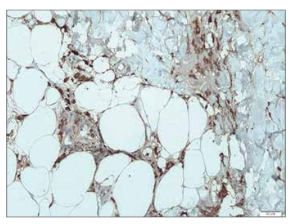
Application: IHC Species: unknown Sample: bone tissue
Restrictive clause
Affinity Biosciences tests all products strictly. Citations are provided as a resource for additional applications that have not been validated by Affinity Biosciences. Please choose the appropriate format for each application and consult Materials and Methods sections for additional details about the use of any product in these publications.
For Research Use Only.
Not for use in diagnostic or therapeutic procedures. Not for resale. Not for distribution without written consent. Affinity Biosciences will not be held responsible for patent infringement or other violations that may occur with the use of our products. Affinity Biosciences, Affinity Biosciences Logo and all other trademarks are the property of Affinity Biosciences LTD.