, using Phospho-TRPV1(Ser502) Antibody at 1/1000 dilution.
5ug/NC membrane strip.
Exposure for 30s with Affinity™ ECL Kit(#KF8001).
Bands result from membrane strip incubation.")

| Product: | Phospho-TRPV1(Ser502) Antibody |
| Catalog: | AF8520 |
| Description: | Rabbit polyclonal antibody to Phospho-TRPV1(Ser502) |
| Application: | WB IHC |
| Cited expt.: | WB |
| Reactivity: | Human, Mouse, Rat |
| Prediction: | Pig, Bovine, Sheep, Rabbit, Dog, Chicken, Xenopus |
| Mol.Wt.: | 95 kDa(Observed); 95kD(Calculated). |
| Uniprot: | Q8NER1 |
| RRID: | AB_2840574 |
Control Products

Related Downloads
Protocols
Product Info
*The optimal dilutions should be determined by the end user. For optimal experimental results, antibody reuse is not recommended.
*Tips:
WB: For western blot detection of denatured protein samples. IHC: For immunohistochemical detection of paraffin sections (IHC-p) or frozen sections (IHC-f) of tissue samples. IF/ICC: For immunofluorescence detection of cell samples. ELISA(peptide): For ELISA detection of antigenic peptide.
Cite Format: Affinity Biosciences Cat# AF8520, RRID:AB_2840574.
Fold/Unfold
Capsaicin receptor; DKFZp434K0220; osm 9 like TRP channel 1; Osm-9-like TRP channel 1; OTRPC1; Transient receptor potential cation channel subfamily V member 1; TRPV 1; Trpv1; TRPV1_HUMAN; Vanilloid receptor 1; Vanilloid receptor subtype 1; VR 1; VR1;
Immunogens
A synthesized peptide derived from human VR1 around the phosphorylation site of Ser502.
Widely expressed at low levels. Expression is elevated in dorsal root ganglia. In skin, expressed in cutaneous sensory nerve fibers, mast cells, epidermal keratinocytes, dermal blood vessels, the inner root sheet and the infundibulum of hair follicles, differentiated sebocytes, sweat gland ducts, and the secretory portion of eccrine sweat glands (at protein level).
- Q8NER1 TRPV1_HUMAN:
- Protein BLAST With
- NCBI/
- ExPASy/
- Uniprot
MKKWSSTDLGAAADPLQKDTCPDPLDGDPNSRPPPAKPQLSTAKSRTRLFGKGDSEEAFPVDCPHEEGELDSCPTITVSPVITIQRPGDGPTGARLLSQDSVAASTEKTLRLYDRRSIFEAVAQNNCQDLESLLLFLQKSKKHLTDNEFKDPETGKTCLLKAMLNLHDGQNTTIPLLLEIARQTDSLKELVNASYTDSYYKGQTALHIAIERRNMALVTLLVENGADVQAAAHGDFFKKTKGRPGFYFGELPLSLAACTNQLGIVKFLLQNSWQTADISARDSVGNTVLHALVEVADNTADNTKFVTSMYNEILMLGAKLHPTLKLEELTNKKGMTPLALAAGTGKIGVLAYILQREIQEPECRHLSRKFTEWAYGPVHSSLYDLSCIDTCEKNSVLEVIAYSSSETPNRHDMLLVEPLNRLLQDKWDRFVKRIFYFNFLVYCLYMIIFTMAAYYRPVDGLPPFKMEKTGDYFRVTGEILSVLGGVYFFFRGIQYFLQRRPSMKTLFVDSYSEMLFFLQSLFMLATVVLYFSHLKEYVASMVFSLALGWTNMLYYTRGFQQMGIYAVMIEKMILRDLCRFMFVYIVFLFGFSTAVVTLIEDGKNDSLPSESTSHRWRGPACRPPDSSYNSLYSTCLELFKFTIGMGDLEFTENYDFKAVFIILLLAYVILTYILLLNMLIALMGETVNKIAQESKNIWKLQRAITILDTEKSFLKCMRKAFRSGKLLQVGYTPDGKDDYRWCFRVDEVNWTTWNTNVGIINEDPGNCEGVKRTLSFSLRSSRVSGRHWKNFALVPLLREASARDRQSAQPEEVYLRQFSGSLKPEDAEVFKSPAASGEK
Predictions
Score>80(red) has high confidence and is suggested to be used for WB detection. *The prediction model is mainly based on the alignment of immunogen sequences, the results are for reference only, not as the basis of quality assurance.
High(score>80) Medium(80>score>50) Low(score<50) No confidence
Research Backgrounds
Ligand-activated non-selective calcium permeant cation channel involved in detection of noxious chemical and thermal stimuli. Seems to mediate proton influx and may be involved in intracellular acidosis in nociceptive neurons. Involved in mediation of inflammatory pain and hyperalgesia. Sensitized by a phosphatidylinositol second messenger system activated by receptor tyrosine kinases, which involves PKC isozymes and PCL. Activation by vanilloids, like capsaicin, and temperatures higher than 42 degrees Celsius, exhibits a time- and Ca(2+)-dependent outward rectification, followed by a long-lasting refractory state. Mild extracellular acidic pH (6.5) potentiates channel activation by noxious heat and vanilloids, whereas acidic conditions (pH <6) directly activate the channel. Can be activated by endogenous compounds, including 12-hydroperoxytetraenoic acid and bradykinin. Acts as ionotropic endocannabinoid receptor with central neuromodulatory effects. Triggers a form of long-term depression (TRPV1-LTD) mediated by the endocannabinoid anandamine in the hippocampus and nucleus accumbens by affecting AMPA receptors endocytosis.
Phosphorylation by PKA reverses capsaicin-induced dephosphorylation at multiple sites, probably including Ser-117 as a major phosphorylation site. Phosphorylation by CAMKII seems to regulate binding to vanilloids. Phosphorylated and modulated by PRKCE, PRKCM and probably PRKCZ. Dephosphorylation by calcineurin seems to lead to receptor desensitization and phosphorylation by CAMKII recovers activity.
Cell junction>Synapse>Postsynaptic cell membrane>Multi-pass membrane protein. Cell projection>Dendritic spine membrane>Multi-pass membrane protein. Cell membrane>Multi-pass membrane protein.
Note: Mostly, but not exclusively expressed in postsynaptic dendritic spines.
Widely expressed at low levels. Expression is elevated in dorsal root ganglia. In skin, expressed in cutaneous sensory nerve fibers, mast cells, epidermal keratinocytes, dermal blood vessels, the inner root sheet and the infundibulum of hair follicles, differentiated sebocytes, sweat gland ducts, and the secretory portion of eccrine sweat glands (at protein level).
The association domain (AD) is necessary for self-association.
Belongs to the transient receptor (TC 1.A.4) family. TrpV subfamily. TRPV1 sub-subfamily.
Research Fields
· Environmental Information Processing > Signaling molecules and interaction > Neuroactive ligand-receptor interaction.
· Organismal Systems > Sensory system > Inflammatory mediator regulation of TRP channels. (View pathway)
References
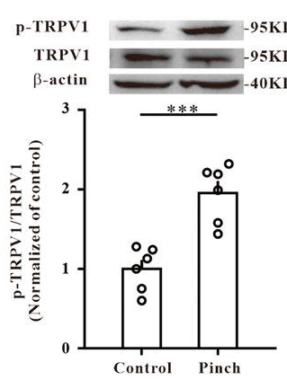
Application: WB Species: Mouse Sample:
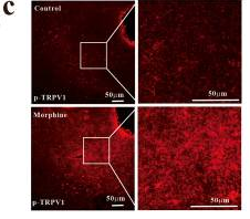
Application: IF/ICC Species: Mouse Sample:
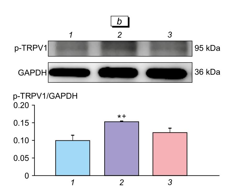
Application: WB Species: Human Sample: U87-MG cells
Restrictive clause
Affinity Biosciences tests all products strictly. Citations are provided as a resource for additional applications that have not been validated by Affinity Biosciences. Please choose the appropriate format for each application and consult Materials and Methods sections for additional details about the use of any product in these publications.
For Research Use Only.
Not for use in diagnostic or therapeutic procedures. Not for resale. Not for distribution without written consent. Affinity Biosciences will not be held responsible for patent infringement or other violations that may occur with the use of our products. Affinity Biosciences, Affinity Biosciences Logo and all other trademarks are the property of Affinity Biosciences LTD.