 Antibody. The lane on the left was treated with blocking peptide.")
, using Phospho-YAP (Ser127) Antibody at 1/1000 dilution.
5ug/NC membrane strip.
Exposure for 10s with Affinity™ ECL Kit(#KF8001).
Bands result from membrane strip incubation.")


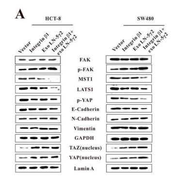
 or both. The expression of FAK, p-FAK(Y397), MST1, LATS1, p-YAP, E-cadherin, N-Cadherin, and Vimentin was then examined by western blot analysis of the whole cell lysates, and the expression of YAP and TAZ in the nucleus was also examined.")
, and treatment with telmisartan or transfection with AT1R siRNA reverses this effect. Grouping: Control group, HAECs only; group 1, HAECs with Ang II (1 µM) treatment; group 2, HAECs with Ang II (1 µM) and siRNA NC transfection; group 3, HAECs with Ang II (1 µM) and AT1R siRNA transfection; group 4, HAECs with Ang II (1 µM) and 1 µl DMSO; and group 5, HAECs with Ang II (1 µM) and the ARB 1 µl (20 mM) telmisartan treatment. (A) Western blot analysis of sets of six independent lysates from HAECs that were untreated, treated with Ang II, treated with Ang II and siRNA NC, treated with Ang II and AT1R siRNA, treated with Ang II and DMSO, or treated with Ang II and telmisartan. Treatment with Ang II upregulated AT1R and p‑YAP, and this effect was alleviated by transfection with an AT1R siRNA or treatment with telmisartan.")
 ST6GAL2 knockdown cells were transfected with ST6GAL2 overexpression vectors, and the proliferation,migration, and invasion capacities of FTC cells were enhanced. (G and H) Western blotting was performed to determine the levels of Hippo signaling molecules in FTC cells. *p < 0.05; scale bars, 20 mm.")
 Expression levels of YAP, TAZ, JNK, NF-κB and TNF-α were monitored by RT-PCR (n = 3). (B–C, F) Proteins (YAP, p-YAP, TAZ, p-TAZ, JNK, Nuclear NF-κB P65, Total P65 and TNF-α) in the pathway were detected by western blot (n = 3).")
 IHC staining of YAP and CK7 in first trimester villi of human placenta. CK7 was regarded as
a positive control of STB and CTB in trophoblasts; scale bars, 100 μm. (b) IHC staining of YAP
and CK7 in Normal and FGR term placenta. CK7 was regarded as a positive control of STB and
CTB in trophoblasts; scale bar, 100 μm. (c) IHC staining of YAP and CK7 in E18.5 C57BL/6J
mouse placenta. CK7 was regarded as a positive control of trophoblasts. (d) Western blotting of
YAP and p-YAP in normal and FGR placentas. **P < 0.01; Mann-Whitney U test. (e) mRNA
levels of CTGF, CYR61 and AMOTL2 were measured by qRT-PCR and normalized to β-actin.
**P < 0.01, *P < 0.05; Mann-Whitney U test. All data are presented as the mean ± SEM.")
 Images of YAP and p-YAP immunofluorescence staining of gradual gestational age (Week-9,
Week-14, Week-17, Week-21, and Week-40) in human placenta. Scale bars, 200 μm. (b)
Expression of p-YAP and YAP in human placenta with progressive gestational age determined by
Western blotting. (c) Scanning images of YAP and p-YAP immunofluorescence staining of
gradual gestational age (E8.5, E9.5, E10.5, E12.5, E13.5, E15.5, E16.5, and E18.5) in mouse
placenta. (d) Expression of p-YAP and YAP in mouse placenta with increasing gestational age
determined by Western blotting.")
 YAP siRNA efficiencies (n = 3, mean and S.D., t-test, &&P < 0.05 compared with siRNA NC;%%P < 0.05 compared with siRNA 860); (B,C) Western Blot of YAP after siRNA transfection *P < 0.05 compared with siRNA NC; (D) Flow cytometry analysis of CD68, CD86, and CD206 expression after 96 h co-culture in both groups. The data in the one-factor histogram represents the cells in the red circle in the upper scatter plot. HAECs + THP-1 + Ang II (1 μM, 24 h) + siRNA NC group: CD68, 41.94%; CD86, 45.73%; CD206, 9.72%. HAECs + THP-1 + Ang II + YAP siRNA group: CD68, 63.13%; CD86, 53.42%; CD206, 7.13%; (E–G) The proportion of CD68 +, CD86 +, and CD206 + cells, respectively (n = 3, mean and S.D., t-test, **P < 0.05 compared with the HAECs + THP-1 + siRNA NC group); (H) Fluorescence staining of adherent macrophages after YAP siRNA transfection. All cells were labeled with DAPI (blue), and the number of adherent macrophages was significantly decreased after transfection with the AT1R siRNA; (I) The ratio between the number of macrophages and the total number of cells (n = 3, mean and S.D., t-test, symbols are the same as in C,D,E).")
 and protein level (b, c) of HSP110 pulmonary arteries in lung tissues in each group (N = 8). d Double immunofluorescence staining of α-SMA (green) and HSP110 (red) in pulmonary arteries (N = 8). White scale bars, 50 μm; Yellow scale bars, 25 μm. White arrows pointed to α-SMA and HSP110 double-positive cells. e Protein levels of LC3II, LC3I, Beclin1, ATG5, ATG7 and p62 in pulmonary arteries (N = 8). f–h Quantitative analysis of relative protein ratio of LC3-II/I and relative protein level of Beclin1, p62, ATG5 and ATG7 (N = 8). i Double immunofluorescence staining of α-SMA (green) and Beclin 1 (red) in pulmonary arteries (N = 8). Scale bars, 50 μm. j Protein levels of p-YAP, YAP, p-TAZ and TAZ in pulmonary arteries (N = 8). k Quantitative analysis of relative protein ratio of p-YAP/t-YAP and p-TAZ/t-TAZ (N = 8). l–n Nuclear protein levels of YAP, TAZ and TEAD4 and quantitative analysis of relative protein level of nuclear YAP, TAZ and TEAD4 (N = 8). o Double immunofluorescence staining of α-SMA (green) and YAP (red) in pulmonary arteries (N = 8). White scale bars, 50 μm; Yellow scale bars, 25 μm. White arrows pointed to α-SMA and YAP double-positive cells. Data are means ± SD from 8 mice per group. *p")
. Representative blots are shown. *P")
 synthesis, and the expression of DNA damage-related proteins were assessed in cholangiocarcinoma cells (CCAs) subjected to YAP1 knockdown or YAP1 overexpression. Western blot was used to measure protein levels (n = 3). With cisplatin incubation, YAP1 knockdown and 1 mg/mL babaodan (BBD) treatment decreased the (a) Bcl-2 level and (b) increased bax level, while the change in (c) cle-caspase-3/caspase-3 levels with BBD treatment was not statistically significant. In the CCAs dealing with cisplatin, the expression levels of (d) p-YAP1/YAP1, (e) ATF4, and (f) SLC1A5 were decreased by YAP1 knockdown and BBD treatment. Additionally, the (g) γH2Ax level was increased and (h) the ERCC1 level was inhibited by YAP1 knockdown and BBD treatment. YAP1 overexpression antagonized the effect of BBD on these proteins. Representative protein bands are shown in (i), (j), and (k). (mean ± standard deviation) +p")
. C. Immunohistochemical staining shows the expression of KI67, YAP and pYAPS127 in mice tumors among sh Control, sh-00525, sh-00525 + GA-017 groups. Scale bar: 100 μm. D. Relative protein expression of YAP, pYAPS127, downstream targets genes (such as CYR61, CTGF and ANKRD1) and LINC00525 in mice tumors among sh Control, sh-00525, sh-00525 + GA-017 groups. E-H. The volume and growth of tumors in the following groups (Control, LINC00525, LINC00525 + Cytochalasin D) and analyzed as in C and D")
 Western blot and qRT-PCR were used to detect the expression of Hippo pathway related proteins. n = 3; (K) miR-302a-3p levels in each group in vitro n = 3. **/*P < 0.01/0.05 vs. Ctrl; #P < 0.05 vs. Hyp.")
 RNF128 was correlated with Hippo pathway proteins (LATS, YAP, and TAZ) in CRC according to StarBase. (B) RNF128 was downregulated in LoVo and HCT116 cells transfected with si-NC and si-RNF128, as shown by western blot analysis. **P")
 The expression level of YAP/TAZ in U373 cells was determined using immunofluorescence assays (*p")
| Product: | Phospho-YAP (Ser127) Antibody |
| Catalog: | AF3328 |
| Description: | Rabbit polyclonal antibody to Phospho-YAP (Ser127) |
| Application: | WB IHC IF/ICC |
| Cited expt.: | WB, IF/ICC |
| Reactivity: | Human, Mouse, Rat, Monkey |
| Prediction: | Pig, Zebrafish, Horse, Sheep, Rabbit, Chicken, Xenopus |
| Mol.Wt.: | 65~78kD(Observed); 54kD(Calculated). |
| Uniprot: | P46937 |
| RRID: | AB_2810276 |
Control Products

Product Info
*The optimal dilutions should be determined by the end user. For optimal experimental results, antibody reuse is not recommended.
*Tips:
WB: For western blot detection of denatured protein samples. IHC: For immunohistochemical detection of paraffin sections (IHC-p) or frozen sections (IHC-f) of tissue samples. IF/ICC: For immunofluorescence detection of cell samples. ELISA(peptide): For ELISA detection of antigenic peptide.
Cite Format: Affinity Biosciences Cat# AF3328, RRID:AB_2810276.
Fold/Unfold
65 kDa Yes associated protein; 65 kDa Yes-associated protein; COB1; YAp 1; YAP 65; YAP; YAP1; YAP1_HUMAN; YAP2; YAP65; yes -associated protein delta; Yes associated protein 1 65kDa; Yes associated protein 1; Yes associated protein 2; yes associated protein beta; YKI; Yorkie homolog;
Immunogens
A synthesized peptide derived from human YAP around the phosphorylation site of Ser127.
Increased expression seen in some liver and prostate cancers. Isoforms lacking the transactivation domain found in striatal neurons of patients with Huntington disease (at protein level).
- P46937 YAP1_HUMAN:
- Protein BLAST With
- NCBI/
- ExPASy/
- Uniprot
MDPGQQPPPQPAPQGQGQPPSQPPQGQGPPSGPGQPAPAATQAAPQAPPAGHQIVHVRGDSETDLEALFNAVMNPKTANVPQTVPMRLRKLPDSFFKPPEPKSHSRQASTDAGTAGALTPQHVRAHSSPASLQLGAVSPGTLTPTGVVSGPAATPTAQHLRQSSFEIPDDVPLPAGWEMAKTSSGQRYFLNHIDQTTTWQDPRKAMLSQMNVTAPTSPPVQQNMMNSASGPLPDGWEQAMTQDGEIYYINHKNKTTSWLDPRLDPRFAMNQRISQSAPVKQPPPLAPQSPQGGVMGGSNSNQQQQMRLQQLQMEKERLRLKQQELLRQAMRNINPSTANSPKCQELALRSQLPTLEQDGGTQNPVSSPGMSQELRTMTTNSSDPFLNSGTYHSRDESTDSGLSMSSYSVPRTPDDFLNSVDEMDTGDTINQSTLPSQQNRFPDYLEAIPGTNVDLGTLEGDGMNIEGEELMPSLQEALSSDILNDMESVLAATKLDKESFLTWL
Predictions
Score>80(red) has high confidence and is suggested to be used for WB detection. *The prediction model is mainly based on the alignment of immunogen sequences, the results are for reference only, not as the basis of quality assurance.
High(score>80) Medium(80>score>50) Low(score<50) No confidence
Research Backgrounds
Transcriptional regulator which can act both as a coactivator and a corepressor and is the critical downstream regulatory target in the Hippo signaling pathway that plays a pivotal role in organ size control and tumor suppression by restricting proliferation and promoting apoptosis. The core of this pathway is composed of a kinase cascade wherein STK3/MST2 and STK4/MST1, in complex with its regulatory protein SAV1, phosphorylates and activates LATS1/2 in complex with its regulatory protein MOB1, which in turn phosphorylates and inactivates YAP1 oncoprotein and WWTR1/TAZ. Plays a key role in tissue tension and 3D tissue shape by regulating cortical actomyosin network formation. Acts via ARHGAP18, a Rho GTPase activating protein that suppresses F-actin polymerization. Plays a key role to control cell proliferation in response to cell contact. Phosphorylation of YAP1 by LATS1/2 inhibits its translocation into the nucleus to regulate cellular genes important for cell proliferation, cell death, and cell migration. The presence of TEAD transcription factors are required for it to stimulate gene expression, cell growth, anchorage-independent growth, and epithelial mesenchymal transition (EMT) induction.
Isoform 2 and isoform 3 can activate the C-terminal fragment (CTF) of ERBB4 (isoform 3).
Phosphorylated by LATS1 and LATS2; leading to cytoplasmic translocation and inactivation. Phosphorylated by ABL1; leading to YAP1 stabilization, enhanced interaction with TP73 and recruitment onto proapoptotic genes; in response to DNA damage. Phosphorylation at Ser-400 and Ser-403 by CK1 is triggered by previous phosphorylation at Ser-397 by LATS proteins and leads to YAP1 ubiquitination by SCF(beta-TRCP) E3 ubiquitin ligase and subsequent degradation. Phosphorylated at Thr-119, Ser-138, Thr-154, Ser-367 and Thr-412 by MAPK8/JNK1 and MAPK9/JNK2, which is required for the regulation of apoptosis by YAP1.
Ubiquitinated by SCF(beta-TRCP) E3 ubiquitin ligase.
Cytoplasm. Nucleus.
Note: Both phosphorylation and cell density can regulate its subcellular localization. Phosphorylation sequesters it in the cytoplasm by inhibiting its translocation into the nucleus. At low density, predominantly nuclear and is translocated to the cytoplasm at high density (PubMed:18158288, PubMed:20048001). PTPN14 induces translocation from the nucleus to the cytoplasm (PubMed:22525271).
Increased expression seen in some liver and prostate cancers. Isoforms lacking the transactivation domain found in striatal neurons of patients with Huntington disease (at protein level).
The first coiled-coil region mediates most of the interaction with TEAD transcription factors.
Belongs to the YAP1 family.
Research Fields
· Environmental Information Processing > Signal transduction > Hippo signaling pathway. (View pathway)
· Environmental Information Processing > Signal transduction > Hippo signaling pathway - multiple species. (View pathway)
References
Application: WB Species: Rat Sample: MSCs

Application: WB Species: rabbit Sample: BMSCs
Restrictive clause
Affinity Biosciences tests all products strictly. Citations are provided as a resource for additional applications that have not been validated by Affinity Biosciences. Please choose the appropriate format for each application and consult Materials and Methods sections for additional details about the use of any product in these publications.
For Research Use Only.
Not for use in diagnostic or therapeutic procedures. Not for resale. Not for distribution without written consent. Affinity Biosciences will not be held responsible for patent infringement or other violations that may occur with the use of our products. Affinity Biosciences, Affinity Biosciences Logo and all other trademarks are the property of Affinity Biosciences LTD.