 Antibody. The lane on the left was treated with blocking peptide.")

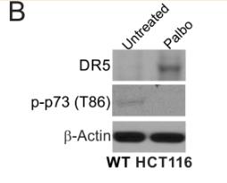
| Product: | Phospho-p73 (Thr86) Antibody |
| Catalog: | AF8296 |
| Description: | Rabbit polyclonal antibody to Phospho-p73 (Thr86) |
| Application: | WB |
| Cited expt.: | WB |
| Reactivity: | Human, Mouse, Rat |
| Prediction: | Pig, Bovine, Sheep, Dog |
| Mol.Wt.: | 80kDa(Observed); 70kD(Calculated). |
| Uniprot: | O15350 |
| RRID: | AB_2840358 |
Control Products

Related Downloads
Protocols
Product Info
*The optimal dilutions should be determined by the end user. For optimal experimental results, antibody reuse is not recommended.
*Tips:
WB: For western blot detection of denatured protein samples. IHC: For immunohistochemical detection of paraffin sections (IHC-p) or frozen sections (IHC-f) of tissue samples. IF/ICC: For immunofluorescence detection of cell samples. ELISA(peptide): For ELISA detection of antigenic peptide.
Cite Format: Affinity Biosciences Cat# AF8296, RRID:AB_2840358.
Fold/Unfold
Delta N p73; DNp73; p53 like transcription factor; p53 related protein; P73; TP73;
Immunogens
A synthesized peptide derived from human p73 around the phosphorylation site of Thr86.
Expressed in striatal neurons of patients with Huntington disease (at protein level). Brain, kidney, placenta, colon, heart, liver, spleen, skeletal muscle, prostate, thymus and pancreas. Highly expressed in fetal tissue.
- O15350 P73_HUMAN:
- Protein BLAST With
- NCBI/
- ExPASy/
- Uniprot
MAQSTATSPDGGTTFEHLWSSLEPDSTYFDLPQSSRGNNEVVGGTDSSMDVFHLEGMTTSVMAQFNLLSSTMDQMSSRAASASPYTPEHAASVPTHSPYAQPSSTFDTMSPAPVIPSNTDYPGPHHFEVTFQQSSTAKSATWTYSPLLKKLYCQIAKTCPIQIKVSTPPPPGTAIRAMPVYKKAEHVTDVVKRCPNHELGRDFNEGQSAPASHLIRVEGNNLSQYVDDPVTGRQSVVVPYEPPQVGTEFTTILYNFMCNSSCVGGMNRRPILIIITLEMRDGQVLGRRSFEGRICACPGRDRKADEDHYREQQALNESSAKNGAASKRAFKQSPPAVPALGAGVKKRRHGDEDTYYLQVRGRENFEILMKLKESLELMELVPQPLVDSYRQQQQLLQRPSHLQPPSYGPVLSPMNKVHGGMNKLPSVNQLVGQPPPHSSAATPNLGPVGPGMLNNHGHAVPANGEMSSSHSAQSMVSGSHCTPPPPYHADPSLVSFLTGLGCPNCIEYFTSQGLQSIYHLQNLTIEDLGALKIPEQYRMTIWRGLQDLKQGHDYSTAQQLLRSSNAATISIGGSGELQRQRVMEAVHFRVRHTITIPNRGGPGGGPDEWADFGFDLPDCKARKQPIKEEFTEAEIH
Predictions
Score>80(red) has high confidence and is suggested to be used for WB detection. *The prediction model is mainly based on the alignment of immunogen sequences, the results are for reference only, not as the basis of quality assurance.
High(score>80) Medium(80>score>50) Low(score<50) No confidence
Research Backgrounds
Participates in the apoptotic response to DNA damage. Isoforms containing the transactivation domain are pro-apoptotic, isoforms lacking the domain are anti-apoptotic and block the function of p53 and transactivating p73 isoforms. May be a tumor suppressor protein.
Isoform alpha (but not isoform beta) is sumoylated on Lys-627, which potentiates proteasomal degradation but does not affect transcriptional activity. Phosphorylation by PLK1 and PLK3 inhibits the transcription regulator activity and pro-apoptotic function.
Higher levels of phosphorylation seen in the brain from patients with Huntington disease.
Polyubiquitinated by RCHY1/PIRH2; leading to its degradation by the proteasome.
Nucleus. Cytoplasm.
Note: Accumulates in the nucleus in response to DNA damage.
Expressed in striatal neurons of patients with Huntington disease (at protein level). Brain, kidney, placenta, colon, heart, liver, spleen, skeletal muscle, prostate, thymus and pancreas. Highly expressed in fetal tissue.
Possesses an acidic transactivation domain, a central DNA binding domain and a C-terminal oligomerization domain that binds to the ABL1 tyrosine kinase SH3 domain.
The PPxY motif mediates interaction with WWOX.
Belongs to the p53 family.
Research Fields
· Cellular Processes > Cell growth and death > p53 signaling pathway. (View pathway)
· Environmental Information Processing > Signal transduction > Hippo signaling pathway. (View pathway)
· Human Diseases > Infectious diseases: Viral > Measles.
· Organismal Systems > Nervous system > Neurotrophin signaling pathway. (View pathway)
References
Application: WB Species: Mice Sample: tumors tissue
Restrictive clause
Affinity Biosciences tests all products strictly. Citations are provided as a resource for additional applications that have not been validated by Affinity Biosciences. Please choose the appropriate format for each application and consult Materials and Methods sections for additional details about the use of any product in these publications.
For Research Use Only.
Not for use in diagnostic or therapeutic procedures. Not for resale. Not for distribution without written consent. Affinity Biosciences will not be held responsible for patent infringement or other violations that may occur with the use of our products. Affinity Biosciences, Affinity Biosciences Logo and all other trademarks are the property of Affinity Biosciences LTD.