Product: Phospho-PPAR alpha (Ser21) Antibody
Catalog: AF8054
Description: Rabbit polyclonal antibody to Phospho-PPAR alpha (Ser21)
Application: WB IF/ICC
Reactivity: Human, Mouse, Rat
Prediction: Bovine, Horse, Sheep, Rabbit, Dog, Xenopus
Mol.Wt.: 52 kDa; 52kD(Calculated).
Uniprot: Q07869
RRID: AB_2840117

View similar products>>

   Size Price Inventory
 100ul $350 In stock
 200ul $450 In stock

Lead Time: Same day delivery

For pricing and ordering contact:
Local distributors

Product Info

Source:
Rabbit
Application:
WB 1:1000-3000, IF/ICC 1:100-1:500
*The optimal dilutions should be determined by the end user.
*Tips:

WB: For western blot detection of denatured protein samples. IHC: For immunohistochemical detection of paraffin sections (IHC-p) or frozen sections (IHC-f) of tissue samples. IF/ICC: For immunofluorescence detection of cell samples. ELISA(peptide): For ELISA detection of antigenic peptide.

Reactivity:
Human,Mouse,Rat
Prediction:
Bovine(100%), Horse(86%), Sheep(100%), Rabbit(100%), Dog(100%), Xenopus(100%)
Clonality:
Polyclonal
Specificity:
Phospho-PPAR alpha (Ser21) Antibody detects endogenous levels of PPAR alpha only when phosphorylated at Ser21.
RRID:
AB_2840117
Cite Format: Affinity Biosciences Cat# AF8054, RRID:AB_2840117.
Conjugate:
Unconjugated.
Purification:
The antibody is from purified rabbit serum by affinity purification via sequential chromatography on phospho-peptide and non-phospho-peptide affinity columns.
Storage:
Rabbit IgG in phosphate buffered saline , pH 7.4, 150mM NaCl, 0.02% sodium azide and 50% glycerol. Store at -20 °C. Stable for 12 months from date of receipt.
Alias:

Fold/Unfold

hPPAR; MGC2237; MGC2452; NR1C1; Nuclear receptor subfamily 1 group C member 1; OTTHUMP00000197740; OTTHUMP00000197741; Peroxisome proliferative activated receptor alpha; Peroxisome proliferator activated receptor alpha; Peroxisome proliferator-activated receptor alpha; PPAR; PPAR-alpha; ppara; PPARA_HUMAN; PPARalpha;

Immunogens

Immunogen:
Uniprot:
Gene(ID):
Expression:
Q07869 PPARA_HUMAN:

Skeletal muscle, liver, heart and kidney. Expressed in monocytes (PubMed:28167758).

Sequence:
MVDTESPLCPLSPLEAGDLESPLSEEFLQEMGNIQEISQSIGEDSSGSFGFTEYQYLGSCPGSDGSVITDTLSPASSPSSVTYPVVPGSVDESPSGALNIECRICGDKASGYHYGVHACEGCKGFFRRTIRLKLVYDKCDRSCKIQKKNRNKCQYCRFHKCLSVGMSHNAIRFGRMPRSEKAKLKAEILTCEHDIEDSETADLKSLAKRIYEAYLKNFNMNKVKARVILSGKASNNPPFVIHDMETLCMAEKTLVAKLVANGIQNKEAEVRIFHCCQCTSVETVTELTEFAKAIPGFANLDLNDQVTLLKYGVYEAIFAMLSSVMNKDGMLVAYGNGFITREFLKSLRKPFCDIMEPKFDFAMKFNALELDDSDISLFVAAIICCGDRPGLLNVGHIEKMQEGIVHVLRLHLQSNHPDDIFLFPKLLQKMADLRQLVTEHAQLVQIIKKTESDAALHPLLQEIYRDMY

Predictions

Predictions:

Score>80(red) has high confidence and is suggested to be used for WB detection. *The prediction model is mainly based on the alignment of immunogen sequences, the results are for reference only, not as the basis of quality assurance.

Species
Results
Score
Bovine
100
Sheep
100
Dog
100
Xenopus
100
Rabbit
100
Horse
86
Pig
0
Zebrafish
0
Chicken
0
Model Confidence:
High(score>80) Medium(80>score>50) Low(score<50) No confidence

PTMs - Q07869 As Substrate

Site PTM Type Enzyme
S6 Phosphorylation
S12 Phosphorylation P28482 (MAPK1) , P27361 (MAPK3)
S21 Phosphorylation P27361 (MAPK3) , P28482 (MAPK1)
S45 Phosphorylation
S179 Phosphorylation P05771-2 (PRKCB) , P17252 (PRKCA)
K185 Sumoylation
S230 Phosphorylation P05771-2 (PRKCB) , P17252 (PRKCA)

Research Backgrounds

Function:

Ligand-activated transcription factor. Key regulator of lipid metabolism. Activated by the endogenous ligand 1-palmitoyl-2-oleoyl-sn-glycerol-3-phosphocholine (16:0/18:1-GPC). Activated by oleylethanolamide, a naturally occurring lipid that regulates satiety. Receptor for peroxisome proliferators such as hypolipidemic drugs and fatty acids. Regulates the peroxisomal beta-oxidation pathway of fatty acids. Functions as transcription activator for the ACOX1 and P450 genes. Transactivation activity requires heterodimerization with RXRA and is antagonized by NR2C2. May be required for the propagation of clock information to metabolic pathways regulated by PER2.

Subcellular Location:

Nucleus.

Extracellular region or secreted Cytosol Plasma membrane Cytoskeleton Lysosome Endosome Peroxisome ER Golgi apparatus Nucleus Mitochondrion Manual annotation Automatic computational assertionSubcellular location
Tissue Specificity:

Skeletal muscle, liver, heart and kidney. Expressed in monocytes.

Subunit Structure:

Heterodimer; with RXRA. This heterodimerization is required for DNA binding and transactivation activity. Interacts with NCOA3 coactivator. Interacts with CITED2; the interaction stimulates its transcriptional activity. Also interacts with PPARBP in vitro. Interacts with AKAP13, LPIN1, PRDM16 and coactivator NCOA6. Interacts with ASXL1 and ASXL2. Interacts with PER2. Interacts with SIRT1; the interaction seems to be modulated by NAD(+) levels. Interacts with CRY1 and CRY2 (By similarity).

Family&Domains:

Belongs to the nuclear hormone receptor family. NR1 subfamily.

Research Fields

· Environmental Information Processing > Signal transduction > cAMP signaling pathway.   (View pathway)

· Human Diseases > Endocrine and metabolic diseases > Insulin resistance.

· Human Diseases > Endocrine and metabolic diseases > Non-alcoholic fatty liver disease (NAFLD).

· Human Diseases > Infectious diseases: Viral > Hepatitis C.

· Organismal Systems > Endocrine system > PPAR signaling pathway.

· Organismal Systems > Endocrine system > Adipocytokine signaling pathway.

· Organismal Systems > Endocrine system > Glucagon signaling pathway.

References

1). Hepatocyte-secreted Autotaxin Exacerbates Nonalcoholic Fatty Liver Disease Through Autocrine Inhibition of the PPARalpha/FGF21 axis. Cellular and Molecular Gastroenterology and Hepatology (PubMed: 35931383) [IF=7.2]

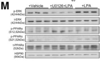
Application: WB    Species: Mouse    Sample:

Figure 7The ATX–LPA axis suppresses hepatic production of FGF21 via ERK-mediated inhibition of PPARα. Six-week-old C57BL/6N male mice (n = 6–8) were fed with either STC or HFD for 8 weeks, and subsequently injected with 1 mg/kg body weight antibody (anti-ATX or nonimmune IgG [control]) every 10 days for another 7 weeks as in Figure 6. Mice were killed at the 15th week. (A) Real-time PCR analysis of mRNA expression levels of several hepatokines in liver (shown as fold change against the STC–Control group). (B) Real-time PCR analysis for mRNA expression levels of several adipokines in epididymal adipose tissue (shown as fold change against the STC–Control group). n = 6–8. Data are presented as the means ± SEM. (C) Serum levels of FGF21. (D) Hepatic mRNA expression levels of Fgf21 determined by real-time PCR analysis. (E) Serum levels of adiponectin. (F–H) Six-week-old C57BL/6N male mice were fed with STC or HFD for 15 weeks after intravenous injection with 1 × 10¹¹ vector genome of AAV encoding shRNA specific to autotaxin (KD) or scramble control (SC) as in Figure 4. (F) Serum levels of FGF21. (G) Hepatic mRNA expression levels of FGF21 determined by real-time PCR analysis. (H) Serum levels of adiponectin. (I–J) Huh7 cells grown in serum-free DMEM were treated with or without the indicated dosage of rhATX, PF8380 (inhibitor of autotaxin), LPA, or Ki16425 (LPA-receptor antagonist) for 24 hours. (I) The mRNA expression levels of FGF21 (expressed as fold changes relative to vehicle [dimethyl sulfoxide]-treated controls) and its (J) protein concentrations in the conditioned medium were determined by real-time PCR and enzyme-linked immunosorbent assay (ELISA), respectively. n = 5. Data are presented as the means ± SEM. (K–L) Huh7 hepatocytes were pretreated with rhATX, LPA, PF8380 (inhibitor of autotaxin), Ki16425 (LPA-receptor antagonist) for 30 minutes, followed by treatment with or without fenofibrate for 24 hours. (K) FGF21 protein was secreted into conditioned medium and (L) hepatic FGF21 mRNA were determined by ELISA and real-time PCR, respectively. Huh7 hepatocytes were pretreated with U0126 (ERK inhibitor), wortmannin (PI3K), or LPA for 30 minutes, (M and N) followed by treatment with or without fenofibrate for 30 minutes to collect cell lysates for Western blot analysis, or (O) 24 hours to measure the relative mRNA abundance of FGF21, PPARA, and VLCAD (very-long-chain acylcoenzyme-A dehydrogenase, a downstream target of PPARα). (P) Relative luciferase activity in Huh7 hepatocytes transfected with a luciferase reporter vector carrying PPAR response element and treated with LPA, U0126, and wortmannin in the presence or absence of fenofibrate for 48 hours. Data are normalized against human β-ACTIN and expressed as the fold change relative to dimethyl sulfoxide–treated controls. (A–L and N–P) Statistical calculations were performed by 1-way analysis of variance followed by Tukey multiple comparisons using Prism 6. ∗P < .05, ∗∗P < .01, and ∗∗∗P < .001. n = 5. Data are presented as the means ± SEM. HSP90, heat shock protein 90; p-ERK, phosphorylated_extracellular signal-related kinase; p-PPARα, phosphorylated peroxisome proliferator–activated receptor α; t-ERK, total extracellular signal-related kinase; t-PPARα, total peroxisome proliferator–activated receptor α.

Restrictive clause


Affinity Biosciences tests all products strictly. Citations are provided as a resource for additional applications that have not been validated by Affinity Biosciences. Please choose the appropriate format for each application and consult Materials and Methods sections for additional details about the use of any product in these publications.

For Research Use Only.
Not for use in diagnostic or therapeutic procedures. Not for resale. Not for distribution without written consent. Affinity Biosciences will not be held responsible for patent infringement or other violations that may occur with the use of our products. Affinity Biosciences, Affinity Biosciences Logo and all other trademarks are the property of Affinity Biosciences LTD.