Cleaved-Caspase 3 (Asp175), p17 Antibody - #BF0711

Product Info
*The optimal dilutions should be determined by the end user.
*Tips:
WB: For western blot detection of denatured protein samples. IHC: For immunohistochemical detection of paraffin sections (IHC-p) or frozen sections (IHC-f) of tissue samples. IF/ICC: For immunofluorescence detection of cell samples. ELISA(peptide): For ELISA detection of antigenic peptide.
Cite Format: Affinity Biosciences Cat# BF0711, RRID:AB_2846190.
Fold/Unfold
A830040C14Rik; Apopain; CASP-3; CASP3; CASP3_HUMAN; Casp3a; Caspase 3; Caspase 3, apoptosis-related cysteine peptidase; Caspase 3, apoptosis-related cysteine protease; Caspase 3, apoptosis-related cysteine protease a; Caspase-3 subunit p12; CC3; CPP-32; CPP32; CPP32B; Cysteine protease CPP32; EC 3.4.22.56; LICE; mldy; OTTHUMP00000165052; OTTHUMP00000165053; OTTHUMP00000165054; PARP cleavage protease; Procaspase3; protein Yama; SCA 1; SCA-1; SREBP cleavage activity 1; Yama;
Immunogens
Purified recombinant fragment of human Caspase 3 expressed in E. Coli.
Highly expressed in lung, spleen, heart, liver and kidney. Moderate levels in brain and skeletal muscle, and low in testis. Also found in many cell lines, highest expression in cells of the immune system.
- P42574 CASP3_HUMAN:
- Protein BLAST With
- NCBI/
- ExPASy/
- Uniprot
MENTENSVDSKSIKNLEPKIIHGSESMDSGISLDNSYKMDYPEMGLCIIINNKNFHKSTGMTSRSGTDVDAANLRETFRNLKYEVRNKNDLTREEIVELMRDVSKEDHSKRSSFVCVLLSHGEEGIIFGTNGPVDLKKITNFFRGDRCRSLTGKPKLFIIQACRGTELDCGIETDSGVDDDMACHKIPVEADFLYAYSTAPGYYSWRNSKDGSWFIQSLCAMLKQYADKLEFMHILTRVNRKVATEFESFSFDATFHAKKQIPCIVSMLTKELYFYH
PTMs - P42574 As Substrate
| Site | PTM Type | Enzyme | Source |
|---|---|---|---|
| M1 | Acetylation | Uniprot | |
| T4 | Phosphorylation | Uniprot | |
| S7 | Phosphorylation | Uniprot | |
| S10 | Phosphorylation | Uniprot | |
| K11 | Acetylation | Uniprot | |
| K11 | Ubiquitination | Uniprot | |
| S12 | Phosphorylation | Uniprot | |
| K14 | Ubiquitination | Uniprot | |
| K19 | Ubiquitination | Uniprot | |
| S24 | Phosphorylation | Uniprot | |
| S26 | Phosphorylation | Uniprot | |
| S29 | Phosphorylation | Uniprot | |
| Y41 | Phosphorylation | Uniprot | |
| K57 | Ubiquitination | Uniprot | |
| S65 | Phosphorylation | Uniprot | |
| T67 | Phosphorylation | Uniprot | |
| K82 | Acetylation | Uniprot | |
| K82 | Ubiquitination | Uniprot | |
| K88 | Ubiquitination | Uniprot | |
| K105 | Ubiquitination | Uniprot | |
| K138 | Ubiquitination | Uniprot | |
| S150 | Phosphorylation | Q16539 (MAPK14) | Uniprot |
| T152 | Phosphorylation | Uniprot | |
| C163 | S-Nitrosylation | Uniprot | |
| T174 | Phosphorylation | Uniprot | |
| S176 | Phosphorylation | Uniprot | |
| K210 | Ubiquitination | Uniprot | |
| K229 | Ubiquitination | Uniprot | |
| S249 | Phosphorylation | Uniprot | |
| K260 | Ubiquitination | Uniprot | |
| T270 | Phosphorylation | Uniprot |
Research Backgrounds
Involved in the activation cascade of caspases responsible for apoptosis execution. At the onset of apoptosis it proteolytically cleaves poly(ADP-ribose) polymerase (PARP) at a '216-Asp-|-Gly-217' bond. Cleaves and activates sterol regulatory element binding proteins (SREBPs) between the basic helix-loop-helix leucine zipper domain and the membrane attachment domain. Cleaves and activates caspase-6, -7 and -9. Involved in the cleavage of huntingtin. Triggers cell adhesion in sympathetic neurons through RET cleavage.
Cleavage by granzyme B, caspase-6, caspase-8 and caspase-10 generates the two active subunits. Additional processing of the propeptides is likely due to the autocatalytic activity of the activated protease. Active heterodimers between the small subunit of caspase-7 protease and the large subunit of caspase-3 also occur and vice versa.
S-nitrosylated on its catalytic site cysteine in unstimulated human cell lines and denitrosylated upon activation of the Fas apoptotic pathway, associated with an increase in intracellular caspase activity. Fas therefore activates caspase-3 not only by inducing the cleavage of the caspase zymogen to its active subunits, but also by stimulating the denitrosylation of its active site thiol.
Cytoplasm.
Highly expressed in lung, spleen, heart, liver and kidney. Moderate levels in brain and skeletal muscle, and low in testis. Also found in many cell lines, highest expression in cells of the immune system.
Heterotetramer that consists of two anti-parallel arranged heterodimers, each one formed by a 17 kDa (p17) and a 12 kDa (p12) subunit. Interacts with BIRC6/bruce.
Belongs to the peptidase C14A family.
Research Fields
· Cellular Processes > Cell growth and death > p53 signaling pathway. (View pathway)
· Cellular Processes > Cell growth and death > Apoptosis. (View pathway)
· Cellular Processes > Cell growth and death > Apoptosis - multiple species. (View pathway)
· Environmental Information Processing > Signal transduction > MAPK signaling pathway. (View pathway)
· Environmental Information Processing > Signal transduction > TNF signaling pathway. (View pathway)
· Human Diseases > Drug resistance: Antineoplastic > Platinum drug resistance.
· Human Diseases > Endocrine and metabolic diseases > Non-alcoholic fatty liver disease (NAFLD).
· Human Diseases > Neurodegenerative diseases > Alzheimer's disease.
· Human Diseases > Neurodegenerative diseases > Parkinson's disease.
· Human Diseases > Neurodegenerative diseases > Amyotrophic lateral sclerosis (ALS).
· Human Diseases > Neurodegenerative diseases > Huntington's disease.
· Human Diseases > Infectious diseases: Bacterial > Epithelial cell signaling in Helicobacter pylori infection.
· Human Diseases > Infectious diseases: Bacterial > Pertussis.
· Human Diseases > Infectious diseases: Bacterial > Legionellosis.
· Human Diseases > Infectious diseases: Parasitic > Toxoplasmosis.
· Human Diseases > Infectious diseases: Parasitic > Amoebiasis.
· Human Diseases > Infectious diseases: Bacterial > Tuberculosis.
· Human Diseases > Infectious diseases: Viral > Hepatitis B.
· Human Diseases > Infectious diseases: Viral > Human papillomavirus infection.
· Human Diseases > Infectious diseases: Viral > Herpes simplex infection.
· Human Diseases > Cancers: Overview > Pathways in cancer. (View pathway)
· Human Diseases > Cancers: Overview > Viral carcinogenesis.
· Human Diseases > Cancers: Overview > Proteoglycans in cancer.
· Human Diseases > Cancers: Overview > MicroRNAs in cancer.
· Human Diseases > Cancers: Specific types > Colorectal cancer. (View pathway)
· Human Diseases > Cancers: Specific types > Small cell lung cancer. (View pathway)
· Human Diseases > Cardiovascular diseases > Viral myocarditis.
· Organismal Systems > Immune system > Natural killer cell mediated cytotoxicity. (View pathway)
· Organismal Systems > Immune system > IL-17 signaling pathway. (View pathway)
· Organismal Systems > Nervous system > Serotonergic synapse.
References

Application: IF/ICC Species: Mice Sample: Tumor
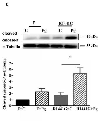
Application: WB Species: mice Sample: R1441G cells and FVBN cells

Application: IF/ICC Species: Mouse Sample:
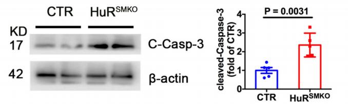
Application: WB Species: mouse Sample: SMCs
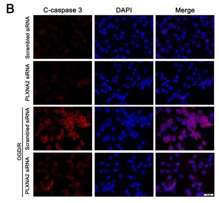
Application: IF/ICC Species: Mice Sample: BV2 microglia cells
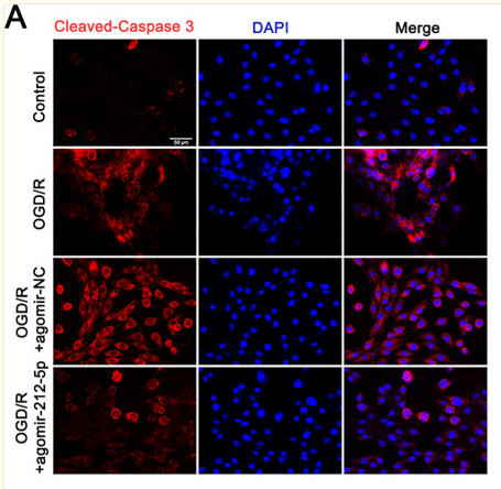
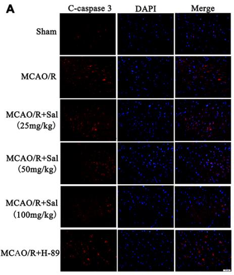
Application: IF/ICC Species: Rat Sample: PC12 cells
Application: IF/ICC Species: rat Sample:

Application: WB Species: rat Sample:
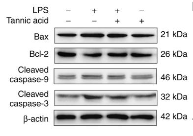
Application: WB Species: rat Sample: H9C2 cells
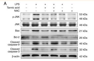
Application: WB Species: Rat Sample: H9C2 cells
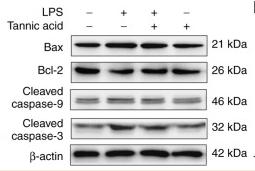
Application: WB Species: Rat Sample: H9C2 cells
Application: WB Species: Rat Sample: H9C2 cells
Restrictive clause
Affinity Biosciences tests all products strictly. Citations are provided as a resource for additional applications that have not been validated by Affinity Biosciences. Please choose the appropriate format for each application and consult Materials and Methods sections for additional details about the use of any product in these publications.
For Research Use Only.
Not for use in diagnostic or therapeutic procedures. Not for resale. Not for distribution without written consent. Affinity Biosciences will not be held responsible for patent infringement or other violations that may occur with the use of our products. Affinity Biosciences, Affinity Biosciences Logo and all other trademarks are the property of Affinity Biosciences LTD.