

, His tag,GST-tagged M (GST-M), and GST tag were expressed in E. coli BL21 (DE3), and the soluble His-chBRD2 and His tag or GST-M and GST tag were purifed on Ni–NTA His*Bind Resin or Glutathione-Sepharose 4B beads, respectively. The bacterial expression purifed proteins were detected by SDS-PAGE along with Coomassie blue staining. Identifcation of the interaction between the M protein and chBRD2 protein by His pull-down assay (D) and GST pull-down assay (E).")
Control Products
Product Info
Source:
Mouse
Application:
WB 1:3000-1:10000, IF/ICC: 1:200, IP 1:100-1:500
*The optimal dilutions should be determined by the end user. For optimal experimental results, antibody reuse is not recommended.
*Tips:
*The optimal dilutions should be determined by the end user. For optimal experimental results, antibody reuse is not recommended.
*Tips:
WB: For western blot detection of denatured protein samples. IHC: For immunohistochemical detection of paraffin sections (IHC-p) or frozen sections (IHC-f) of tissue samples. IF/ICC: For immunofluorescence detection of cell samples. ELISA(peptide): For ELISA detection of antigenic peptide.
Reactivity:
All
Clonality:
Monoclonal [T113]
Specificity:
His-Tag Mouse Monoclonal antibody detects endogenous levels of internal, C-terminal, or N-terminal 6His -tagged proteins.
RRID:
AB_2839416
Cite Format: Affinity Biosciences Cat# T0009, RRID:AB_2839416.
Cite Format: Affinity Biosciences Cat# T0009, RRID:AB_2839416.
Conjugate:
Unconjugated.
Purification:
Affinity-chromatography.
Storage:
Mouse IgG1 in phosphate buffered saline (without Mg2+ and Ca2+), pH 7.4, 150mM NaCl, 0.02% sodium azide and 50% glycerol. Store at -20 °C. Stable for 12 months from date of receipt.
Alias:
Fold/Unfold
his tag
Immunogens
Immunogen:
A synthetic peptide HHHHHH coupled to KLH.
Description:
N/A
References
1). Discovery of a potent allosteric activator of DGKQ that ameliorates obesity-induced insulin resistance via the sn-1,2-DAG-PKCε signaling axis. Cell metabolism, 2020
(PubMed: 36525963)
[IF=27.7]
2). Identification of human LDHC4 as a potential target for anticancer drug discovery. Acta Pharmaceutica Sinica B, 2022
(PubMed: 35646544)
[IF=14.7]
Application: WB Species: Mice Sample: A549 cells
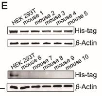
Figure 2
Overexpression of LDHC promotes tumor growth in mice. (A) The nude mice inoculated with A549 cells overexpressing LDHC (upper) and with control A549 cells (lower), the picture was taken on the 45th day after inoculation. (B) Tumor growth curves of the LDHC-overexpression group (red) and the control group (black). The volumes of the tumors overexpressing LDHC are significantly higher than that of the control tumors (n = 5, data are mean ± SD, ∗∗∗P < 0.001). (C) The picture of the tumors from the LDHC-overexpression group and the control group with a ruler to show the diameters of the tumors. (D) Statistical analysis of tumor mass. The masses of the tumors overexpressing LDHC are significantly higher than that of the control tumors (n = 5, data are mean ± SD, ∗∗∗P < 0.001). (E) Detection of His6-tag of the recombinant LDHC in tumors by Western blot. Mice 1 to 5 were inoculated with the LDHC-overexpression A549 cells, while mice 6 to 10 were inoculated with the control A549 cells. HEK 293 cells were used as the positive control.
3). pH-responsive nanocomposite hydrogel for simultaneous prevention of postoperative adhesion and tumor recurrence. Acta Biomaterialia, 2023
(PubMed: 36563777)
[IF=9.4]
4). Multifunctional human serum albumin fusion protein as a docetaxel nanocarrier for chemo-photothermal synergetic therapy of ovarian cancer. ACS Applied Materials & Interfaces, 2022
(PubMed: 35441508)
[IF=8.3]
5). Staphylococcal superantigen-like protein 10 induces necroptosis through TNFR1 activation of RIPK3-dependent signal pathways. Communications Biology, 2022
(PubMed: 35962126)
[IF=5.9]
6). Discovery of a new function of human butyrylcholinesterase and the catalytic activity of its natural variants toward homocysteine thiolactone hydrolysis. Chemico-biological interactions, 2025
(PubMed: 40744384)
[IF=4.7]
7). The 1316T> C missenses mutation in MTHFR contributes to MTHFR deficiency by targeting MTHFR to proteasome degradation. Aging-US, 2021
(PubMed: 33290257)
[IF=3.9]
8). Improving glucose oxidase catalysis in Aspergillus niger via Vitreoscilla hemoglobin fusion protein. Applied microbiology and biotechnology, 2024
(PubMed: 38183481)
[IF=3.9]
9). Recombinant expression a novel fibronectin—collage fusion peptide modulating stem cell stemness via integrin β3. APPLIED MICROBIOLOGY AND BIOTECHNOLOGY, 2022
(PubMed: 35590080)
[IF=3.9]
10). Aedes albopictus salivary adenosine deaminase is an immunomodulatory factor facilitating dengue virus replication. Scientific reports, 2023
(PubMed: 37794048)
[IF=3.8]
Restrictive clause
Affinity Biosciences tests all products strictly. Citations are provided as a resource for additional applications that have not been validated by Affinity Biosciences. Please choose the appropriate format for each application and consult Materials and Methods sections for additional details about the use of any product in these publications.
For Research Use Only.
Not for use in diagnostic or therapeutic procedures. Not for resale. Not for distribution without written consent. Affinity Biosciences will not be held responsible for patent infringement or other violations that may occur with the use of our products. Affinity Biosciences, Affinity Biosciences Logo and all other trademarks are the property of Affinity Biosciences LTD.