

 Silver staining of precipitations by BioID. LO2 cells were stably transfected with pCDH–SENP3–BirA (named LO2–SENP3–BirA). LO2–SENP3–BirA cells were untreated or treated with DEN. (B) A pie chart showing the number of proteins identified by MS. (C) The interaction of SENP3 with TKT, NPM1, and ATP5F1A was verified by co-IP. LO2 cells were transfected with pCDH–SENP3–BirA and treated with DEN. (D) The in vitro interaction of SENP3 with TKT was validated by GST pull-down assay. Flag-SENP3 was transfected into HEK293T cells, and SENP3 was purified from cell lysates with anti-Flag M2 gels after 24 h of transfection. GST–TKT was obtained from prokaryotic expression system. (E) The physical interaction of SENP3 with TKT or ATP5F1A was measured by FRET with acceptor photobleaching method. After treatment with DEN for 2 h, LO2 cells were fixed and stained with primary antibodies and fluorescence-conjugated secondary antibodies. Scale bar: 10 μm. The efficiency of FRET from 10 cells are shown. (F) Transient SUMOylated proteins that interact with SENP3 were evaluated. co-IP was performed in HEK293T cells transfected with HA–SENP3.")
 or Sls1-FLAG (positive control), and Δmtf2 cells expressing Sls1-FLAG were subjected to anti-FLAG co-immunoprecipitation (A). Mitochondrial extracts prepared from WT cells expressing untagged Mrh5 or Mrh5-Myc, and Δmtf2 cells expressing Mrh5-Myc were subjected to anti-Myc co-immunoprecipitation (B). C, loss of Sls1 disrupts Mrh5C. WT cells expressing untagged Mrh5 or Mrh5-Myc and Δmtf2 cells expressing Mrh5-Myc were subjected to anti-Myc co-immunoprecipitation. D, loss of Mrh5 does not affect interactions among other members of Mrh5C. WT cells expressing untagged Sls1 or Sls1-FLAG, and Δmrh5 cells expressing Sls1-FLAG were subjected to anti-FLAG co-immunoprecipitation. E, loss of Ppr4 does not affect interactions among other members of Mrh5C. WT cells expressing untagged Sls1 or Sls1-FLAG, and Δppr4 cells expressing Sls1-FLAG were subjected to anti-FLAG co-immunoprecipitation. F, Mtf2 and Sls1 form a complex independently of Mrh5 and Ppr4. WT cells expressing untagged Sls1 or Sls1-FLAG, and Δmrh5Δppr4 cells expressing Sls1-FLAG were subjected to anti-FLAG co-immunoprecipitation. Mitochondrial extracts (IN) and immunoprecipitates (IP) were analyzed by immunoblotting using specific anti-tag Abs. Genes encoding the Mrh5C subunits were endogenously tagged to facilitate their detection.")
Control Products
Product Info
Source:
Mouse
Application:
WB 1:3000-1:10000, IF/ICC: 1:200, IP 1:200
*The optimal dilutions should be determined by the end user. For optimal experimental results, antibody reuse is not recommended.
*Tips:
*The optimal dilutions should be determined by the end user. For optimal experimental results, antibody reuse is not recommended.
*Tips:
WB: For western blot detection of denatured protein samples. IHC: For immunohistochemical detection of paraffin sections (IHC-p) or frozen sections (IHC-f) of tissue samples. IF/ICC: For immunofluorescence detection of cell samples. ELISA(peptide): For ELISA detection of antigenic peptide.
Reactivity:
All
Clonality:
Monoclonal [T80]
Specificity:
HA-Tag Mouse Monoclonal antibody detects endogenous levels of internal, C-terminal, or N-terminal HA-tagged recombinant proteins.
RRID:
AB_2839415
Cite Format: Affinity Biosciences Cat# T0008, RRID:AB_2839415.
Cite Format: Affinity Biosciences Cat# T0008, RRID:AB_2839415.
Conjugate:
Unconjugated.
Purification:
Affinity-chromatography.
Storage:
Mouse IgG1 in phosphate buffered saline (without Mg2+ and Ca2+), pH 7.4, 150mM NaCl,. Store at -20 °C. Stable for 12 months from date of receipt.
Alias:
Fold/Unfold
ha tag
Immunogens
Immunogen:
A synthetic peptide YPYDVPDYA coupled to KLH.
Description:
N/A
References
1). The m6A reader YTHDF1 promotes ovarian cancer progression via augmenting EIF3C translation. NUCLEIC ACIDS RESEARCH, 2020
(PubMed: 31996915)
[IF=16.6]
Application: WB Species: Human Sample: in A2780 or SKOV3 cells
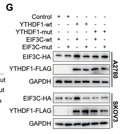
Figure 6. YTHDF1 regulates EIF3C translation in an m6A-dependent manner. (A) Relative RNA level of EIF3C in A2780 and SKOV3 upon YTHDF1
knockdown. (B) Western blot detected the protein level of EIF3C in A2780 and SKOV3 cells upon YTHDF1 knockdown. (C) Gene-specific m6A qPCR
validation of m6A levels in A2780 and SKOV3 cells. Primers to m6A negative region of EEF1A as the negative control and primers to m6A postive region of
EEF1A as the positive control. (D) YTHDF1 RIP followed by RT-qPCR confirmed the interaction between YTHDF1 and EIF3C mRNA. (E) Schematic
representation of wild-type (YTHDF1-wt) and mutant (YTHDF1-mut) YTHDF1 constructs. (F) RIP-derived RNA and protein in A2780 cells were
measured by RT-qPCR and western blot, respectively. GAPDH was used as the negative control in western blot assays. (G) Western blot confirmed HAtagged EIF3C expression in A2780 or SKOV3 cells co-transfected with empty vector, wild-type or mutant Flag-tagged YTHDF1 and wild-type or mutant
HA-tagged EIF3C. (H) Nascent protein synthesis was detected by HPG incorporation upon YTHDF1 knockdown or EIF3C knockdown in A2780 cells.
Scale bar, 100 m. Data are shown as means ± S.D. *P < 0.05, **P < 0.01, ***P < 0.001, NS, not significant.
2). Bunyavirus SFTSV exploits autophagic flux for viral assembly and egress. Autophagy, 2021
(PubMed: 34747299)
[IF=14.6]
3). TOR3A represses type I interferon production and limits viral clearance during respiratory syncytial virus infection. Emerging microbes & infections, 2026
(PubMed: 41739568)
[IF=13.2]
4). WWP2 drives the progression of gastric cancer by facilitating the ubiquitination and degradation of LATS1 protein. Cell Communication and Signaling, 2023
(PubMed: 36803368)
[IF=8.4]
5). Aberrant serum-derived FN1 variants bind to integrin β1 on glomerular endothelial cells contributing to thin basement membrane nephropathy. International journal of biological macromolecules, 2024
(PubMed: 39368581)
[IF=7.7]
6). Up-regulated lncRNA CYLD as a ceRNA of miR-2383 facilitates bovine viral diarrhea virus replication by promoting CYLD expression to counteract RIG-I-mediated type-I IFN production. International journal of biological macromolecules, 2023
(PubMed: 37839600)
[IF=7.7]
7). Oryza sativa POSITIVE REGULATOR OF IRON DEFICIENCY RESPONSE 2 (OsPRI2) and OsPRI3 are involved in the maintenance of Fe homeostasis. PLANT CELL AND ENVIRONMENT, 2020
(PubMed: 31674679)
[IF=6.0]
Application: WB Species: yeast Sample: yeast cells
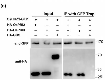
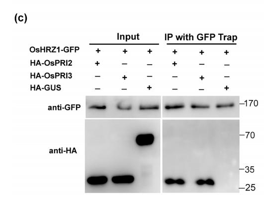
FIGURE 1| Interaction of OsHRZ1 with OsPRI2 and OsPRI3.(c) CoIP assay. Total proteins from different combinations with OsHRZ1‐GFP and HA‐OsPRI2/HA‐OsPRI3/HA‐GUS were immunoprecipitated with GFP‐Trap followed by immunoblotting with the indicated antibodies. HRZ1‐GFP/HA‐GUS, negative control. Protein molecular weight (in kDa) is indicated.
Application: WB Species: Plant Sample: Root
FIGURE 1 Interaction of OsHRZ1 with OsPRI2 and OsPRI3. (a) Yeast two‐hybrid assays. Yeast co‐transformed with different BD and AD
plasmid combinations were spotted in parallel in 10‐fold dilution series on synthetic dropout medium lacking Leu/Trp/His/Ade. The C‐terminal
truncated OsHRZ1 and full‐length OsPRI2/3 were cloned into pGBKT7 and pGADT7, respectively. OsHRZ1‐C/OsPRI1, positive control. OsHRZ1‐
C/Empty, negative control. (b) Pull‐down assay. OsHRZ1 was fused with the GST tag and OsPRI2/3 were fused with the His tag. Recombinant
proteins were expressed in E. coli. Proteins were pulled down by glutathione Sepharose 4B and detected using the anti‐His antibody. Protein
molecular weight (in kDa) is indicated. (c) CoIP assay. Total proteins from different combinations with OsHRZ1‐GFP and HA‐OsPRI2/HA‐OsPRI3/
HA‐GUS were immunoprecipitated with GFP‐Trap followed by immunoblotting with the indicated antibodies. HRZ1‐GFP/HA‐GUS, negative
control. Protein molecular weight (in kDa) is indicated. (d) Degradation of OsPRI2 or OsPRI3 was carried out by detecting the OsPRI2/3‐GFP
protein level in co‐infiltration experiments with increasing amounts of OsHRZ1‐GFP. GFP proteins were used as an internal control. Anti‐GFP
antibody was used in western blot. Protein molecular weight (in kDa) is indicated. Stars indicate the non‐specific bands. Numbers indicate the ratio
of the concentrations of agrobacteria used in co‐infiltration. Empty vector, a binary vector pOCA30 with a 35S promoter; GFP, 35S:GFP in
pOCA30; OsPRI2‐GFP, 35S:OsPRI2‐GFP in pOCA30; OsPRI3‐GFP, 35S:OsPRI3‐GFP in pOCA30; OsHRZ1‐GFP, 35S:OsHRZ1‐GFP in pOCA30.
(e) Cell‐free degradation. Ten‐day‐old roots grown in Fe‐sufficient solution were harvested and used for protein extraction. Incubation with or
without MG132 was performed over the indicated time course
8). IL-1β/IL-1R1 signaling induced by intranasal lipopolysaccharide infusion regulates alpha-Synuclein pathology in the olfactory bulb, substantia nigra and striatum. Brain Pathology, 2020
(PubMed: 32678959)
[IF=5.8]
9). Mutation of Basic Residues R283, R286, and K288 in the Matrix Protein of Newcastle Disease Virus Attenuates Viral Replication and Pathogenicity. International Journal of Molecular Sciences, 2023
(PubMed: 36674496)
[IF=5.6]
10). RNF149 modulates the type I IFN innate antiviral immune responses through degrading IRF3. PLoS pathogens, 2025
(PubMed: 40245000)
[IF=5.5]
Restrictive clause
Affinity Biosciences tests all products strictly. Citations are provided as a resource for additional applications that have not been validated by Affinity Biosciences. Please choose the appropriate format for each application and consult Materials and Methods sections for additional details about the use of any product in these publications.
For Research Use Only.
Not for use in diagnostic or therapeutic procedures. Not for resale. Not for distribution without written consent. Affinity Biosciences will not be held responsible for patent infringement or other violations that may occur with the use of our products. Affinity Biosciences, Affinity Biosciences Logo and all other trademarks are the property of Affinity Biosciences LTD.