Product Info

Source:
Mouse
Application:
WB 1:3000-1:10000, IHC 1:50-1:200, IF/ICC: 1:100-1:500, IP 1:100-1:500
*The optimal dilutions should be determined by the end user.
*Tips:

WB: For western blot detection of denatured protein samples. IHC: For immunohistochemical detection of paraffin sections (IHC-p) or frozen sections (IHC-f) of tissue samples. IF/ICC: For immunofluorescence detection of cell samples. ELISA(peptide): For ELISA detection of antigenic peptide.

Reactivity:
All
Clonality:
Monoclonal [5F67]
Specificity:
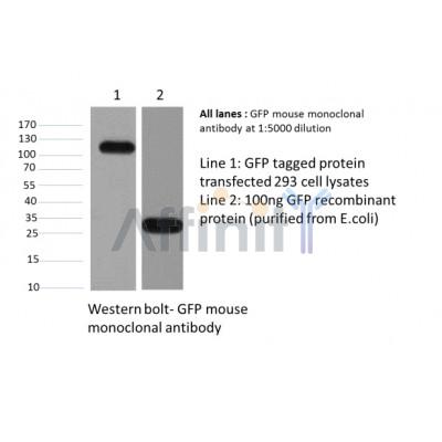
GFP-Tag Mouse Monoclonal antibody detects endogenous levels of C-terminal, internal, and N-terminal GFP-tagged proteins.
RRID:
AB_2839413
Cite Format: Affinity Biosciences Cat# T0005, RRID:AB_2839413.
Conjugate:
Unconjugated.
Purification:
Affinity-chromatography.
Storage:
Mouse IgG1 in phosphate buffered saline (without Mg2+ and Ca2+), pH 7.4, 150mM NaCl, 0.02% sodium azide and 50% glycerol. Store at -20 °C. Stable for 12 months from date of receipt.

Immunogens

Immunogen:

Full-length GFP protein.

Description:
N/A

References

1). Discovery of a potent allosteric activator of DGKQ that ameliorates obesity-induced insulin resistance via the sn-1,2-DAG-PKCε signaling axis. Cell metabolism, 2020 (PubMed: 36525963) [IF=29.0]

2). CPT1A induction following epigenetic perturbation promotes MAVS palmitoylation and activation to potentiate antitumor immunity. Molecular cell, 2023 (PubMed: 38016475) [IF=16.0]

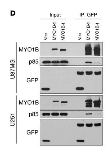
3). Splicing factor SRSF1 promotes gliomagenesis via oncogenic splice-switching of MYO1B. The Journal of Clinical Investigation, 2019 (PubMed: 30481162) [IF=15.9]

Application: WB    Species: human    Sample: U87MG and U251 cells

Figure 10.| SRSF1-guided MYO1B splicing determines cell fate through the PDK1/AKT and PAK/LIMK pathways. (D) Co-IP confirmation of the interaction between EGFP-fused MYO1B proteins (MYO1B-fl and MYO1B-t) and endogenous p85 PI3K in U87MG and U251 cells.

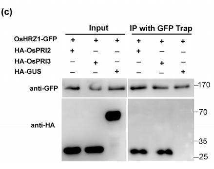
4). Oryza sativa POSITIVE REGULATOR OF IRON DEFICIENCY RESPONSE 2 (OsPRI2) and OsPRI3 are involved in the maintenance of Fe homeostasis. PLANT CELL AND ENVIRONMENT, 2020 (PubMed: 31674679) [IF=7.3]

Application: WB    Species: yeast    Sample: yeast cells

FIGURE 1| Interaction of OsHRZ1 with OsPRI2 and OsPRI3.(c) CoIP assay. Total proteins from different combinations with OsHRZ1‐GFP and HA‐OsPRI2/HA‐OsPRI3/HA‐GUS were immunoprecipitated with GFP‐Trap followed by immunoblotting with the indicated antibodies. HRZ1‐GFP/HA‐GUS, negative control. Protein molecular weight (in kDa) is indicated.

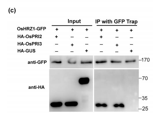
Application: WB    Species: Plant    Sample: Root

FIGURE 1 Interaction of OsHRZ1 with OsPRI2 and OsPRI3. (a) Yeast two‐hybrid assays. Yeast co‐transformed with different BD and AD plasmid combinations were spotted in parallel in 10‐fold dilution series on synthetic dropout medium lacking Leu/Trp/His/Ade. The C‐terminal truncated OsHRZ1 and full‐length OsPRI2/3 were cloned into pGBKT7 and pGADT7, respectively. OsHRZ1‐C/OsPRI1, positive control. OsHRZ1‐ C/Empty, negative control. (b) Pull‐down assay. OsHRZ1 was fused with the GST tag and OsPRI2/3 were fused with the His tag. Recombinant proteins were expressed in E. coli. Proteins were pulled down by glutathione Sepharose 4B and detected using the anti‐His antibody. Protein molecular weight (in kDa) is indicated. (c) CoIP assay. Total proteins from different combinations with OsHRZ1‐GFP and HA‐OsPRI2/HA‐OsPRI3/ HA‐GUS were immunoprecipitated with GFP‐Trap followed by immunoblotting with the indicated antibodies. HRZ1‐GFP/HA‐GUS, negative control. Protein molecular weight (in kDa) is indicated. (d) Degradation of OsPRI2 or OsPRI3 was carried out by detecting the OsPRI2/3‐GFP protein level in co‐infiltration experiments with increasing amounts of OsHRZ1‐GFP. GFP proteins were used as an internal control. Anti‐GFP antibody was used in western blot. Protein molecular weight (in kDa) is indicated. Stars indicate the non‐specific bands. Numbers indicate the ratio of the concentrations of agrobacteria used in co‐infiltration. Empty vector, a binary vector pOCA30 with a 35S promoter; GFP, 35S:GFP in pOCA30; OsPRI2‐GFP, 35S:OsPRI2‐GFP in pOCA30; OsPRI3‐GFP, 35S:OsPRI3‐GFP in pOCA30; OsHRZ1‐GFP, 35S:OsHRZ1‐GFP in pOCA30. (e) Cell‐free degradation. Ten‐day‐old roots grown in Fe‐sufficient solution were harvested and used for protein extraction. Incubation with or without MG132 was performed over the indicated time course

5). Identification and functional analysis of two new de novo KCNMA1 variants associated with Liang–Wang syndrome. Acta Physiologica, 2022 (PubMed: 35156297) [IF=6.3]

6). SALL4 promotes angiogenesis in gastric cancer by regulating VEGF expression and targeting SALL4/VEGF pathway inhibits cancer progression. Cancer Cell International, 2023 (PubMed: 37525212) [IF=5.8]

7). Mutated SASH1 promotes Mitf expression in a heterozygous mutated SASH1 knock‑in mouse model. International Journal of Molecular Medicine, 2020 (PubMed: 32582980) [IF=5.4]

8). Orf virus ORF120 protein positively regulates the NF-κB pathway by interacting with G3BP1. Journal of Virology, 2021 (PubMed: 34287041) [IF=5.4]

Application: IF/ICC    Species: Human    Sample: OFTu cells

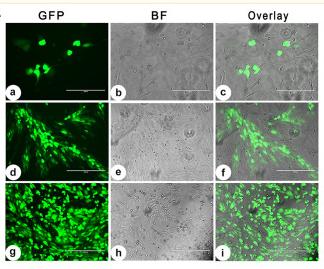
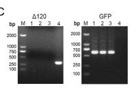
FIG 1 Generation of the recombinant viruses and their growth properties in vitro. (A) Fluorescence microscopy showing the cytopathic effects (CPEs) of the OFTu cells infected with OV-SY17Δ120 deletion mutant virus by plaque purification. The plaques with a strong fluorescent signal in the OFTu cells infected with OV-SY17Δ120 48 hpi during the second round of plaque purification. (b, e, and h) The same fields as shown in a, d, and g by bright-field microscopy. (c, f, and i) Overlay of the a and b, d and e, or g and h images. The cells were visualized under a microscope at 10× magnification. Bar, 400 μm. (B) A revertant mutant OV-SY17-RV120 was produced using red fluorescent protein (RFP) as a selection marker and obtained after two rounds of plaque assay purification on monolayers of OFTu cell cultures 48 hpi. (b, e, and h) The same fields as in a, d, and g by bright-field microscopy. (c, f, and i) Overlay of the a and b, d and e, or g and h images. The cells were visualized under a microscope at 10× magnification. Bar, 400 μm. (C) PCR was performed to confirm the absence of ORFV ORF120 and the presence of the EGFP reporter gene sequences in the OV-SY17Δ120 genome. The PCR analysis showed that the ORF120 gene was successfully deleted in the OV-SY17Δ120 mutant. M, molecular marker; lanes 1 to 3, three OV-SY17Δ120 recombinants from plaque purification; lane 4, wild-type OV-SY17. (D) PCR was performed to confirm the presence of both the ORF120 and RFP reporter genes in the OV-SY17-RV120 genome. The PCR analysis showed that the corresponding revertant virus OV-SY17-RV120 was successfully rescued. M, molecular marker; lanes 1 to 3, three OV-SY17-RV120 recombinants from plaque purification; lane 4, wild-type OV-SY17. (E) In vitro growth kinetics of the OV-SY17Δ120 mutant virus, OV-SY17 parental virus, and OV-SY17-RV120 revertant virus. Primary OFTu cells were infected (MOI, 10 or 0.1) with either the OV-SY17Δ120 mutant virus, parental virus OV-SY17, or revertant virus OV-SY17-RV120 and the virus titers were determined by TCID50 assays at the indicated postinfection time points. The growth curves of the OV-SY17Δ120 mutant virus were similar to those of the OV-SY17 parental virus and the OV-SY17-RV120 revertant virus, indicating that the deletion of ORF120 did not affect virus replication.

Application: WB    Species: Human    Sample: OFTu cells

FIG 1 Generation of the recombinant viruses and their growth properties in vitro. (A) Fluorescence microscopy showing the cytopathic effects (CPEs) of the OFTu cells infected with OV-SY17Δ120 deletion mutant virus by plaque purification. The plaques with a strong fluorescent signal in the OFTu cells infected with OV-SY17Δ120 48 hpi during the second round of plaque purification. (b, e, and h) The same fields as shown in a, d, and g by bright-field microscopy. (c, f, and i) Overlay of the a and b, d and e, or g and h images. The cells were visualized under a microscope at 10× magnification. Bar, 400 μm. (B) A revertant mutant OV-SY17-RV120 was produced using red fluorescent protein (RFP) as a selection marker and obtained after two rounds of plaque assay purification on monolayers of OFTu cell cultures 48 hpi. (b, e, and h) The same fields as in a, d, and g by bright-field microscopy. (c, f, and i) Overlay of the a and b, d and e, or g and h images. The cells were visualized under a microscope at 10× magnification. Bar, 400 μm. (C) PCR was performed to confirm the absence of ORFV ORF120 and the presence of the EGFP reporter gene sequences in the OV-SY17Δ120 genome. The PCR analysis showed that the ORF120 gene was successfully deleted in the OV-SY17Δ120 mutant. M, molecular marker; lanes 1 to 3, three OV-SY17Δ120 recombinants from plaque purification; lane 4, wild-type OV-SY17. (D) PCR was performed to confirm the presence of both the ORF120 and RFP reporter genes in the OV-SY17-RV120 genome. The PCR analysis showed that the corresponding revertant virus OV-SY17-RV120 was successfully rescued. M, molecular marker; lanes 1 to 3, three OV-SY17-RV120 recombinants from plaque purification; lane 4, wild-type OV-SY17. (E) In vitro growth kinetics of the OV-SY17Δ120 mutant virus, OV-SY17 parental virus, and OV-SY17-RV120 revertant virus. Primary OFTu cells were infected (MOI, 10 or 0.1) with either the OV-SY17Δ120 mutant virus, parental virus OV-SY17, or revertant virus OV-SY17-RV120 and the virus titers were determined by TCID50 assays at the indicated postinfection time points. The growth curves of the OV-SY17Δ120 mutant virus were similar to those of the OV-SY17 parental virus and the OV-SY17-RV120 revertant virus, indicating that the deletion of ORF120 did not affect virus replication.

9). Neuropilin-2 Signaling Modulates Mossy Fiber Sprouting by Regulating Axon Collateral Formation Through CRMP2 in a Rat Model of Epilepsy. MOLECULAR NEUROBIOLOGY, 2022 (PubMed: 36044155) [IF=5.1]

10). Chicken speckle-type POZ protein (SPOP) negatively regulates MyD88/NF-κB signaling pathway mediated proinflammatory cytokine production to promote the replication of Newcastle disease virus. Poultry science, 2024 (PubMed: 38290339) [IF=4.4]

Load more

Restrictive clause


Affinity Biosciences tests all products strictly. Citations are provided as a resource for additional applications that have not been validated by Affinity Biosciences. Please choose the appropriate format for each application and consult Materials and Methods sections for additional details about the use of any product in these publications.

For Research Use Only.
Not for use in diagnostic or therapeutic procedures. Not for resale. Not for distribution without written consent. Affinity Biosciences will not be held responsible for patent infringement or other violations that may occur with the use of our products. Affinity Biosciences, Affinity Biosciences Logo and all other trademarks are the property of Affinity Biosciences LTD.