Flag-Tag Antibody - #T0003

 was observed in most of Ngn3-Cre;CAT-Pdx1 insulin + (blue) and glucagon + (green) cells at P0.5 and 6W; the Crepenetrance rate was a maintained at approximately 95%. Scale bars: 50 mm.")
 ACHN cells were transfected with scramble shRNA or SIRT5 shRNA plasmid, SDHA was precipitated and its succinylation level was probed by anti-pan-succinylation antibody. SIRT5 silencing led to hypersuccinylation of SDHA in ccRCC cells and increased activity of SDHA. (C-E) SIRT5 silencing decreased cellular NADPH/NADP+and GSH/GSSG ratio and increased cellular ROS level. (F) Co-IP of SIRT5 and FLAG-tagged WT SDHA in HEK293A cells revealed SIRT5 coimmunoprecipitated with SDHA. (G-J) SIRT5 silencing repressed ACHN and 769-P cell proliferation and colony formation. Methods followed Fig. 5G-J.")
FBXW7-185aa-FLAG-overexpressing BT549 cells were cotransfected with sh-FBXW7 or USP28 vectors. The FBXW7-185aaFLAG, FBXW7, USP28, and c-Myc expression levels were quantified by western blot analysis.")

 and STAT5b (B) by Cbl-b and c-Cbl in HEK293T cells.")
 at 48 h after transfection for 24 h. Axons and their collaterals were detected by immunofluorescent staining with GFP and Tau-1. Arrows indicate main axons and stars indicate axon collaterals. Scale bar, 40 μm. F, G Quantitation of axon collateral number and length in E. Sema3F treatment reduced the number and length of axon collaterals in neurons transfected with shCtrl but not in neurons with shNpn-2 transfection, which was rescued by hNpn-2 co-transfection. *P < 0.05, **P < 0.01. H Quantitation of main axon length in E. Main axon elongation in neurons transfected with shCtrl was inhibited upon Sema3F treatment, but not in neurons with shNpn-2 transfection, which was rescued by hNpn-2 expression. **P < 0.01, ***P < 0.001. One-way ANOVA, post hoc Tukey test. Error bars represent SEM")
, Δsls1 (B), Δppr4 (C), and Δmtf2 (D) cells were diluted in fresh YES to an OD600 of 0.2 and continued to grow to log phase. Cells were harvested, and mitochondria were prepared using the spheroplast method. Mitochondrial extracts were analyzed by immunoblotting using indicated Abs. Protein levels were quantified and expressed as percentage change with respect to WT. Data were normalized to Mcp60 and presented as mean ± S.D. of three independent experiments. Statistically significant differences were determined by Student′s t test (∗, p < 0.05; ∗∗, p < 0.01; ∗∗∗, p < 0.001).")
Product Info
Source:
Mouse
Application:
WB 1:3000-1:10000, IF/ICC: 1:200, IP 1:200, IHC 1:50-1:200
*The optimal dilutions should be determined by the end user. For optimal experimental results, antibody reuse is not recommended.
*Tips:
*The optimal dilutions should be determined by the end user. For optimal experimental results, antibody reuse is not recommended.
*Tips:
WB: For western blot detection of denatured protein samples. IHC: For immunohistochemical detection of paraffin sections (IHC-p) or frozen sections (IHC-f) of tissue samples. IF/ICC: For immunofluorescence detection of cell samples. ELISA(peptide): For ELISA detection of antigenic peptide.
Reactivity:
All
Clonality:
Monoclonal [T177]
RRID:
AB_2839412
Cite Format: Affinity Biosciences Cat# T0003, RRID:AB_2839412.
Cite Format: Affinity Biosciences Cat# T0003, RRID:AB_2839412.
Conjugate:
Unconjugated.
Purification:
Affinity-chromatography.
Storage:
Mouse IgG1 in phosphate buffered saline (without Mg2+ and Ca2+), pH 7.4, 150mM NaCl, 0.02% sodium azide and 50% glycerol. Store at -20 °C. Stable for 12 months from date of receipt.
Alias:
Fold/Unfold
DDDDK epitope tag; DDDK; ddk; DYKDDDDK; DYKDDDDK epitope tag; DYKDDDDK tag; ECS epitope tag; ECS tag; Enterokinase Cleavage Site epitope tag; Enterokinase Cleavage Site tag; FLAG; FLAG tag antibody
Immunogens
Immunogen:
A synthetic peptide DYKDDDDK coupled to KLH.
Description:
FLAG tag Mouse mAb is part of the series of Tag antibodies, the excellent quality in the research. FLAG tag antibody is a highly sensitive and affinity PAB applicable to FLAG-tagged fusion protein detection. FLAG tag antibody can detect FLAG tags in internal, C-terminal, or N-terminal recombinant proteins.
References
1). Young LINE-1 transposon 5′ UTRs marked by elongation factor ELL3 function as enhancers to regulate naïve pluripotency in embryonic stem cells. Nature Cell Biology, 2023
(PubMed: 37591949)
[IF=17.3]
2). An alternatively spliced STING isoform localizes in the cytoplasmic membrane and directly senses extracellular cGAMP. The Journal of clinical investigation, 2022
(PubMed: 34905508)
[IF=13.3]
3). Ultrasound Guided Local Delivery of Bioorthogonal PDL1 Degrader for Enhanced Immunotherapy. Small (Weinheim an der Bergstrasse, Germany), 2025
(PubMed: 39511869)
[IF=13.0]
4). Psychologic Stress Drives Progression of Malignant Tumors via DRD2/HIF1α Signaling. CANCER RESEARCH, 2021
(PubMed: 34321238)
[IF=12.5]
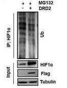
Application: WB Species: mouse Sample:
Fig. 4 |DRD2 overexpression inhibited the ubiquitin-dependent degradation of HIF1α. N, Effect of DRD2 on the content of ubiquitin binding to HIF1α. DRD2-overexpressing cells treated with MG132, and cellular extracts were prepared for co-IP assay with anti–HIF1α antibody, followed by Western blotting detection of ubiquitin. siRNA, small interfering RNA. ns, nonsignificant; *, P < 0.05; **, P < 0.01
5). Nucleolar protein PEXF controls ribosomal RNA synthesis and pluripotency exit. Developmental cell, 2025
(PubMed: 39729985)
[IF=10.7]
6). Origin of the OAS-RNase L innate immune pathway before the rise of jawed vertebrates via molecular tinkering. Proceedings of the National Academy of Sciences of the United States of America, 2023
(PubMed: 37487089)
[IF=9.4]
7). Micheliolide Ameliorates Diabetic Kidney Disease by Inhibiting Mtdh-mediated Renal Inflammation in Type 2 Diabetic db/db Mice. PHARMACOLOGICAL RESEARCH, 2019
(PubMed: 31669149)
[IF=9.1]
8). Voluntary exercise alleviates neural functional deficits in Parkinson's disease mice by inhibiting microglial ferroptosis via SLC7A11/ALOX12 axis. NPJ Parkinson's disease, 2025
(PubMed: 40122927)
[IF=8.7]
9). WWP2 drives the progression of gastric cancer by facilitating the ubiquitination and degradation of LATS1 protein. Cell Communication and Signaling, 2023
(PubMed: 36803368)
[IF=8.4]
10). SPINK13 acts as a tumor suppressor in hepatocellular carcinoma by inhibiting Akt phosphorylation. Cell death & disease, 2024
(PubMed: 39537605)
[IF=8.1]
Restrictive clause
Affinity Biosciences tests all products strictly. Citations are provided as a resource for additional applications that have not been validated by Affinity Biosciences. Please choose the appropriate format for each application and consult Materials and Methods sections for additional details about the use of any product in these publications.
For Research Use Only.
Not for use in diagnostic or therapeutic procedures. Not for resale. Not for distribution without written consent. Affinity Biosciences will not be held responsible for patent infringement or other violations that may occur with the use of our products. Affinity Biosciences, Affinity Biosciences Logo and all other trademarks are the property of Affinity Biosciences LTD.