EDG1 Antibody - #DF2785
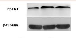
 Western blot analysis of Sphk1 and S1PR1 in the cerebral cortex and hippocampus of rats at 24h after MCAO. (C,D) Representative photographs represented the expression of CD62P in the cerebral cortex and hippocampus at 24h reperfusion after MCAO. The positive products were brown granules after immunostaining, which was remarkably increased followed MCAO and was significantly decreased after treatment with DSCXQ. Representative images of Sphk1 and S1PR1 were presented, and the protein levels were expressed as a ratio of the β-tubulin levels (n = 3). Data were expressed with mean values ± standard deviation (SD). ##p < 0.01 vs. Sham group, **p < 0.01 *p < 0.05 vs. Model group.")
| Product: | EDG1 Antibody |
| Catalog: | DF2785 |
| Description: | Rabbit polyclonal antibody to EDG1 |
| Application: | WB IHC |
| Reactivity: | Human, Mouse |
| Prediction: | Pig, Bovine, Horse, Sheep, Rabbit, Dog |
| Mol.Wt.: | 43 kDa; 43kD(Calculated). |
| Uniprot: | P21453 |
| RRID: | AB_2839991 |
Product Info
*The optimal dilutions should be determined by the end user.
*Tips:
WB: For western blot detection of denatured protein samples. IHC: For immunohistochemical detection of paraffin sections (IHC-p) or frozen sections (IHC-f) of tissue samples. IF/ICC: For immunofluorescence detection of cell samples. ELISA(peptide): For ELISA detection of antigenic peptide.
Cite Format: Affinity Biosciences Cat# DF2785, RRID:AB_2839991.
Fold/Unfold
CD363; CHEDG 1; CHEDG1; D1S3362; ECGF 1; ECGF1; EDG 1; EDG1; endothelial differentiation G protein coupled receptor 1; Endothelial differentiation G-protein coupled receptor 1; Endothelial differentiation sphingolipid G protein coupled receptor 1; FLJ58121; G protein coupled sphingolipid receptor; g protein-coupled receptor edg-1; S1P receptor 1; S1P receptor Edg 1; S1P receptor Edg-1; S1P receptor Edg1; S1P(1) receptor; S1P1; s1pr1; S1PR1_HUMAN; sphingolipid g-protein-coupled receptor 1; Sphingosine 1 phosphate receptor Edg 1; Sphingosine 1 phosphate receptor EDG1; sphingosine 1- phosphate receptor 1; Sphingosine 1-phosphate receptor 1; Sphingosine 1-phosphate receptor Edg-1; Sphingosine 1phosphate receptor type 1 S1P1;
Immunogens
Endothelial cells, and to a lesser extent, in vascular smooth muscle cells, fibroblasts, melanocytes, and cells of epithelioid origin.
- P21453 S1PR1_HUMAN:
- Protein BLAST With
- NCBI/
- ExPASy/
- Uniprot
MGPTSVPLVKAHRSSVSDYVNYDIIVRHYNYTGKLNISADKENSIKLTSVVFILICCFIILENIFVLLTIWKTKKFHRPMYYFIGNLALSDLLAGVAYTANLLLSGATTYKLTPAQWFLREGSMFVALSASVFSLLAIAIERYITMLKMKLHNGSNNFRLFLLISACWVISLILGGLPIMGWNCISALSSCSTVLPLYHKHYILFCTTVFTLLLLSIVILYCRIYSLVRTRSRRLTFRKNISKASRSSEKSLALLKTVIIVLSVFIACWAPLFILLLLDVGCKVKTCDILFRAEYFLVLAVLNSGTNPIIYTLTNKEMRRAFIRIMSCCKCPSGDSAGKFKRPIIAGMEFSRSKSDNSSHPQKDEGDNPETIMSSGNVNSSS
Predictions
Score>80(red) has high confidence and is suggested to be used for WB detection. *The prediction model is mainly based on the alignment of immunogen sequences, the results are for reference only, not as the basis of quality assurance.
High(score>80) Medium(80>score>50) Low(score<50) No confidence
PTMs - P21453 As Substrate
| Site | PTM Type | Enzyme | Source |
|---|---|---|---|
| T4 | O-Glycosylation | Uniprot | |
| S5 | O-Glycosylation | Uniprot | |
| N30 | N-Glycosylation | Uniprot | |
| S129 | Phosphorylation | Uniprot | |
| Y143 | Phosphorylation | Uniprot | |
| T236 | Phosphorylation | P31749 (AKT1) | Uniprot |
| K330 | Acetylation | Uniprot | |
| K341 | Ubiquitination | Uniprot | |
| S351 | Phosphorylation | Uniprot | |
| S353 | Phosphorylation | Uniprot | |
| S355 | Phosphorylation | Uniprot | |
| S358 | Phosphorylation | Uniprot | |
| S359 | Phosphorylation | Uniprot |
Research Backgrounds
G-protein coupled receptor for the bioactive lysosphingolipid sphingosine 1-phosphate (S1P) that seems to be coupled to the G(i) subclass of heteromeric G proteins. Signaling leads to the activation of RAC1, SRC, PTK2/FAK1 and MAP kinases. Plays an important role in cell migration, probably via its role in the reorganization of the actin cytoskeleton and the formation of lamellipodia in response to stimuli that increase the activity of the sphingosine kinase SPHK1. Required for normal chemotaxis toward sphingosine 1-phosphate. Required for normal embryonic heart development and normal cardiac morphogenesis. Plays an important role in the regulation of sprouting angiogenesis and vascular maturation. Inhibits sprouting angiogenesis to prevent excessive sprouting during blood vessel development. Required for normal egress of mature T-cells from the thymus into the blood stream and into peripheral lymphoid organs. Plays a role in the migration of osteoclast precursor cells, the regulation of bone mineralization and bone homeostasis (By similarity). Plays a role in responses to oxidized 1-palmitoyl-2-arachidonoyl-sn-glycero-3-phosphocholine by pulmonary endothelial cells and in the protection against ventilator-induced lung injury.
S1P-induced endothelial cell migration requires the PKB/AKT1-mediated phosphorylation of the third intracellular loop at the Thr-236 residue.
Cell membrane>Multi-pass membrane protein. Endosome. Membrane raft.
Note: Recruited to caveolin-enriched plasma membrane microdomains in response to oxidized 1-palmitoyl-2-arachidonoyl-sn-glycero-3-phosphocholine. Ligand binding leads to receptor internalization.
Endothelial cells, and to a lesser extent, in vascular smooth muscle cells, fibroblasts, melanocytes, and cells of epithelioid origin.
Interacts with GNAI1 and GNAI3.
Belongs to the G-protein coupled receptor 1 family.
Research Fields
· Environmental Information Processing > Signal transduction > FoxO signaling pathway. (View pathway)
· Environmental Information Processing > Signal transduction > Sphingolipid signaling pathway. (View pathway)
· Environmental Information Processing > Signaling molecules and interaction > Neuroactive ligand-receptor interaction.
References
Application: WB Species: Rat Sample: cerebral cortex and hippocampus tissue

Application: WB Species: rat Sample: cerebral cortex and hippocampus
Restrictive clause
Affinity Biosciences tests all products strictly. Citations are provided as a resource for additional applications that have not been validated by Affinity Biosciences. Please choose the appropriate format for each application and consult Materials and Methods sections for additional details about the use of any product in these publications.
For Research Use Only.
Not for use in diagnostic or therapeutic procedures. Not for resale. Not for distribution without written consent. Affinity Biosciences will not be held responsible for patent infringement or other violations that may occur with the use of our products. Affinity Biosciences, Affinity Biosciences Logo and all other trademarks are the property of Affinity Biosciences LTD.