Phospho-NF-kB p65 (Ser536) Antibody - #AF2006
- WB
- IHC
- IF/ICC
- pElisa
- Cites
 Antibody.
Lane 1: 293 cells, blocked with antigen-specific peptides,
Lane 2: 293 cells,
Lane 3: COS-7 cells.")
 Antibody.
Lane 1: HepG2 cells(serum starvation treatment), blocked with antigen-specific peptides,
Lane 2: HepG2 cells(serum starvation treatment),
Lane 3: RAW264.7 cells(serum starvation treatment).")
 Antibody.
Lane 1: Rat heart, blocked with antigen-specific peptides,
Lane 2: Rat heart,
Lane 3: HaLa(heat-shock treatment),
Lane 4: RAW264.7 cells(heat-shock treatment).")
, using Phospho-NF-kB p65 (Ser536) Antibody. The lane on the left was treated with blocking peptide.")
, using Phospho-NF-kB p65 (Ser536) Antibody. The lane on the left was treated with blocking peptide.")
 Antibody.
Lane 1: A549 cells(heat-shock treatment), blocked with antigen-specific peptides,
Lane 2: A549 cells(heat-shock treatment),
Lane 3: RAW264.7 cells(H2O2 treatment).")
 Antibody.
Lane 1: Hepg2 cells(serum starvation treatment), blocked with antigen-specific peptides.
Lane 2: Hepg2 cells(serum starvation treatment).
Lane 3: Hela cells(heat-shock treatment).")


, diluted 1/600 was used as secondary antibody.")
 by IF/ICC. The samples were fixed with PFA and permeabilized in 0.1% Triton X-100,then blocked in 10% serum for 45 minutes at 25°C. Samples were then incubated with primary Ab(AF2006) and mouse anti-beta tubulin Ab(T0023) for 1 hour at 37°C. An AlexaFluor594 conjugated goat anti-rabbit IgG(H+L) Ab(Red) and an AlexaFluor488 conjugated goat anti-mouse IgG(H+L) Ab(Green) were used as the secondary antibody.
The nuclear counter stain is DAPI(blue).")

- or vehicle-treated cystic kidneys. (A) TNF-α, MCP-1, CFB and SOD2 were analyzed by western blot in 9-week-old +/+ and Cy/+ kidneys. (B–E) Immunohistochemical staining for oxidative stress markers 8-OHdG and nitrotyrosine in the tubulointerstitial area. Computer-assisted morphometry was used to quantify changes of 8-OHdG and nitrotyrosine in each group. Scale bar = 50 µm. (F) NF-κB pathway ( p-p65, p65, p105 and p50) and mTOR pathway ( p-S6K and total S6K) were analyzed by western blot in 9-week-old +/+ and Cy/+ kidneys. Blots are representative of three independent experiments.")
 Eca109 and (C and D) KYSE150 cells were transfected with GV142-survivin overexpression plasmid (Eca109 survivin OE and KYSE150 survivin OE), GV142-control plasmid (Eca109 control and KYSE150 control), and mock transfection. Expression levels of survivin, NF-κB p65, IΚΚβ, and IΚΚα were analyzed by (A and C) RT‑qPCR and (B and D) western blotting of total protein. Transcript levels were measured by RT-qPCR of total isolated RNA, with GADPH serving as an internal control. Columns indicate the mean values from triplicate experiments and the error bars indicate standard deviation. * P<0.05; **P")

 Western blotting analyses of total NF-κ B p65 and phospho-NF-κB p65 in primary murine chondrocytes with or without IL-1β (10 ng/ml) for 24h. Increased phospho-NF-κB p65 protein expression in chondrocytes from AMPKα cDKO mice compared with their WT littermates was observed. GAPDH served as a loading control.")
 Representative IHC images of MMP-3, MMP-13 and phospho-NF-κB p65 in the medial tibial plateau in AMPKα1α2 conditional double knockout (AMPKα cDKO) mice and their WT littermates 2 weeks post-sham operation and DMM surgery or in mice at 9 months of age. Scale bars=20μm. The cellularity of the section was confirmed with haematoxylin staining.")
 The protein expressions of the purified GST-FCN-A and GST were determined by SDS-PAGE. (b) The extracted membrane proteins from RAW264.7 cells were incubated with the purified GST-FCN-A or GST proteins. Co-IP analysis of the interaction between TLR4 of macrophage and GST-FCN-A was performed by using anti-TLR4. Rabbit IgG was used as a negative control in co-IP. (c, e) BMDMs, isolated from WT, TLR4-/- or MyD88-/- mice, were stimulated with FCN-A (10g/mL) for 24 h, then the expressions of iNOS and Arg-1 from BMDMs were examined by Western blot analysis. (d, f) The levels of pro-inflammatory cytokines IL-1in cell lysates, and secreted IL-6, TNF- were detected by ELISA. (g, h) Western blot analysis of p-IRAK1, p-p65, p-ERK1/2, and p-JNK in the BMDM lysates of TLR4-/-, MyD88-/- or WT after stimulation with FCN-A for 0-45 min. In d and f, values are mean ± [SEM] from three independent experiments.")
 in U2-OS cells treated by LY294002 or/and Estrogen receptor β (ERβ) siRNA in the presence of 10-10 M E2.")

.")
 The cloned lncRNA uc.48+ was inserted in the pcDNA3.0 at EcoR I and Kpn I sites. The expression of uc.48+ was recorded by real‐time PCR. (b) The left ventricular function including LVSP, LVEDP, and ±dp/dtmax was assessed by BL‐420F. (c) The myocardial enzymes including AST, CK, and LDH release were measured. (d) The infarct size was evaluated through TTC staining. The white area represented the infarct size, which was evaluated and calculated as a percentage of the risk area.(e) P2X7R mRNA was recorded by real‐time PCR. (f) Representative immunoblots and the relative protein levels of P2X7R, p‐NF‐κB, Bax,Bcl‐2, and Cyt were analyzed.")
. The total and phosphorylated expression of p65,IKKα and IKKβ after the above treatment was examined by WB and demonstrated by histogram. (a and b).The levels of inflammatory cytokines and chemokines were then measured by ELISA. (c and d) The expression of adhesion molecules (ICAM‐1 and VCAM‐1) was detected by WB (e). *p < 0.05, **p < 0.01")
 fed with normal diet and six ApoE mice(10 months old, male) fed with high‐fat diet respectively, representing the control and hypercholesterolemic groups. (a)Histological alternation was evaluated with hematoxylin and eosin staining. (c,e,g) The proteins as indicated were detected by immunohistochemistry. (b,d,f,h) All quantitative data are expressed as mean ±SEM. *p < 0.05 versus control. Scale bar=100 μm. ApoE: apolipoprotein E;CAT: catalase; NF‐κB: nuclear factor ‐κB;SEM: standard error of the mean")
 TDSCs were pretreated with 10 μM BAY 11‐7082(BAY) for 1 hr, followed by incubation with 10 mg/dl CHO and BAY for the indicated times. NF‐κB pathway was assessed by western blot analysis for the proteins indicated (a,e), and the tendon‐related gene expressions of TDSCs were western blotted for the proteins indicated (g).")
 MSCs were cocultured with ACs at a ratio of 1:20 for the indicated time. The expressions of IκBα, p65 and their phosphorylated forms in MSCs were analysed by western blot.")
 The ovarian expression of NF-κB, p-NF-κB, p38 MAPK, and p-p38 MAPK, and the relative NF-κB and p38 MAPK activity were derived as p-NF-κB and p-p38 MAPK, and are expressed as fold changes over Ctrl. HUVECs were treated with condition culture medium from KGN(10 IU/ml hCG with or without 100 ng/ml CD200Fc) for 24 h.")
 The
effect of SCVP-1 on the level of phosphorylation of p65 in RAW264.7 cells by Western blotting.
(B) Relative quantitative analysis of p-p65/p65 protein phosphorylation levels. (C-F) Effect of
specific NF-κB inhibitors (BAY11-7082) on SCVP-1/LPS-induced NO, IL-1β, IL-6 and TNF-α
production of RAW264.7 cells. The results were expressed as means ± SD (n = 3). (B) *p<0.05,
**p<0.01 vs. control group. (C-F) *p<0.05, **p<0.01 vs. without anti- NF-κB antibodies treated
group")
 The protein expressions of cleaved-IL-1β, IL-6, pro-caspase 1 Caspase 1, NF-
κB, phospho-NF-κB, TLR4, NLRP3, ASC, phospho-IκBα and IκBα, (B–D) Expression levels of Cleaved-IL-1β, IL-6 and ASC protein were analyzed. (E–G) Protein expression levels ratio of
cleaved-caspase-1 to pro-caspase-1, phospho-IκBα to IκBα and, phospho-NF-кB to NF-κB. (H–I) Expression levels of NLRP3 and TLR4 protein were analyzed. The location assay of by
laser scanning confocal microscopy. β-Actin was used as control. (J) Expression of IL-6 were observed by fluorescence microscopy, red indicated positive immunofluorescence results
for IL-6, blue indicated the result of DAPI. The data were presented as the mean ± SD from three independent experiments (n = 3). Compared to the control group, #p b 0.05, ##p b 0.01; compared to the model group *p b 0.05, **p b 0.01.")
 The mRNA expression levels of
TIFA gene in BSR-T7/5 cells infected with rSS1GFP and rSS1GFP-M/NLSm were verified by qRT-PCR. (B) The protein expression levels of TIFA in
BSR-T7/5 cells infected with rSS1GFP and rSS1GFP-M/NLSm were examined by Western blotting. The relative expression levels of TIFA were compared
with the control GAPDH expression. (C) The subcellular localization of EGFP-M or EGFP-M/NLSm and HA-TIFA in plasmids co-transfected BSR-T7/5
cells. Original magnification was 1 × 200. (D) The effect of different dosage EGFP-M or EGFP-M/NLSm on the expression level of endogenous TIFA in
plasmid transfected BSR-T7/5 cells. The relative expression levels of TIFA were compared with the control GAPDH expression. (E) The expression patterns
of TIFA, pTIFA, TRAF6, NF-κB p65, pNF-κB p65, and IL-2 in BSR-T7/5 cells infected with rSS1GFP and rSS1GFP-M/NLSm at 12 and 24 hpi. The
relative expression levels of these proteins were compared with the control GAPDH expression. (F) The effect of TIFA overexpression on the expression of
IL-2 and virus titers of rSS1GFP and rSS1GFP-M/NLSm at 12 and 24 hpi. (G) The effect of siRNA-mediated knockdown of TIFA on the expression of IL-2
and virus titers of rSS1GFP and rSS1GFP-M/NLSm at 12 and 24 hpi. (H) The schematic diagram illustrated that the M protein in the cytoplasm inhibited
host cell immune response by down-regulating TIFA/TRAF6/NF-κB signaling pathway")
 HEK293T cells transfected with either HA empty vector or UL2-HA expression
plasmid were stimulated with TNF-α (20 ng/mL) for the indicated times (0, 30, and 60 min) according to previous studies (59, 77), and then equal amounts of cell
lysates were analyzed by WBs with phospho-NF-κB–p65 (Ser536) Ab (A), phospho-NF-κB–p65 (Ser276) Ab (C) (top panel), or anti-p65 pAb (second panel). Protein
levels of UL2 (third panel) and β-actin (bottom panel) in the same cell lysates were also determined. (B,D) Densitometry of phospho-NF-κB–p65 Ser536 (B) and
Ser276 bands (D) from (A,C), respectively, were normalized to loading control β-actin. Data were expressed as means ± SD from three independent experiments.
ns, not significant and ***P < 0.001.")

. Significance: *P < 0.05 vs. the HC group, and #P < 0.05 vs. the KD group. f, g Effects of IL-1R8 silencing on the
activation of ERK and NFκB p65 were observed. Significance: *P < 0.05 vs. the HC group, #P < 0.05 vs. the KD group, and &P < 0.05 vs. the KD + IL-37b
group. i The nuclear translocation of NFκB p65 was observed after silencing IL-1R8 expression (n = 3). Magnification: ×200, Scale bar = 100 μm.
Experiments were done at least three times in triplicate.")
 Representative IHC images of MMP-3, MMP-13 and phospho-NF-κB p65 in the medial tibial plateau in AMPKα1α2 conditional double knockout (AMPKα cDKO) mice and their WT littermates 2 weeks post-sham operation and DMM surgery or in mice at 9 months of age. Scale bars = 20 μm. The cellularity of the section was confirmed with haematoxylin staining.")
 LNCaP cells and (B) PC3 cells were cultured in 6‑well plates and treated with different concentrations of TCM‑1 (0, 2, 5 and 10 mg/ml). At 24 h, cells were harvested and lysed for western blot analysis to assess the protein expression levels of p‑PI3K/p85(Y458), PI3K/p85, p‑AKT(S473), p‑AKT(T308), AKT, p‑p65(S536), p65 and β‑actin (loading control).")
. The ratio of epididymal fat weight to the total body weight (B). The mRNA expression of F4/80 (C). The mRNA and
protein expressions of IL-6, IL-1β, TNF-α and MCP-1 (E, F). The protein levels of p-IKKβ/IKKβ, p-P65/P65 (G, H). The data are expressed as means ± SEM. *P < 0.05,
**P < 0.01. ns: not significant.")
 rats. (A) Western blot was used to detect the protein levels in the brain
tissues. The full-length gels are presented in Supplementary Fig. S2. The protein expression was
evaluated by proteomic analysis. (B) Changes in the retinol-binding protein 4 (RBP4); (C) Changes in
serum albumin. *p < 0.05 and ***p < 0.001 vs. I/R.")
 immunoblotting analysis of p65 and p-p65 (Ser536) after LPS/TNF-α and wogonin treatment with indicated concentrations in AML12
cells. C, Representative flow cytometry images and analysis of cell apoptosis after indicated treatment in AML12 cells. D, Immunoblotting
analysis of pro-apoptotic Bax, Cleaved-Caspase3, Cleaved-PARP and anti-apoptotic Bcl2 expression related to (C). E, Densitometry analysis
of Cleaved-caspase3, Cleaved-PARP, Bax and Bcl2 related to (D). NC, the negative control group. The results are presented as mean ± SD
through at least 3 independent experiments and were analysed by one-way analysis of variance (ANOVA). *P < .05, **P < .01, ***P < .001")
 Cell viability was detected by MTT assay. Cells were treated with different concentrations of BR (10–200 nM) for 24 h. TNF-α (B), pro-IL-1β (C), PGE2 (D) and NO (E) levels in culture media were determined by kits. Cells were pretreated for 2 h in the presence or absence of BR (25, 50, or 100 nM) or indomethacin(positive control, 10 μM) and then stimulated with LPS (100 ng/ml) for 24 h. The values are expressed as mean ± SD (n = 6) of three independent experiments.Western blotting was performed to analyze the protein expression of total NF-κB p65, phosphorylation of NF-κB p65 (F), NF-κB p65 in nuclear (G) and in cytosol (H).")
 The expression of phospho-NF-κB p65 (Ser 536) was analyzed by immunohistochemistry (IHC) and semi-quantified in the fructose-fed mice, and representative images are shown (× 500 magnification).Values are shown as means ± SEM (n=10). Results were statistically analyzed using one-way ANOVA coupled with Newman-Keuls’s multiple-comparison test, and values having different superscripts are significantly different (p < 0.05).")
 Protein levels of NF‑κB P65, p‑P65, IKKβ and IκBα in the total cell fraction of control and co‑cultured Jurkat cells and (B) densitometric analysis of the western blot data.")
 Representative immunoblots and the relative protein levels of P2X7R, p‐NF‐κB, Bax,Bcl‐2, and Cyt were analyzed.")


 (5A).")


 Representative western blot results for AMPK/NF-κB p65/NLRP3 signaling pathway proteins in AGEs/salidroside treated-HUVECs with or without compound C.GAPDH was used as a loading control. (B) Histograms analysis of the western blot results. The data are representative of one experiment performed in triplicate and are expressed as the mean ± S.D.")
. A: representative western blotting band; B: The expressions of MyD88(a),IRAK-4(b), FADD (c), TAK-1(d), p-p38/p38 (e) and p-p65/p65 (f).")
 Representative Western blot results for IκBα, p-IκBα, p65, and p-p65 in the testis of rats from all four groups.")
 on colonic expression levels of nuclear factor-kappa B (NF-kB) pathway proteins assessed by Western blot in dextran sulfate sodium (DSS)-induced colitis mice (A).")
 The ability of Exos-26a to generate neurofilament (red fluorescent dye) and inhibit glial fibrillary acidic protein (green fluorescent dye) in PC12 cells. (b, c) Representative images of western blots used to determine the expression levels of NF, GFAP, IKB, p-IKB, p65, and p-p65 and semiquantification of the data. *P < 0.05, **P < 0.01, and ***P < 0.001 compared with the control group by t test or ANOVA test. #P < 0.05 and ##P < 0.01 compared with the NF-κB inhibitor group by t test. N.S., not significant. n = 3 for each group.")

 HEK293T cells transfected with either HA empty vector or UL2-HA expression plasmid were stimulated with TNF-α (20 ng/mL) for the indicated times (0, 30, and 60 min) according to previous studies (59, 77), and then equal amounts of cell lysates were analyzed by WBs with phospho-NF-κB–p65 (Ser536) Ab (A), phospho-NF-κB–p65 (Ser276) Ab (C) (top panel), or anti-p65 pAb (second panel). Protein levels of UL2 (third panel) and β-actin (bottom panel) in the same cell lysates were also determined. (B,D) Densitometry of phospho-NF-κB–p65 Ser536 (B) and Ser276 bands (D) from (A,C), respectively, were normalized to loading control β-actin.")
. (A)and (B) statistical schematics represented the reduction of protein expression induced by CUS and RAGE knockdown. (C) Representative Western blots.")
. Cells were administrated by dioscin (15 ng/mL)for 24 h, and then treated with MSU (100 μg/mL) for 12 h.d The protein expressions of TLR4,MyD88, p-IKKβ, IKKβ, p-p65 in cytoplasm, and NF-κB p65 innuclei were measured utilizing western blotting. GAPDH and Histone H3 were acted as internal references, respectively.")
 for 12 h, BALB/c mice were treated with or without lycorine (20 mg/kg or 40 mg/kg). 1, control group; 2, LPS group; 3,LPS+lycorine (20 mg/kg) group; 4, LPS+lycorine (40 mg/kg) group. bRelative protein expression levels of HMGB1, TLR4, TLR5, Myc88,P65, P-P65, IκBα, and P-IκBα in the lung tissues were determined by Western blot analysis. β-actin as an internal control.")
 and ZN (0, 1, 10, and 20 μg/ml). (A–C) p-IκB and p-p65 protein levels in HUVECs were determined (n=5).")
 Protein expression level of IκB-α, p-IκB-α, NF-κB, p-NF-κB 24 h after HI brain injury.")
. (A) Western Blot images of relative
protein; (B) p-IκB/IκB ratio; (C) p-p65/p65 ratio; (D) p65 cytosolic/nuclear ratio. ##P < 0.01 versus intact group; *P < 0.05 versus vehicle group; **P < 0.01
versus vehicle group; Δ
P < 0.05 versus SASP group; ΔΔP < 0.01 versus SASP group.")
 and intestine (B, E) by western blot analysis. The blots of p-p6, p65, p-IκBα and IκBα (C, F). The nuclear translocation of p-p65 in liver
(G) and intestine (H) by EMSA. Data are represented as means ± SD (n = 6). **p < 0.01 versus the control. #p < 0.05 and ##p < 0.01 versus the GVHD group. &
&p < 0.01 versus the GVHD + BBR group. †
p < 0.05 and ††p < 0.01 versus the GVHD + CsA group.")
 Western blot analysis of the expression of PI3K/Akt/NF-κB signaling pathway proteins after treatment with eriodictyol (0, 25, 50, or 100 μM) for 48 h. (B) Viability of U87MG and CHG-5 cells as measured by CCK-8 assay. Cells were treated with or without eriodictyol (100 μM) for 48 h after PI3K agonist (740 Y-P, 25 μg/ml) or inhibitor (LY294002, 30 μM) pretreatment for 2 h. (C) The number of apoptotic cells was examined by flow cytometry after treatment as in (B). (D) Quantification of the apoptotic cells. (E) Expression levels of Bax, PARP, and PI3K/Akt/NF-κB signaling pathway proteins were measured by Western blot after treatment as in (B). The data are shown as the mean ± SD of three experiments. *P < 0.01, ***P < 0.001 compared with the control group; # P < 0.05, ## P < 0.01, ### P < 0.001 compared with the eriodictyol group.")
 activation and translocation. (A) Immunoblot analysis of phospho-NF-κB p65 (Ser536) protein in liver tissues from WT and CD47KO mice fed LFD or HFD (upper) and band density normalized to β-actin [lower; data are means (±SDs) of three independent experiments]. (B) Confocal images showing subcellular distribution of phosphorylated NF-κB (pNF-κB) (Ser536); pNF-κB (Ser536); and nucleus were stained in red by AF594 and in blue by DAPI, respectively. The representative images from four independent samples are shown. Scale bar represents 10 μm. Data are presented as mean ± SD. *p < 0.05 and ****P < 0.0001.")
 and IL-6 (D) in
liver tissues was examined by immunohistochemical analysis (400×, brown yellow granules indicate positive reaction), B. Quantitative analysis of the immunohistochemical expression of p-NF-кB, C. Quantitative analysis of the immunohistochemical expression of IL-6.")
 and quantified (b) after knockdown of MARK4 using siMARK4–1. c, d Protein levels of IL-6 and
phospho-p65 were measured by Western blotting (c) and quantified (d) after knockdown of MARK4 using siMARK4–2. e Immunofluorescence analysis of
the expression and localization of phospho-p65 in SMSCs after knockdown of MARK4 using siMARK4-1 and siMARK4-2. The data shown represent the
means ± standard deviations (SDs) of at least three independent experiments. *P < 0.05, **P < 0.01, ***P < 0.001 compared with the NC group; #
P < 0.05, ##P < 0.01, ###P < 0.001 compared with the NC + IL-1β group. MARK: microtubule affinity-regulating kinase; IL: interleukin; NF-κB: nuclear factor-κB;
SMSC: synovium-derived mesenchymal stem cell.")
. d The phosphorylation of IκBα and P65 in the control group, ACLT, and glycitin treatment
group were tested by western blotting (relative quantitation was shown in Fig. S4D). e Immunofluorescent staining was performed in chondrocytes to further
investigate nuclear translocation of NF-κB-P65. f (Relative quantitation was shown in Fig. S4E), the expression of P-IκB in the ACLT mouse model were
assayed by (relative quantitation was shown in Fig. S4F). The values are mean ± S.E.M of at least three independent experiments. *P < 0.05; **P < 0.01.")
 in the
human intervertebral disc tissues of the IVDD group and the control group (Control group, n = 4; IVDD group, n = 20). HNPCs were cultured with PBS, TNF-α (5 ng/
ml), TNF-α (5 ng/ml) + IL-38 (10 ng/ml), TNF-α (5 ng/ml) + IL-38 (20 ng/ml) for 1 h, the total protein of each group was extracted, and the level of NF-κB P-P65
(b-c) was detected by western blot. The levels of NF-κB P-P65 (d-e) in the HNPCs treated with TNF-α (5 ng/ml) and different concentrations of IL-38 for 1 h were
detected by IF staining. Scale bar, 50 μm. Each experiment was repeated at least three times. The values are the mean ± SD. Significant differences are indicated in
the above assays. **P < 0.01, ***P < 0.001.")
 The representative graphs of the expressions of TRAF2, p38, P-p38, p65, P-p65 in different groups. (B) The cartogram of the expressions of p65 and P-p65 in different groups. (C) The cartogram of the expressions of TRAF2, p38, and P-p38 in different groups. **p < 0.01 vs Control group. ##p < 0.05 vs BAFF + TNF-α group.")
 Relative mRNA expression of intestinal inflammation factors. (B). Relative mRNA expression of intestinal anti-inflammation factors. (C)Western blots of total and phosphorylated protein expressions of p38MAPK and NF-κBp65 in intestine. Statistical significance was determined using the unpaired, two-tailed Student’s t-test. *, **, represented significant difference at P < 0.05, P <0.01 level, respectively.")
 Immunoblot and (B) quantitative analysis of MMP9 and MMP13 expression in Raw 264.7 cells in response to S. aureus infection. Two-tailed Student’s t test, n = 3/group. (C) Immunoblot and (D) quantitative analysis of the effect of knocking down TWIST1 on the expression of MMP9 and MMP13 in Raw 264.7 cells after S. aureus infection. Two-tailed Student’s t test, n = 3/group. (E) Immunoblot and quantitative analysis (F) of NF-κB in Raw 264.7 cells at 0, 1, 3, 6, 12, and 24 h after S. aureus infection. ANOVA followed by Dunnett’s test, n = 3/group. (G) Immunoblot and quantitative analysis (H) of the effect of NF-κB inhibitor (Bay11-7082) on the expression of MMP9 and MMP13 in Raw 264.7 cells after S. aureus infection. Two-tailed Student’s t test, n = 3/group. *p < 0.05, and **p < 0.01.")

ELISA. Besides, its expression was elucidated in HepG2 cells and liver tissue by (B) qPCR (n = 6). Expressions were normalized by GAPDH. The phospho and total NFκBp65 protein expression was determined in (C) cells and (D) tissue by immunoblotting (n = 3). Expressions were normalized by total NF-κBp65. NF-κB expression
was further validated by immunofluorescence in (E) cells, (F) its quantification (n = 5), and (G) tissue and (H) its quantification (n = 5) by using Alexa Fluor 594
(red), DAPI (blue). Magnification 40×. Inflammatory markers, encompassing (I) IL6, IL1β, and TNF-α were estimated in serum using ELISA kits as per manufacturer's
instructions. Inflammatory cytokines were analyzed further by qPCR in (J) cells and (K) tissue (n = 6). Expressions were normalized by GAPDH. Data are shown as
Mean ± S.E.M (n = 8), *Control vs NASH; #NASH vs NAR. *P < 0.05, **P < 0.01, ***P < 0.001; #P < 0.05, ##P < 0.01, ###P < 0.001. (For interpretation
of the references to color in this figure legend, the reader is referred to the web version of this article.)")
 Relative expression of miR-7, ESAM, and RELA were analyzed in breast tumor tissues and adjacent normal tissues of 12 patients by qRT-PCR. (B) Relative expression of miR-7 and ESAM. (C) Relative expression of RELA and ESAM. (D) Western blotting analysis of the expression of RELA, p-RELA, and ESAM. N, noncancerous tissues; T, tumor tissues. (E) Semiquantitative analysis of the protein expression of RELA, p-RELA, and ESAM; refer to the differences as indicated.")
 on the protein expression of NLRP3 (a), cleaved caspase-1 (b), and p(Ser536)-NF-κB p65 (c) in the renal cortex of normal rats,
diabetic rats, and diabetic rats treated with Sar at low and high (20, 60 mg/kg) doses, respectively. Mean ± SEM, n =6-7. * p < 0.05, vs. Normal group; # p < 0.05,
## p < 0.01, vs. DM group")
 at 1 nmol/min for
20 min was used to induce excitatory renal reflex. PVN microinjection of PBS, 1% DMSO,
losartan (10 nmol), captopril
(10 nmol), tempol (20 nmol), or
apocynin (1 nmol) was carried out
10 min before the renal infusion.
The measurements were carried
out at the end of the renal infusion. *P < 0.05 vs Veh; †P < 0.05
vs PBS or DMSO. Values are
mean ± SE. n = 5 per group")
 TLR4 expression in the lung was evaluated using (B) immunohistochemistry. Scale bar, 25 µm. Representative western blotting images and statistical analysis of (C) TLR4, (D) p-NF-κB and NF-κB expression levels in the lung. Data are presented as the mean ± SD. N=8 in each group. *P<0.05 vs. sham; #P<0.05 vs. I/R. EA, electroacupuncture; I/R, ischemia/reperfusion; SEA, sham electroacupunture, p, phosphorylated; TLR4, Toll-like receptor 4.")
. NO levels in the cell culture media of
Raw264.7 cells were determined using a Griess assay. B–E). Western blot analysis of iNOS, COX-2, NF-κB and p-NF-κB expression. These assays were performed in
three in dependent experiments. ANOVA followed by Bonferroni’s multiple comparison tests was used. (*) p < 0.05, (***) p < 0.001 and (****) p < 0.0001 versus
control group treated with vehicle; (##) p < 0.01, (###) p < 0.001 and (####) p < 0.0001 versus model group.")

 Putative miR-214-3p binding and mutated sites in DNAJC3-AS1. (b) Dual-luciferase reporter assay was performed to determine the luciferase activity of 293 T cells. (c) The expression of miR-214-3p was evaluated after the down-regulation of DNAJC3-AS1 in LoVo cells. (d-f) The protein levels of LIVIN (d), p-IκB (e), IκB (e), p-P65 (f) and P65 (f) in LoVo cells were detected by western blot. Data is shown as mean ± SD, ** p < 0.01, *** p < 0.001.")
. a The staining of p-BTK and p-NF-κB p65 in peritumoral normal renal tissues and IgAN renal tissues. b The count of pBTK and p-NF-κB p65 in renal kidney (*p < 0.05)")
, p‐NF‐κB p65, ICAM‐1, TNF‐α and IL‐1β. A is the Western Blot strip of ANX‐A1, p‐NF‐κB p65 and ICAM‐1. B is the histogram of ANX‐A1, p‐NF‐κB p65 and ICAM‐1, compared with IR‐I group (P < 0.05). C is the histogram of TNF‐α and IL‐1β, compared with IR‐I group (P < 0.05)")
 Representative photographs of PPAR-γ, P-P65, P65, P-IκBα, and IκBα. The relative protein
levels of (B) PPAR-γ, (C) P-P65/P65 ratio, and (D) P-IκBα/IκBα ratio were measured. Data are expressed as the mean ± SEM. ##p < 0.01 versus the
normal group; **p < 0.01 versus the DSS group. Δp < 0.05, ΔΔp < 0.01 versus the SASP group.")
 for
24 h followed by further stimulations with LPS (1 μg/mL) for 12 h. (A–C) Protein quantification of pIkB α, p-p65, was determined by western blotting, quantified by
densitometry and was normalized to IkB α, p65. Data as determined by ANOVA followed by Tukeys multiple comparisons are presented as means ± SD, * P < 0.05, **
P < 0.01.")
 Relative mRNA expression levels of apoptosis-related genes (P65 and IL-17) were measured in Dami cells by qPCR analysis; (B) protein levels of IL-17 were detected in culture supernatation of Dami cells by ELISA analysis; (C) protein levels of total P65, p-P65, p-IKBα, IL-17 and GAPDH were detected in Dami cells by Western blot analysis; (D) flow cytometric analysis of apoptotic Dami cells. The difference of apoptosis rates among four groups was analyzed; (E) TEM analysis of apoptotic Dami cells; Scale bars: 1 um; (F) TUNEL staining of apoptotic Dami cells; Scale bars: 100 um; (G) protein levels of IL-17 were detected in culture supernatation of Dami cells by ELISA analysis; (H) relative mRNA expression levels of apoptosis-related genes (Bax and Bcl-2) were measured in Dami cells by qPCR analysis; (I) protein levels of total P65, p-P65, p-IKBα, IL-17 and GAPDH were detected in Dami cells by Western blot analysis; All data are expressed as mean ± SEM (n≥3). *p<0.05, **p<0.01, ***p<0.001 versus vehicle. Analysis of Variance (ANOVA) and Student’s t-test (two-tailed).")
 Representative Western blotting images of pIκBα, IκBα, cytoplasmic p65 and nuclear p65 (B–E) The protein expression levels of pIκBα, IκBα, cytoplasmic p65 and nuclear p65 in the NF-κB pathway. Data are expressed as the means ± SD (n = 3). # p < 0.05, ## p < 0.01 vs. control group; *p < 0.05, **p < 0.01 vs. DSS group; & p < 0.05, && p < 0.01 vs. COP group.")
 Real-time PCR analysis showing the mRNA expression of IL-6, Nampt and Sirt1 normalized to β-actin. (B) ELISA result showing the IL-6 levels. (C) NAD+/NADH analysis showing intracellular content of NAD+ and NADH, and their ratio. (D) Western blot analysis showing the protein expression of Nampt, Sirt1, TAK1 and p-NF-κB p65. (E) The nuclear translocation of NF-κB p65 was measured by fluorescence immunocytochemistry (200×). Red, NF-κB p65; blue, DAPI. The data are expressed as the mean ± SD. *, p<0.05; **, p<0.01. #, p<0.05 vs. con; ##, p<0.01 vs. con.")
-mediated activation of the PI3K/Akt pathway is involved in the modulation of DcR3 levels in HCT116 cells. (A) HCT116 cells were transfected with TIPE or an empty vector (control) for 24 h with lipopolysaccharide (LPS) stimulation for the indicated time (0, 15, 30, and 60 min). Cells were harvested, and whole-cell extracts were prepared for Western blot analysis of the indicated proteins. The blots shown are representative of those obtained in three separate experiments. (B) HCT116 cells were cultured in 6-well plates and transfected with TIPE or an empty vector (control) for 24 h, then qRT-PCR assays were performed to measure the relative DcR3 mRNA expression of HCT116 cells after treatment with LY294002 (50 μM), NSC23766, U0126, and BAY 11-7082. Expression was normalized to that in the control cells. (C) The culture medium was collected, and DcR3 protein levels were measured by ELISA. (D) HCT116 cells were transfected with tumor necrosis factor-induced protein-8 (TIPE) after treatment with lipopolysaccharide (LPS) for the indicated time (0, 30, and 60 min), and Western blot analysis of whole-cell lysates was performed to examine the indicated proteins in HCT116 cells after treatment with or without LY294002 (50 μM). ns, not significant, *p < 0.05, ***p < 0.001.")
 Expression of TLR4, phosphorylated p38 (P-p38), p38, P-ERK1/2, ERK1/2, P-JNK, JNK, P-p65, p65 was shown. (B) The analysis of grey scale value. Con was control group, M was model group, H was the high dose OPA group, L was low dose OPA group, P was positive group. Data were expressed as the mean ± SD, the statistical analyses were done with t-test; *** p < 0.001, ** p < 0.01, * p < 0.05 compared with the model group; ### p < 0.001, ## p < 0.01, # p < 0.05 compared with the control group.")

 The protein expression levels of TLR4, MyD88, and p-p65 (Ser536) in nasal mucosa. (b) Immunofluorescence staining of p65 in nasal mucosa. Scale bar=50 µm. (c) The protein levels of p65 in cytoplasm and nucleus were measured by Western blot. Data represent means ± SD (n=6). ***P<0.001 compared with sham group. ###P<0.001 compared with AR+NC group. Sham group (normal mice), AR group (OVA-induced AR mice), AR+NC group (AR mice treated with empty lentivirus), AR+miR-224-5p group (AR mice treated with lentiviral-mmu-miR-224-5p). TLR4, toll-like Receptor 4; OVA, ovalbumin; NC, negative control; AR, allergic rhinitis.")
 repressed the timosaponin BII-induced inhibition of mTOR/NFκB and activation of autophagy. Western blot images and the
analyses for mTOR/NFκB signaling and its downstream effectors in the high glucose-induced osteoblasts with the treatment of timosaponin BII (10 μmol/L) and in
those cells with the cotreatment of timosaponin BII (10 μmol/L) and MHY1485 (10 μmol/L). **P < 0.01 compared with the high glucose group.")
 and the quantification of the p-NFκB expression in tibias of different groups of rats.
Error bars indicated SEM (n = 6). **P < 0.01 compared with the Mod group. The arrows denoted positive staining. B: Representative western blot images and the
quantification of p-NFκB/NFκB and p-IκB/IκB in the high glucose-induced osteoblasts with treatment with timosaponin BII (0.1 μmol/L, 1 μmol/L, 10 μmol/L) and in
those cells with NAC (25 μmol/L) incubation for 48 h **P < 0.01 compared with the high glucose group. C: Translocation of NFκB by confocal microscopy. Scale bar:
10 μm. D: Representative immunoblotting (upper panel) of protein expression of NFκB in nucleus and cytoplasm and the quantitate data (bottom panel) in osteoblasts
with treatment with timosaponin BII (10 μmol/L). **P < 0.01 compared to the treatment with high glucose alone.")
 iNOS, (B) TNF-α, (C) IL-6, (D) IFN-γ, (E) IL-18 and (F) IL-10, were determined by ELISA. (G) Nuclear translocation of p-NF-κB was determined by immunofluorescence staining. Scale bar, 100 µm. (H and I) p-NF-κB expression in the nucleus were determined by western blotting. (J and K) Ratio of whole cell p-NF-κB/total NF-κB expression was determined by western blotting. Data are presented as the mean ± standard error of the mean (n=3). **P<0.01, ***P<0.001 vs. sham group; #P<0.05, ##P<0.01, ###P<0.001 vs. LPS+ATP group. DI, dimethyl itaconate; iNOS, inducible nitric oxide synthase; LPS, lipopolysaccharide; p-, phosphorylated.")
. The relative protein expression of p-p65 (b) in colon tissue was detected by Western Blot. All values are presented as the mean ± SEM. ##P < 0.01 versus normal group; ∗P < 0.05, ∗∗P < 0.01, and ∗∗∗P < 0.001 versus DSS group.")
. The transfection efficiency of bovine miR-125b mimic or inhibitor in MAC-T cells as measured by fluorescence micrograph (A) and qPCR (B). At 48 h post-transfection, MAC-T cells were challenged with LPS for 3 h. Cells were then collected to measure mRNA expression and production of NKIRAS2 (C), NF-κB (D), IL-6 (E), and TNF-α (F). Expression levels of mRNA and protein were determined by qPCR and Western blot, respectively. *p < 0.05, **p < 0.01, ***p < 0.001")
 Representative photomicrographs of Iba1 (specificity marker of microglia) and HMGB1 plus DAPI staining in the dentate gyrus caused by the indicated stimuli. Scale bar = 50 μm. (B) The rate of HMGB1- and Iba1-positive cells in the dentate gyrus caused by the indicated stimuli. (C) Representative photomicrographs of Iba1 (specificity marker of microglia) and phosphor-NF-κB plus DAPI staining in the dentate gyrus caused by the indicated stimuli. Scale bar = 50 μm. (D) The rate of phosphor-NF-κB- and Iba1-positive cells in the dentate gyrus caused by the indicated stimuli. Data are presented as the mean ± SD (n = 6). ****P <0.0001. Vehicle, POCD, EA, and α-BGT are described above.")
 The contents of MDA, SOD, mn-SOD, GSH-Px and CAT in lumbar
spinal cord tissues with correspondingly commercial kits. (F-H) Relative protein levels of Nrf2 and HO-1 in lumbar spinal
cord tissues using western blotting. (I) Double immunofluorescence of Nrf2 and Iba-1 in lumbar spinal cord tissues. Scale
bar = 50 μm. (J and K) Relative protein levels of total NF-κB p65 and p-NF-κB p65 (ser536) in lumbar spinal cord tissues using
western blotting. n = 6 rats per group; ∗∗∗ P< 0.001, ∗∗ P< 0.01, ∗ P< 0.05. PAH = perillaldehyde; MDA = malondialdehyde;
SOD = superoxide dismutase; mn-SOD = manganese-dependent superoxide dismutase; GSH-Px = glutathione peroxidase;
CAT = catalase; Nrf2 = nuclear factor E2-related factor 2; HO-1 = heme oxygenase-1; Iba-1 = ionized calcium binding
adaptor protein 1; NF-κB = nuclear factor-kappa B.")
, DSS group (n = 5) and DSS+ICA group (n = 5). B. Statistical data and comparison for p-p65 expression in 3 groups. C. Statistical data and comparison for p65 expression in 3 groups. D. Statistical analysis for ratios of p-p65/p65 in all 3 groups. * P < .05 vs.DSS group at the same time point. # P < .05 vs. Control group.")
 NLRP3 and ASC, (B) cleaved-caspase-1 and caspase-1, (C) iNOS, (D) p-P65, P65, p-IκBα, and IκBα. The images (in gray) were analyzedusing Image J software. Data were expressed as mean ± SEM (n = 6).")
 Phosphorylation and acetylation levels of p65 and expressions of SIRT1, SIRT6 and SIRT7 protein (normalized to that of β-tubulin) detected by western blotting.")
 LPS. (B) LBP. (C) TNF-α. (D) IL-1β. (E) IL-6. (F) MPO. (G) Western blot bands. (H) The
protein expression levels of CD14, MD2, TLR4, MyD88, p-p65, and p-IκBα. The contents of target proteins were normalized to β-actin. The results are presented as the
mean ± SD (ELIS aassay: n = 10, protein expression: n = 3, mRNA expression: n = 6). ## p < 0.01 vs. NC group; * p < 0.05, ** p < 0.01 vs. model group.")
. (A) and (B) TF procoagulant activity was detected by chromogenic substrate
method in (A) HUVECs and (B) PBMCs, including the LPS induced group, the LPS plus NC mimic group, the LPS plus the miR-19a-3p mimic group, the LPS plus TNFα
group, the LPS plus NC mimic and TNFα group, and the LPS plus the miR-19a-3p mimic and TNFα group. (C) and (D) Western blot analysis was performed to detect
the protein expression of TF, NF-κB p65, p-P65, IκB-a, p-IκB-a, and GAPDH in (C) HUVECs and (D) PBMCs in the six above mentioned groups. (E) and (F) Comparison
of TF protein levels among the six above mentioned groups. (G) and (H) Comparison of NF-κB p65 and p-P65 protein levels among the six above mentioned groups.
(I) and (J) Comparison of IκB-a and p-IκB-a protein levels among the six above mentioned groups. Significant differences among these groups were assessed by oneway ANOVA; * p < 0.05, ** p < 0.01, *** p < 0.001. NS means nonsignificant.")
 through the activation of proliferator-activated receptor-γ (PPAR-γ) to inhibit the nuclear factor-kappa B (NF-κB)/nod-like receptor protein 3 (NLRP3) inflammatory axis in spinal cord neurons. (a–c) Tumor cell inoculation significantly induced the activation of NF-κB and NLRP3 in the spinal cord of BCP rats (∗∗∗p < 0.001 vs. the sham group; n = 4, one-way ANOVA (a–c)). (d, e) Immunofluorescence results showed that in the dorsal horn of the spinal cord of BCP rats, p-NF-κB (red) and NLRP3 (red) were primarily expressed in neurons (green) rather than astrocytes (green) or microglia (green). Lumbar enlargements were collected on day 18 after the operation or cancer cell inoculation. Sections were counterstained with DAPI (blue) to label cell nuclei. The white arrows indicate colocalization of p-NF-κB and NLRP3 with NeuN (neuronal nuclei, neuronal-specific marker), GFAP (glial fibrillary acidic protein, astrocyte specific marker), and IBA-1- (ionized calcium binding adapter molecule 1, microglial specific marker) immunoreactive cells in the spinal dorsal horn, respectively; n = 4. Scale bar = 50 μm. n represents the number of experimental animals in each group. (f–i) Repeated intrathecal injection with PDTC, an NF-κB inhibitor, significantly inhibited the established BCP (∗∗p < 0.01 and ∗∗∗p < 0.001 vs. the vehicle control group; n = 6, two-way repeated measures ANOVA (i)) and BCP-induced NF-κB/NLRP3 activation (∗∗∗p < 0.001 vs. the vehicle control group; n = 4, one-way ANOVA (f–h)). (j–l) Repeated intrathecal injection with MCC950, an NLRP3 inhibitor, significantly inhibited the established BCP (∗∗p < 0.01 and ∗∗∗p < 0.001 vs. the vehicle control group; n = 6, two-way repeated measures ANOVA (l)) and BCP-induced NLRP3 activation (∗∗∗p < 0.001 vs. the vehicle control group; n = 4, one-way ANOVA (j, k)). (m–o) Rosiglitazone inhibits the BCP-induced activation of the NF-κB/NLRP3 inflammatory axis, whereas GW9662 reversed this effect (∗p < 0.05 and ∗∗∗p < 0.001 vs. the vehicle control group; ∗p < 0.05 and ∗∗∗p < 0.001 vs. the BCP+RG group; n = 4, one-way ANOVA (m–o)). N.S: not statistically significant.")

 (A). The expression of NF-κB-P65 and IL-1β mRNA in NRK-52e cells were quantified by qRT-PCR and normalized (n = 3) (B). The levels of TNF-α in NRK-52e cells were determined by ELISA method (n = 6) (C). Compared with NC or NC-sicD36 group, ## p < 0.01; compared with M or M-sicD36 group, * p < 0.05, ** p < 0.01.")
 Western blot analysis of p-NF-κB/NF-κB and p-IκBα/IκBα proteins")

 as well as NF-κB p65 (b) and p-NF-κB p65 (c) by
immunofluorescence in SH-SY5Y cells treated with high glucose. HG, HG + Sar5, shF2R HG, shF2R HG + Sar5 represent cells infected
with lentivirus containing F2R negative control or F2R shRNA (F2R, the gene symbol of PAR-1) subsequently cultured with HG or HG
plus 5 μmol/L Sar, respectively. Mean ± S.E.M, n = 7. *p < .05, **p < .01. Scale bar: 20 μm [Colour figure can be viewed at
wileyonlinelibrary.com]")
 Western blotting and (B) semi-quantification of protein expression levels. (C) Quantification of mRNA expression levels. *P<0.05 vs. control; #P<0.05 vs. siRNA NC; ^P<0.05 vs. overexpression NC. AAV, adeno-associated virus; KDM2B, lysine-specific demethylase 2B; NC, negative control; NLRP3, NOD-, LRR- and pyrin domain-containing protein 3; p-, phosphorylated; si/siRNA, small interfering RNA; si-KDM2B, AAV-siKDM2B; siRNA NC, AAV-siRNA scrambled negative control; TLR4, toll-like receptor 4.")
 Western blot analysis revealed that the transfection of RBFOX1 plasmid inhibits the upregulation of p-p65 and the downregulation of NRF2/HO-1 induced by H/R. #P<0.05 vs. the control and &P<0.05 vs. the H/R + vector NC. RBFOX1, RNA binding fox-1 homolog 1; H/R, hypoxia/reoxygenation; NC, negative control.")
 on T. gondii proliferation through the NF-κB pathway. Detection of socs1 transcription with qRT-PCR at 24 h post-transfection and translation with western blot at 48 h post-transfection in RAW264.7 cells. a, b Transfection with miR-155-5p mimics or normal control miRNA (mimic-NC). c, d Transfection with the siRNA targeting socs1 (si-socs1) and the normal control siRNA (si-NC). e, f Detection of the phosphorylation levels of p65 and IKBα in the NF-κB pathway in the RAW264.7 cells transfected with miR-155-5p mimics, normal control siRNA (mir-NC), si-socs1, or si-NC for 36 h. g Detection of the T. gondii B1 gene copies in the RAW264.7 cells transfected with si-socs1 or si-NC for 24 h, and stimulated with IFN-γ for 24 h, then infected with T. gondii for 24 h. Densitometric quantitation of each band in b, d, and e was applied using ImageJ software. One-way ANOVA was used for between-group comparisons, and Tukey’s multiple-group test was used for multiple-group comparisons. Each experiment was carried out three times (**P < 0.01, ***P < 0.001)")
. ##p < 0.01, ###p < 0.001 vs. control group. *p < 0.05, **p < 0.01, ***p < 0.001 vs. LPS alone group. +p < 0.05, ++p < 0.01, +++p < 0.001 vs. combined treatment group (5 + 20 mg/kg)")
 Cardiac function of mice after AAV administration and MI induction(n = 10); (B) Levels of SPI1, TLR4, p-NFκB, and c-caspase 3 in infarcted border zone in mouse cardiac tissues examined by IHC staining (n = 5)")
 on T. gondii proliferation through the NF-κB pathway. Detection of socs1 transcription with qRT-PCR at 24 h post-transfection and translation with western blot at 48 h post-transfection in RAW264.7 cells. e, f Detection of the phosphorylation levels of p65 and IKBα in the NF-κB pathway in the RAW264.7 cells transfected with miR-155-5p mimics, normal control siRNA (mir-NC), si-socs1, or si-NC for 36 h.")
 The relative mRNA level of IL-1β in different groups. Here we used IL-17A (25 ng/ml) and TNF-α (25 ng/ml) to stimulate HaCaT cells. Simultaneously, we also added 2.5/5/10/20 μM PUN into these IL-17A and TNF-α-stimulated HaCaT cells. The total RNA was subsequently extracted after treatment with IL-17A (25 ng/ml) + TNF-α (25 ng/ml) and 2.5/5/10/20 μM PUN for 24 h. (B) The pro- and mature expression of IL-1β in different groups. The total protein was collected after treatment with IL-17A (25 ng/ml) + TNF-α (25 ng/ml) and 2.5/5/10/20 μM PUN for 48 h. (C) The expression of phosphorylation (Ser536) and total p65 in the cytoplasm and nucleus after exposure of IL-17A (25 ng/ml) + TNF-α (25 ng/ml) and 2.5/5/10/20 μM PUN for 24 h. (D) Immunostaining with an anti-p65 antibody showed that 2.5/5/10/20 μM PUN functionally blocks TNF-α and IL-17A-induced nuclear translocation of p65. Scale bar, 50 μm. (E) The expression of cleaved and total caspase-1 after exposure of IL-17A (25 ng/ml) + TNF-α (25 ng/ml) and 2.5/5/10/20 μM PUN for 48 h. (F) The relative mRNA level of CXCL-1 (left) and CCL-20 (right) in HaCaT cells after treatment with IL-1β and IL-1β + PUN for 48 h *p < 0.05, **p < 0.01, ***p < 0.001. One-way ANOVA with Student-Newman-Keuls method for (A, F). Data represent the mean ± S.E.M.")
 The protein expression of p-p65, p65, p-ERK1/2, ERK1/2, p-p38, p38, p-JNK, and JNK were determined by Western blot and quantified. GAPDH was conducted as a loading control. (c) The nuclei translocation of p65 (red) was detected by the immunofluorescence assay. Nuclei (blue) were stained with DAPI. One-way ANOVA, ##p < 0.01, compared with control cells; $p < 0.05, $$p < 0.01, compared with IL-1β-treated cells.")
 After the last 4-OHT injection, Rosa26LSL-MLS-Stat3 and Rosa26LSL-MLS-Stat3;UbcERT2Cre/+ mice were injected with curcumin prior to LPS. Survival was monitored every 6 - 12 h after injection. The data shown represent a combination of two independent experiments. Statistical comparison of survival was performed with a log-rank test. **, P < 0.01, *, P < 0.05. (B) Serum samples were obtained from these mice and subjected to ELISA to examine MCP-5 levels. (C-D) The serum levels of LDH and albumin were assessed. (E) Lung tissues were removed, and the percentages of macrophages were analyzed by flow cytometry. (B-E) The data are representative of one of two independent experiments. Significance was calculated using a two-way ANOVA. **, P < 0.01, * P < 0.05, n.s., not significant. (F) Rosa26LSL-MLS-Stat3 and Rosa26LSL-MLS-Stat3;LyzCre/+ mice were injected with 10 mg/kg curcumin prior to LPS (20 mg/kg). Survival was monitored every 6 - 12 h after injection. Statistical comparison of survival was performed with a log-rank test. **, P < 0.01, *, P < 0.05. (G) BMDMs of the indicated genotypes were treated with 8 μM curcumin for 20 h and incubated with either 100 ng/mL LPS (30 min) or 100 ng/mL IFN-α (1 h) before harvesting. The cells were harvested, lysed, and subjected to a Western blot analysis. The membranes were blotted with the indicated antibodies. (H) Cells were treated with 8 μM curcumin for 20 h and incubated with 100 ng/mL LPS for another 15 min. Nuclei and cytoplasmic fractions were extracted. The levels of the indicated proteins were analyzed by a Western blot analysis. (I) BMDMs were treated with curcumin. After 20 h, these cells were stimulated with or without 100 ng/mL LPS for 6 h before loading. BMDMs were then treated sequentially with BSA-conjugated palmitate, oligomycin, FCCP, and ETO as indicated. The data are representative of one of two independent experiments performed with n = 5. (J) 4-OHT-treated BMDMs were treated with curcumin for 20 h, and then, the BMDMs were exposed to 100 ng/mL LPS and 10 μmol/L MG132 for an additional 6 h before harvesting. The cell lysates were isolated, IP with an anti-CPT1a antibody and analyzed by a Western blot analysis using an anti-ubiquitin antibody. (G, H and J) The results shown are representative of one of three independent experiments. (K) Schematic representation of the mitochondrial STAT3-mediated increase in FAO in a USP50-dependent manner in the LPS-induced sepsis model. Upon the LPS stimulation, mitochondrial STAT3 could induce CPT1a stabilization by promoting USP50 expression and facilitating CPT1a-USP50 binding, leading to a switch from glucose to an increased reliance on FAO for ATP production, which, in turn, further enhanced NF-κB nuclear localization, leading to a great amount of cytokine production.")
, p65 and phosphorylated p65 were detected, and their expressions relative to GAPDH were shown. B HK-2 cells were stained via immunofluorescence. MIF was tagged by FITC (green) and NLRP3 was tagged by Cy3 (red). The white arrow pointed to the overlapped area of high fluorescence. Scale bar = 100 μm. C Cell viability of HK-2 cells was evaluated via CCK-8 method. D HK-2 cells were stained by annexin-V-FITC and propidium iodide(PI). E PI stained HK-2 cells were counted by flow cytometry. Data were mean ± SD from three independent experiments. Each point represented an independent data. Group comparisons were performed by (A) & (E) one-way ANOVA followed by Tukey’s post hoc test, and (C) two-way ANOVA followed by Tukey’s post hoc test. (N = 3/group, #P < 0.05 vs vector + PBS group, *P < 0.05 vs vector + LPS group, NS means no significant difference).")
 Expression of α7nAChR, SIRT1, ac-p65, p-p65 and p65 in macrophages investigated by immunoblotting; (B–E) quantitative result of (A). #P<0.05, ##P<0.01, ###P<0.001 compared with normal, *P<0.05, **P<0.01, ***P<0.001 compared with AIA rats, &P<0.05 compared with MG (early).")
 Western blotting was performed to detect the expression of p65 and p-p65 in TNF-α-stimulated FLSs. (B) Expression levels of ERK1/2, p-ERK1/2, JNK, p-JNK, p38 and p-p38. NF-κB, nuclear factor-kappa B; MAPK, mitogen-activated protein kinase. The data represent the mean ± SD (n=3). ##P<0.01 vs. Control; **P<0.01 vs. TNF-α+NC siRNA.")
 qRT-PCR analysis was performed to detect the relative mRNA expression of cathepsin K, TRAP, NFATc1, and MMP-9 in BMSCs. (b) Western blot analysis was performed to detect the relative protein expression of IκBα, p-IκBα, p65, p-p65, IKKα, p-IKKα, NFATc1, and cathepsin K in BMSCs (n = 3); results are represented as the mean ± SD. ▲P < 0.05, compared to the Con group; ▲▲P < 0.01, compared to the Con group; ∗P < 0.05, compared to the Ti group; ∗∗P < 0.01, compared to the Ti group. BMSCs: bone marrow stromal cells; Ti: titanium particles; Con: control.")
 and microglia (IBA-1, red) in different groups. Scale bar = 50 μm. D Quantitative PCR analysis of cytokines (TNF-α and IL-6) on 3 days post-CVST. n = 5 to 6 per group. *P < 0.05, **P < 0.01. E Representative images of TNF-α expression in diverse groups. Scale bar = 50 μm. F Flow cytometry assay of brain-infiltrating immune cells including neutrophils (CD11b+CD45highLy6G+), monocyte/macrophages (CD11b+CD45highF4/80+) and microglia (CD11b+CD45int) in the damaged cortex on 3 days after CVST. G Quantitative analysis of infiltrated immune cells. n = 5 per group. *P < 0.05")
 Western blot analysis of Bcl-2, Bax, caspase3 and caspase9. (B–E) Relative protein abundance of Bcl-2, Bax, caspase3 and caspase9. Data are presented as means ± SE. *P < 0.05, **P < 0.01.")
 Western blot analyses and quantitative data. The impact of FPR2 activation by WKYMVm on phosphorylation of p38, ERK1/2, NF-κB p65, and IκBα protein level in LPS-treated HAPI microglia with or without pretreatment of WKYMVm. (d–f) Western blot analyses and quantitative data for p-p38, p-ERK1/2, p-NF-κB p65, and IκBα from each group in primary microglia. SB203580: p38-specific inhibitor; U0126: ERK-specific inhibitor; BAY11-7082: NF-κB p65-specific inhibitor. ∗p < 0.05 vs. the control group; #p < 0.05 vs. the LPS group.")

@zeolite imidazolate framework-8 (ZIF-8) administration counteracted inflammation in the cochlea caused by noise exposure. (A,B) Protein expression level and statistical analysis of phosphonuclear factor kappa B (p-NF-κB), interleukin (IL)-6, and IL-1β in the cochleae of various groups. (C) Representative images of IL-1 staining in the middle–basal cochlear turns from three groups (StV: Stria Vascularis; SL: Spiral Ligament; SLB: Spiral Limbus; OC: Organ of Corti; SGNs: Spiral Ganglion Neurons). Data are presented as means ± standard deviation (SD) (**p < 0.01, ***p < 0.001, ns: no statistical significance).")
 p-p65 and (B) IκBα were evaluated by (C) western blot analysis. n=5/group. **P<0.01 vs. the NC group/ IL-1β group. IκBα, inhibitor of NF-κB α; p-p65, phosphorylated p65; NC, negative control; IL-1β, interleukin-1β.")
 Immunofluorescence staining of CD68 and Gal-3 in heart tissue at 16 weeks after STZ injection. (D–H) Western bloting analysis of Gal-3, p-IκB, IκB and p- NF-κB p65. Data are shown as mean ± SD. n = 3, *P < 0.05 vs. Control; # P < 0.05 vs. DCM+ AAV9-NC.")
, or transfected with vehicle or mtDNA (10 μg/ml) for 24 h, then were harvested for western blot and qPCR. (A) Representative blots and relative quantification protein levels of STING pathway (n = 3). (B) qPCR quantitation of IFN‐β (n = 7). (C) Representative images of nuclear translocation of IRF3 in BMDC treated as indicated. Scale bar: 5 μm. (D) Representative blots and relative quantification protein levels of TLR9 pathway (n = 3). (E) qPCR quantitation of TNF‐α, IL‐6 and VCAM‐1 (n = 6–7). (F) Representative blots and relative quantification protein levels of NLRP3 pathway (n = 3). (G) qPCR quantitation of IL‐1β, IL‐18 (n = 7). Data are shown as the mean ± SEM. *p < 0.05; **p < 0.01; ***p < 0.001. BMDC, bone marrow derived dendritic cell; IFN, interferon; mtDNA, mitochondrial DNA; mtDNA (T), mtDNA transfection; Veh (T), vehicle transfection.")
. The images shown are representatives (Scale bar = 100 μm). The protein expression levels of p65 and IκBα were analyzed using specific antibodies by western blot (C). The black bars and white bars denote 786-O cells and ACHN cells respectively. Data are presented as means ± SD (n ≥ 3). *P < 0.05 and **P < 0.01")
")
. (a, b) The level of HMGB1 protein was measured by immunohistochemistry (n = 6). (c–h) The expression degrees of HMGB1, TLR4, NF-κB, p-38 MAPK, and ERK1/2 proteins analysis by Western blot (n = 3). GA: glycyrrhizic acid, CLP: cecal ligation and puncture, HMGB1: high mobility group box-1, TLR4: toll-like receptor4, NF-κB: nuclear factor-κB, p-38 MAPK: p-38 mitogen-activated protein kinase, ERK1/2: extracellular signal-regulated kinase l/2. ##p < 0.01vs. the Sham group; ∗p < 0.05, ∗∗p < 0.01vs. the CLP group.")
 Representative western blotting band, (b) TLR4, (c) MYD88, (d) IRAK-1, (e) the p-IκBα/IκBα ratio, and (f) the p-p65/p65 ratio. The results were expressed as mean ± SEM (n = 3). ##P < 0.01 vs. the control group; ∗P < 0.05 and ∗∗P < 0.01 vs. the 5-FU group; &P < 0.05 and &&P < 0.01vs. the BBR group.")

 and histology score for colon tissue in the Abx and NC group. B Immunohistochemistry (scale bar, 50 μm) for LPS in subcutaneous and orthotopic tumor tissues and western blot of intratumoral LPS levels from three biological duplications for the Abx and NC group. C Transcription levels of cytokines by RT-qPCR and protein levels of IL6 in cell supernatant by ELISA in RM-1 cultured with LPS (100 μg/ml) for 24 h. D, E Immunofluorescence (scale bar, 100 μm) for p-p65 and p-STAT3 in RM-1 cultured with or without LPS for 24 h; western blot of relative proteins for RM-1 cultured with LPS at different concentrations for 24 h; transcription levels of IL6 by RT-qPCR and western blot of relative proteins in RM-1 cultured with LPS or LPS with BAY-11-7082 for 24 h; protein levels of p-STAT3 and STAT3 in RM-1 cultured with CM or CM with antibody-IL6 for 24 h. F, G IL6 levels in tumor tissue lysate and serum by ELISA. Western blot of relative proteins in tumor from three biological duplications. Immunohistochemistry of tumor tissues for p-p65- and p-STAT3-positive cell (scale bar, 50 μm). Statistical significance was assessed by unpaired Student’s T-test or LSD in one-way ANOVA. *p < 0.05, **p < 0.01, and ***p < 0.001: compared to the NC group; #p < 0.05, ##p < 0.01, and ###p < 0.001: compared to the LPS or CM group")
 and histology score for colon tissue in the Abx and NC group. B Immunohistochemistry (scale bar, 50 μm) for LPS in subcutaneous and orthotopic tumor tissues and western blot of intratumoral LPS levels from three biological duplications for the Abx and NC group. C Transcription levels of cytokines by RT-qPCR and protein levels of IL6 in cell supernatant by ELISA in RM-1 cultured with LPS (100 μg/ml) for 24 h. D, E Immunofluorescence (scale bar, 100 μm) for p-p65 and p-STAT3 in RM-1 cultured with or without LPS for 24 h; western blot of relative proteins for RM-1 cultured with LPS at different concentrations for 24 h; transcription levels of IL6 by RT-qPCR and western blot of relative proteins in RM-1 cultured with LPS or LPS with BAY-11-7082 for 24 h; protein levels of p-STAT3 and STAT3 in RM-1 cultured with CM or CM with antibody-IL6 for 24 h. F, G IL6 levels in tumor tissue lysate and serum by ELISA. Western blot of relative proteins in tumor from three biological duplications. Immunohistochemistry of tumor tissues for p-p65- and p-STAT3-positive cell (scale bar, 50 μm). Statistical significance was assessed by unpaired Student’s T-test or LSD in one-way ANOVA. *p < 0.05, **p < 0.01, and ***p < 0.001: compared to the NC group; #p < 0.05, ##p < 0.01, and ###p < 0.001: compared to the LPS or CM group")
 and histology score for colon tissue in the Abx and NC group. B Immunohistochemistry (scale bar, 50 μm) for LPS in subcutaneous and orthotopic tumor tissues and western blot of intratumoral LPS levels from three biological duplications for the Abx and NC group. C Transcription levels of cytokines by RT-qPCR and protein levels of IL6 in cell supernatant by ELISA in RM-1 cultured with LPS (100 μg/ml) for 24 h. D, E Immunofluorescence (scale bar, 100 μm) for p-p65 and p-STAT3 in RM-1 cultured with or without LPS for 24 h; western blot of relative proteins for RM-1 cultured with LPS at different concentrations for 24 h; transcription levels of IL6 by RT-qPCR and western blot of relative proteins in RM-1 cultured with LPS or LPS with BAY-11-7082 for 24 h; protein levels of p-STAT3 and STAT3 in RM-1 cultured with CM or CM with antibody-IL6 for 24 h. F, G IL6 levels in tumor tissue lysate and serum by ELISA. Western blot of relative proteins in tumor from three biological duplications. Immunohistochemistry of tumor tissues for p-p65- and p-STAT3-positive cell (scale bar, 50 μm). Statistical significance was assessed by unpaired Student’s T-test or LSD in one-way ANOVA. *p < 0.05, **p < 0.01, and ***p < 0.001: compared to the NC group; #p < 0.05, ##p < 0.01, and ###p < 0.001: compared to the LPS or CM group")
 p65. (B) p-p65. (C) IκB-α. (D) p-IκB-α. (E) TLR4. (F) The expression of proteins. Staining intensity was assessed with Image J. Data are represented as mean ± SD (n=3). **P<0.01 and ***P<0.001 mean significance of model group compared with control group. #P<0.05, ##P<0.01 and ###P<0.001 mean significance of drug treatments compared with the model group.")
 The mRNA expression levels of NLRP3, ASC, GSDMD, and caspase 1 in CPB-induced lung injury rats treated with XFZYD, Ac-YVAD-CMK, and Bay-11-7082 were measured via RT-qPCR. (b and c) western blots were performed to determine NLRP3, ASC, Caspase-1 p20, Pro-GSDMD, GSDMD p30, IL-18, IL-1β p-P65, P65, p-IKBα, and IKBα levels in lung tissues of rats with CPB-induced acute lung injury. β-actin was used as a loading control for the blots. All the data were presented as the means ± SD. from independent experiments performed in triplicate. ∗P < 0.05; ∗∗P < 0.01, vs. CPB group.")
 Relative mRNA levels of targets genes in macrophages that phagocytosed control erythrocytes or As-exposed erythrocytes were determined by qPCR (n = 3 per group) and data are presented. (C) Protein levels of targets in splenic macrophages phagocytosed control erythrocytes or As-exposed erythrocytes were analyzed by western blotting. (D) The levels of p-NFκB, iNOS and Cox-2 in mouse spleen sections were analyzed by immunohistochemistry. Differences between groups were analyzed by using the unpaired Student’s t tests. ***p < 0.001, **p < 0.005 or *p < 0.05 indicates significant difference.")
 Relative mRNA levels of targets genes in macrophages that phagocytosed control erythrocytes or As-exposed erythrocytes were determined by qPCR (n = 3 per group) and data are presented. (C) Protein levels of targets in splenic macrophages phagocytosed control erythrocytes or As-exposed erythrocytes were analyzed by western blotting. (D) The levels of p-NFκB, iNOS and Cox-2 in mouse spleen sections were analyzed by immunohistochemistry. Differences between groups were analyzed by using the unpaired Student’s t tests. ***p < 0.001, **p < 0.005 or *p < 0.05 indicates significant difference.")
 The expression of Nlrp3 and p-NF-κB at protein level in lung tissue, n = 6. (B) The mRNA expression of Nlrp3 in vivo, n = 8. (C) The mRNA expression of Nlrp3 in vitro, n = 3. (D) The mRNA expression of IL-1β and IL-18 in lung tissue, n = 8. (E) The mRNA expression of IL-1β and IL-18 in HPMECs, n = 3. Data were expressed as mean ± SD. Statistical significance is indicated as *P < 0.05 vs. the sham + vehicle groups, #P < 0.05 vs. the LPS + vehicle groups.")

, Observation of BMSCs labeled with BrdU under microscope (100 μm, 20 μm). (B), Immunohistochemical staining of NF-κB-P65 and p-NF-κB-P65 expression in joints of CIA rat’s knee (200 μm, 100 μm). (C), NF-κB-P65 and p-NF-κB-P65 expression. (D), IL-1β,TNF-α and NF-κB-P65 mRNA expression in the spleen. Data are presented as the mean ± SD (n = 5/group); ** p < 0.01 vs. PBS rats. Different letters mean significant difference (p < 0.05).")
 Volcano plots of comparative RNA-seq data between the CON group and the LPS group. (B) GO analysis based on the RNA-seq in the biological process of differential transcripts. (C) Representative KEGG analysis with the transcript corresponding to different genes. (D) The mRNA levels of TLR4, MyD88, NOD1, RIPK2, NFKBIA, NF-κB p65 were evaluated by RT-qPCR. (E) Hepatic protein levels of NOD1, RIPK2, IκB-α, and P-p65 were assessed by western blot, quantified using ImageJ analysis, and normalized to β-actin. n = 4−6 per group. Results are showed as means with SEM represented by a vertical bar. * represents significant differences between the CON group and the LPS group; * p < 0.05, ** p< 0.01, *** p<0.001.")
")
 Effects of commutator on the NF-κB signaling pathway in rats. (h) The levels of NF-κB p65 and p-NF-κBp65. (i) The relative value of p-NF-κB p65/NF-κBp65. ∗∗P < 0.01 vs. NC group; #P < 0.05 and ##P < 0.01 vs. EC group.")


 mHSCs were co-cultured with the CMs from RAW264.7 cells with LPS/IFNγ and PHI treatment for 12 h. (b-g) The expression of MMP2, TIMP1, TGF-β, α-SMA, COL1 and NF-κB mRNA in mHSCs after co-culture with macrophage-derived CMs for 24 h was detected by RT-qPCR (n = 3). (h) The expression of TGF-β, α-SMA, COL1, P65 and P-P65 proteins in mHSCs after co-culture with macrophage-derived CMs for 24 h was detected by western blotting. (i) The relative quantification of TGF-β, α-SMA, COL1 and P-P65/P65 protein expression in western blotting results was analyzed by ImageJ software (n = 3). (j) The expression of cytoplasm and nucleus NF-κB P65 proteins in mHSCs after co-culture with macrophage-derived CMs for 24 h was detected by western blotting. (k, l) The relative quantification of cytoplasm and nucleus NF-κB P65 protein expression in western blotting results was analyzed by ImageJ software (n = 3). (m) Immunofluorescence staining of α-SMA in mHSCs after co-culture with CMs from RAW264.7 cells with different treatments for 24 h (Scale bar=100 µm). Results are presented as mean ± SD. ###P")
 Western blot and quantitative analysis of CD68 in THP-1 cells under PMA treatment. §")
. β-actin was used as loading control. Data represent means ± SD of five independent experiments, *p < 0.05, **p < 0.01 by one-way ANOVA with Dunnett’s post-hoc test . Tf: transferrin.")
 The protein expression levels of p-IκBα and p-p65 in different groups of transfected VSMCs. The expression levels of (B) p-IκBα and (C) p-p65 were compared among different groups. *P")
 Western blotting analysis of the expression levels of P65 and p-P65 in the SHFM1 silenced or overexpressed TE-1 cells. The relative expression was analyzed. (C and D) Immunofluorescence analysis indicated the location of P65 in the SHFM1 silenced or overexpressed TE-1 cells. Data are presented as the mean ± standard deviation. **P")
 Relative expression levels of p-NF-κB p65. (b) Relative expression levels of p-IκBα. (c) Relative expression levels of caspase-3. (NO, normoxia group; NA, normoxia + artesunate group; HO, hyperoxia group; HA, hyperoxia + artesunate group. P value was calculated by one-way ANOVA and SNK test.")
 treatment increased the expression of autophagy related protein in HGFs. Data were expressed as mean ± SD, n = 3. Compared to the control group, *P")
, B Interleukin 1 beta (il1β) and C Caspase3 (casp3) (n = 6), D Western blot analysis of intestinal P65 and IL1β (n = 3), E Quantitation of the levels of P65 and IL1β normalized to that of GAPDH (n = 3). Data was expressed as mean ± SEM. SM, fish fed with soybean diet; SMC, fish fed with soybean diet supplemented with 3,000 U/kg xylanase. The significant differences between two group were presented at P")
 and TAPI‐1 (1 μM) for 24 h. (A) and (B) mTIMD4 expression was detected by immunofluorescent staining (n = 5). (C–F) The protein expression of mTIMD4, sTIMD4 in medium, TLR‐4, p‐NF‐κB, NF‐κB, IL‐6, caspase‐1, pro‐IL‐1β, TGF‐β1, and IL‐10 were detected by Western blotting (n = 3). (G–J) The mRNA expression mTIMD4, TLR‐4, IL‐6, and TGF‐β1 was determined by qRT‐PCR (n = 3–4). (K–N) RAW264.7 cells were treated with LPS (1 μg/mL) and TAPI‐1 (1 μM) for 24 h, and the protein expression of mTIMD4, sTIMD4 in medium, TLR‐4, p‐NF‐κB, NF‐κB, IL‐6, caspase‐1, pro‐IL‐1β, TGF‐β1, and IL‐10 were detected by Western blotting (n = 3). (O–P) RAW264.7 cells were transfected with 20 nM control si‐NC or si‐TIMD4 for 48 h and incubated with ox‐LDL (80 μg/mL) and TAPI‐1 (1 μM) for 24 h, followed by determination of mTIMD4, TLR‐4, p‐NF‐κB, NF‐κB, IL‐6, IL‐10, TGF‐β1, caspase‐1, pro‐IL‐1β protein expression by Western blotting (n = 3). HSP90 or GAPDH was used as a loading control. ns, not significant.")
 and hippocampus (b). Quantitative protein levels of p-NF-κB/NF-κB and p-IκB-α/IκB-α in the cortex (c) and hippocampus (d). Cor: cortex; Hip: hippocampus. Data are expressed as mean ± standard deviation. Multiple comparison analysis is based on analysis of variance. P < 0.05 is considered statistically significant. ∗P < 0.05 and ∗∗P < 0.01, versus sham group; ##P < 0.01, versus PSD group.")
 Protein levels of p-NF-κB, NF-κB, IL-6, TNF-α, NLRP3, pro-caspase1, cleaved-caspase1, ASC, pro-IL-1β, and IL-1β were determined using Western blot method (n = 6). (E–G) mRNA levels of Nlrp3, Caspase1, and Asc were determined by qPCR (n = 5). (H–J) Serum levels of IL-1β, IL-6, and TNF-α (n = 5). ns, not significant; * p < 0.05; ** p < 0.01 vs. “Con” group; # p < 0.05, ## p < 0.01 vs. “EtOH” group.")
 Protein levels of p-NF-κB, NF-κB, IL-6, TNF-α, NLRP3, pro-caspase1, cleaved-caspase1, ASC, pro-IL-1β, and IL-1β were determined using Western blot method (n = 6). (E–G) mRNA levels of Nlrp3, Caspase1, and Asc were determined by qPCR (n = 5). (H–J) Serum levels of IL-1β, IL-6, and TNF-α (n = 5). ns, not significant; * p < 0.05; ** p < 0.01 vs. “Con” group; # p < 0.05, ## p < 0.01 vs. “EtOH” group.")
 HSP90AA1 over-expression affected the phosphorylation of STAT1 and nuclear translocation of p-STAT1 in PK-15 and 3D4/2 cells were observed by laser confocal microscopy. (B) HSP90AA1 over-expression affected the nuclear translocation of p-P65 in PK-15 and 3D4/2 cells were observed by laser confocal microscopy.")
 Protein expression levels of p-IκBα, IκBα, p-p65 and p65 were determined by western blotting and GAPDH was used as a loading control. (B) Immunofluorescence assay for p65. Scale bar, 25 µm. The ratio for nuclear p65 was quantified. Data were analyzed with unpaired t-test or one-way ANOVA followed by Tukey's test. Data are presented as the mean ± SD. **P")
 Pyroptosis of BXPC-3 and Patu-8988t cells after knockdown of GALNT6 detected by SEM. Red arrows point to pyrosomes formed in cells and ruptured cell membranes. (B, C) Western blot assay was used to detect the changes of NLRP3, cleaved-caspase-1, caspase-1, cleaved-caspase-3, caspase-3, GSDMD-N, GSDMD, GSDME-N and GSDME expressions in BXPC-3 and Patu-8988t cells after knockdown of GALNT6. The asterisk indicates a heterozygous band. (D) The expression levels of p-NF-κB, NF-κB, NLRP3, cleaved-caspase-1, cleaved-caspase-3, GSDMD, GSDMD-N, GSDME and GSDME-N in overexpression GALNT6 Patu-8988t cells were measured by western blot assay. The asterisk indicates a heterozygous band. (E, F) ACS in BXPC-3 and Patu-8988t cells was analyzed by immunofluorescence. (G) The mRNA expression of IL-1β, IL-6 and TNF-α in BXPC-3 and Patu-8988t cells were detected by qRT-PCR. (H) ELISA was used to detect the levels of IL-1β, IL-6, TNF-α and IL-18 in BXPC-3 and Patu-8988t cells. Each experiment was repeated at least three times independently, and the experimental data were expressed as mean standard deviation (X ± SD).")
 (100 μg/mL) for 24 h. The content of RANTES in the supernatant of the four groups of cells was determined by ELISA. Data are presented as the mean ± SD; the comparison between different groups was performed using the LSD method in one-way ANOVA (bP < 0.01); B: The cells were pretreated with magnolol or solvent for 8 h and then with LPS (100 μg/mL) for 24 h. The expression of RANTES protein in Caco2 cells in each group was detected by Western blot analysis. 0 or Solvent: Treated with solvent only; 10 μmol/L magnolol: Treated with 10 μmol/L magnolol; LPS: Treated with solvent and LPS (100 μg/mL); LPS + magnolol 10 μmol/L: Treated with magnolol 10 μmol/L and with LPS (100 μg/mL); GAPDH: Glyceraldehyde-3-phosphate dehydrogenase; RANTES: Regulated on activation, normal T-cell expressed and secreted.")
 and MMP9 (c). Statistical analysis was performed using ANOVA. d A498 cells were transfected with pcDNA3.1-Flag, Flag-MTA1, siNC, and si-MTA1. After 48 h, cells were subjected to western blotting analysis using antibodies targeting p-p65, p65 and ACTB. The bands of p-p65, p65 and ACTB were from Additional file 1: Fig. 3. e, f Image J was used to calculate the gray scanned bands of p-p65 (b) and p65 (C). *p")
 and upregulated (c) KEGG pathway in overexpression zebrafish bmp8a 3T3-L1 cells. b, d After induction of adipogenic differentiation, the downregulated (b) and upregulated (d) KEGG pathway in overexpression mouse Bmp8a 3T3-L1 cells. e, f Immunoblot analysis and quantification of p-IKKα/β and p-p65 in Mock, LV-ZsGreen1, and LV-bmp8a 3T3-L1 cells (n = 3). g, h Immunoblot analysis and quantification of p-IKKα/β and p-p65 in Mock, LV-ZsGreen1, and LV-Bmp8a 3T3-L1cells. Protein expression levels were quantified by ImageJ software and normalized to total protein (n = 3). i Co-immunoprecipitation and immunoblot analysis of co-transfected with PPARγ and p65 (n = 3). j Schematic drawing of predicted PPRE site in Fabp4 promoter region. k Schematic drawing of WT and PPRE site mutation Luc-report plasmids. l, m Quantification of the activity of Fabp4-promoter (l) and Fabp4-promoter-ΔPPRE (m) luciferase reporters in mouse HEK293T cells transfected with Vector, pCMV-Pparγ, or co-transfected pCMV-Pparγ and pCMV-p65, respectively. Renilla luciferase was used as the internal control (n = 3). Data were from three independent experiments and were analyzed by One-way ANOVA and were presented as mean ± SD (ns not significant, **p")
. *P < 0.05 compared to the normal saline-treated group.")
 Two specific regions (76–127 nt and 209–260 nt) of CPAL that were found to bind with NFκB using the online prediction website. (b) RIP analysis for CPAL-NFκB interaction. Immunoprecipitation of NFκB acquired a large amount of CPAL (**P < 0.01, compared with anti-IgG; n = 3). (c) Expression of P-NFκB and NFκB at the protein levels in each group (**P < 0.01, compared with sham group; ##P < 0.01, compared with +AAV9-sh-NC group; n = 4 to n = 6). (d) Immunohistochemical staining of P-NFκB protein in the infarct border zones of the left ventricular (**P < 0.01, compared with sham group; ##P < 0.01, compared with +AAV9-sh-NC group; n = 3 to n = 4). (e) Expression of P-NFκB and NFκB at the protein level in cardiomyocytes of control, LPS, +si-NC, and +si-CPAL groups (**P < 0.01, compared with control; #P < 0.05, compared with LPS group; n = 9). (f) Staining for P-NFκB (green) and α-actinin (red) proteins in the cardiomyocytes of control, LPS, +si-NC, and +si-CPAL groups (blue: DAPI; n = 4). (g) P-NFκB expression in the neonatal mouse cardiomyocytes of pcDNA3.1-OE-NC, pcDNA3.1-OE-CPAL, +ODNs-CPAL, and +ODNs-NC groups (**P < 0.01, compared with pcDNA3.1-OE-NC; ##P < 0.01, compared with pcDNA3.1-OE-CPAL; n = 5).")
, ERK (B), JNK (C), and P65 (D) were quantified by western blotting in macrophages as indicated treatment. (E) Immunofluorescent staining of P65 was performed in macrophages as indicated treatment. The scar bar indicated 50 μm. N = 3.")
 The effect of hypoxia treatment for 4 weeks on Cul5/TRAF6/NF-κB/HIF-1α/VEGF pathway expression in lung tissues of mice, and reverse effect of pevonedistat. (a) Western blot analysis of TRAF6/NF-κB/HIF-1α/VEGF pathway expression in N4W and H4W group and statistical analysis of the proteins relative expression normalized to GAPDH. (b) Western blot analysis of TRAF6/NF-κB/HIF-1α/VEGF pathway expression in N4W + DMSO, N4W + pevonedistat, H4W+ DMSO, and H4W + pevonedistat group, and statistical analysis of the proteins relative expression normalized to GAPDH. (B) The effect of hypoxia and/or Cul 5 treatment on TRAF6/NF-κB/HIF-1α/VEGF pathway expression in PAECs. Western blot analysis of TRAF6/NF-κB/HIF-1α/VEGF pathway expression in endothelial cells exposed to normoxia and hypoxia with or without Cul 5, and statistical analysis of the proteins relative expression normalized to GAPDH. ∗p < 0.05, ∗∗p < 0.01, ∗∗∗p < 0.001. Student’s t test was used for statistical analysis in Figure 7A(a). Data are represented as the means ± SD, one-way ANOVA was used for statistical analysis in Figure 7A(b) and 7B.")
 Representative images of immunohistochemical staining of NLRP3, ASC and Caspase-1 in kidney tissues . 200×, Scale bar = 50 μm. 400×, Scale bar = 25 μm. (B) Quantitative analysis of NLRP3, ASC and Caspase-1 in (A). (C) Representative images of Western blots showing the protein expression of NLRP3, ASC and Caspase-1 in kidney tissues. (D) Quantitative analysis of protein expression of NLRP3, ASC and Caspase-1 in (C). (E) Representative images of immunohistochemical staining of pp65 in kidney tissues. 200×, Scale bar = 50 μm. 400×, Scale bar = 25 μm. (F) Quantitative analysis of pp65 in (E). (G) Representative images of Western blots showing the protein expression of pp65 and p65 in kidney tissues. (H) Quantitative analysis of protein expression of pp65 in (C). Data presented as mean ± SD. n = 3. *p < 0.05, **p < 0.01, ***p < 0.001 vs. WT CI-AKI mice.")
 Representative images of immunohistochemical staining of NLRP3, ASC and Caspase-1 in kidney tissues . 200×, Scale bar = 50 μm. 400×, Scale bar = 25 μm. (B) Quantitative analysis of NLRP3, ASC and Caspase-1 in (A). (C) Representative images of Western blots showing the protein expression of NLRP3, ASC and Caspase-1 in kidney tissues. (D) Quantitative analysis of protein expression of NLRP3, ASC and Caspase-1 in (C). (E) Representative images of immunohistochemical staining of pp65 in kidney tissues. 200×, Scale bar = 50 μm. 400×, Scale bar = 25 μm. (F) Quantitative analysis of pp65 in (E). (G) Representative images of Western blots showing the protein expression of pp65 and p65 in kidney tissues. (H) Quantitative analysis of protein expression of pp65 in (C). Data presented as mean ± SD. n = 3. *p < 0.05, **p < 0.01, ***p < 0.001 vs. WT CI-AKI mice.")
 regulating enzymes (ACE, Ang II, ACE2, and Ang 1-7) in the lung tissue (n = 6 per group). E Protein levels of important RAS-regulating enzymes (ACE, AT1R, ACE2, and MasR) (n = 3 per group). The relative protein levels are displayed. F Levels of NF-κB phosphoproteins (p-P65 and p-IκB) (n = 3 per group). The relative protein levels are displayed. The immunofluorescence of HS, SDC-1, and surfactant protein D in the lungs is shown in G in representative images and densitometry (scar bar, 20 mm, n = 3 per group). H Evans Blue staining tested lung permeability (n = 3 for each group). Using ELISA, we assessed the BALF concentrations of HS I and HPA J; SP-D in serum K and BALF L (n = 5 for each group). M Relative protein levels of SDC-1, HPA, EXT-1, and SP-D in The lung tissues (n = 3 for each group). All data were shown as mean ± SD.")
 Relative protein expression of p-ERK1/2, p-JNK, and p-p38 in vitro. (B) Relative mRNA expression levels of MAPK1, MAPK8, and MAPK14 in vitro. (C) Relative protein expression of p-c-Fos and p-p65 in vitro. (D) Relative mRNA expression levels of FOS, JUN, and NFKB1A in vitro. *P")
 phosphorylation under proinflammatory conditions. (a) Time-course changes in phos-phorylation of p65 (p-p65) relative to p65 level. Human IVD NP cells were stimulated with 10 ng/mL IL-1β, and the levels of p-p65 and p65 were assessed at different time points, i.e., 0 (set as 1.0), 15, 30, 60, 120, and 240 min. (b,c) Expression of p-p65 relative to that of p65 and phosphorylated AMPK (p-AMPK) relative to that of AMPK in IVD NP cells treated with phosphate-buffered saline (PBS) (group C; set as 1.0), AdipoRon (2 μM, group A), IL-1β (10 ng/mL, group I), or both (group A + I) for 15 min. Data are presented as dot and box plots. Results from six independent experiments were analyzed using one-way repeated-measures ANOVA with the Tukey–Kramer post hoc test (n = 6). Significant differences were set as * p < 0.05 and ** p < 0.01.")
. Immunofluorescence analysis indicated that P-P65 expression was upregulated in HG and LPS states but was reduced after transfection of miR-212-3p mimic and si-SNHG16. However, treatment with the miR-212-3p inhibitor reversed this response (b, d). WB analysis indicated that P65 and P-P65 were upregulated in HG states but downregulated after si-SNHG16 (c)")
. Immunofluorescence analysis indicated that P-P65 expression was upregulated in HG and LPS states but was reduced after transfection of miR-212-3p mimic and si-SNHG16. However, treatment with the miR-212-3p inhibitor reversed this response (b, d). WB analysis indicated that P65 and P-P65 were upregulated in HG states but downregulated after si-SNHG16 (c)")
 Western blot and analysis of HDAC3, p-p65/p65, and COX1 expressions in the cortex of PSA mice (n = 3 mice/group). (G) Brain PGE2 levels from mice with indicated treatment were measured after PTS + CRS administration by ELISA kit (n = 4 mice/group). (H)Immunofluorescence of COX1 and Iba1 in the vehicle-treated and esketamine-treated PSA mice groups. (Scale bar = 20 μm). Arrows represent randomly selected COX1 positive points. The data mentioned above are presented as the means ± SEM and were analyzed by one-way ANOVA with the Bonferroni post hoc test. Ns means no significance")
 Representative bands of NF-κB, p-NF-κB, p-IKK-α, p-IKK-β, IκB, p-IκB, TNF and IL-6wereshown.")
. B. The mRNA expression levels of P65 NF-κB, NLRP3, IL-1β, and IL-18 in each group were analyzed by qRT-qPCR (n = 3). Data are presented as the mean ± SD.")
 for 2 h and S. aureus treatment (105 CFU) for 24 h (n = 5). E. Immunofluorescence staining was performed to detect TJs protein ZO-1, Occludin, and Claudin-3 expressions in MMECs (scale bars, 10 μm). F-H. Protein levels in the NF-κB signaling pathway, including IκB, p65, and phosphorylation levels of IκB and p65, were detected with HEX pretreatment (0, 2.5, and 5 μM) for 2 h and S. aureus treatment (105 CFU) for 24 h (n = 5). One-way ANOVA (B-D and G-H) was used, and values are presented as the mean ± SD (B-D and G-H). *p < 0.05, **p < 0.01, and ***p < 0.001 indicate significantly different from each group. ns, no significance.")
 Schematic of the experimental procedure showing the non-contact co-culture model of RAW264.7 and PC12 cells to study microglial activation after TBI (above) and a contact co-culture model to investigate neuronal apoptosis (below). (B) Reverse transcription quantitative polymerase chain reaction assessing the relative mRNA expression levels of surface antibodies in RAW264.7 cells in the TBI and Sham groups. (C) Immunofluorescence experiments depicting the levels and distribution of P-P65 in RAW264.7 cells. (D) Comparison of the average fluorescence intensity of P-P65 in immunofluorescence experiments. Enzyme-linked immunosorbent assay (ELISA) measuring cytokine expression levels of (E) TNF-α, (F) CXCL-8 and (G) CCL2 in the PC12 and RAW264.7 cell co-culture systems, with paired comparisons to the TBI group. (H) Western blotting detected TYROBP expression and NF-κB pathway activation status via P65 phosphorylation under varying conditions. (I) CCK-8 assays comparing cell viability in each group, with the average OD of the NC group as 100% survival for reference calculated as, cell viability=(ODsample-ODblank)/(ODcontrol-ODblank). (J) Annexin V-PI assay determining cell apoptosis proportions, with the bar chart in the lower right corner summarizing the proportions of early-stage (annexin V positive, PI negative) and late-stage apoptotic cells (both annexin V and PI positive). *P")
 Schematic of the experimental procedure showing the non-contact co-culture model of RAW264.7 and PC12 cells to study microglial activation after TBI (above) and a contact co-culture model to investigate neuronal apoptosis (below). (B) Reverse transcription quantitative polymerase chain reaction assessing the relative mRNA expression levels of surface antibodies in RAW264.7 cells in the TBI and Sham groups. (C) Immunofluorescence experiments depicting the levels and distribution of P-P65 in RAW264.7 cells. (D) Comparison of the average fluorescence intensity of P-P65 in immunofluorescence experiments. Enzyme-linked immunosorbent assay (ELISA) measuring cytokine expression levels of (E) TNF-α, (F) CXCL-8 and (G) CCL2 in the PC12 and RAW264.7 cell co-culture systems, with paired comparisons to the TBI group. (H) Western blotting detected TYROBP expression and NF-κB pathway activation status via P65 phosphorylation under varying conditions. (I) CCK-8 assays comparing cell viability in each group, with the average OD of the NC group as 100% survival for reference calculated as, cell viability=(ODsample-ODblank)/(ODcontrol-ODblank). (J) Annexin V-PI assay determining cell apoptosis proportions, with the bar chart in the lower right corner summarizing the proportions of early-stage (annexin V positive, PI negative) and late-stage apoptotic cells (both annexin V and PI positive). *P")
 Western blot protein bands; (B-C) Relative protein levels. The data are presented as means ± SD (n = 3).")
, p-IκBα ‾(χ±s, n = 3)in hippocampi of rats in each group. Protein expressions of p65, p-p65 (S536), and p-IκBα increased in PPD animals, which reversed by SJF, AUDA, and paroxetine hydrochloride.")
 The serum inflammatory cytokines (i.e., TNF-α, IL-1β, IL-6, and IL-10) in serum were measured by ELISA. (E–J) Representative images of phosphorylation levels of P38, Erk1/2, JNK, NF-κB and IKK α/β were examined by western blot and the fold activation data analysis. Data are expressed as mean ± SD (at least n = 6/group), #p < 0.05 (vs. Sham group), *p < 0.05 (vs. CLP group).")
 NLRP3 (A), ASC (B), GSDMD (C), and Caspase-1 (D) mRNA expression levels were measured by quantitative reverse transcription-polymerase chain reaction. (E) Western blot analysis of inflammation- and pyroptosis-associated protein expression levels. β-Actin was used as an internal reference for NLRP3, ASC, GADMD, IL-18, and IL-1β. GAPDH was used as an internal reference for TLR4, P65, p-P65, IκBα, and p-IκBα. (F–M) NLRP3 (F), ASC (G), GSDMD (H), IL-18 (I), IL-1β (J), TLR4 (K), p-P65/P65 (L), and p-IκBα/IκBα (M) protein expression levels were measured by Western blot assay. All western blot data were normalized to the sham group. Data are expressed as mean ± SD (n = 3). *P < 0.05, **P < 0.01, vs. sham group; #P < 0.05, ##P < 0.01, vs. model group (one-way analysis of variance followed by Tukey's post hoc test). ASC: Apoptosis-associated speck-like protein containing CARD; BA: Biochanin A; GAPDH: glyceraldehyde-3-phosphate dehydrogenase; GSDMD: gasdermin D; IL-18: interleukin-18; IL-1β: interleukin-1β; NLRP3: NOD-like receptor thermal protein domain associated protein 3; PC: positive control; p-P65: phospho-NF-κB P65; p-IκBα: phospho-NF-kappa-B inhibitor alpha; SCI: spinal cord injury; TLR4: Toll-like receptor 4.")

 or vehicle along with ACSS2i (10 μmol/L) for 24 hours. (green, p65; blue, DAPI; scale bars, 100 μm). B The effects of ACSS2 siRNA (siACSS2) on the protein expression and phosphorylation of p65 in HK-2 cells stimulated with LPS (1 μg/mL) were analyzed by Western blotting (n = 3). C and D Western blotting and immunofluorescence analyses of NLRP3 in HK-2 cells which were stimulated with LPS (1 μg/mL) or vehicle and co-incubated with ACSS2i (10 μmol/L) for 24 or 48 hours (red, NLRP3; blue, DAPI, scale bars, 100 μm; n = 3). E and F Western blotting analyses of the protein expression of (E) NLRP3 or (F) pro-caspase-1, caspase-1, pro-IL-1β, and IL-1β in HK-2 cells which were treated with LPS (1 μg/mL) or vehicle and ACSS2 siRNA (siACSS2) for 24 hours (n = 3). G The expression of IL-1β in mouse kidney was determined by immunofluorescence (green, IL-1β; blue, DAPI; scale bars, 50 μm). Data were presented as mean ± SD. *P")
 RAGE expression in the CAG disease database. (B) Western blot. The protein expression levels of AGE and RAGE were determined. (C) Western blot assay was used to detect the protein expression levels of NF-κB p65 and p-NF-κB p65.")
 and d corresponding quantification to assess the membrane potential. e Chondrocyte staining with MitoSOX probe and f corresponding quantification to assess the level of endogenous ROS. g Chondrocyte staining with Fluo-4 AM probe and h corresponding quantification to assess the disturbance of Ca2+ efflux. i) ATP assay kit to assess ATP levels in chondrocytes after different treatments. j 7-AAD/Annexin V-APC apoptosis kit to assess apoptosis in chondrocytes after different treatments. k Western blotting and l quantitative analysis to assess Bax, Caspase-3, and Bcl-2 protein levels. Concentration: 50 μg/mL; NIR parameters: 808 nm, 1.0 W/cm2, 5 min. Data are expressed as mean ± SD (n = 3). * and # for P")
 293T cells grown in 12-well plate (3 × 10⁵ cells/well) were transfected with A528R plasmid (0.25 µg, 0.5 µg), pTLR8 (0.5 µg) as indicated. Twenty-four hours post transfection, cells were stimulated with R848 (5 µg/mL) for 12 h, the expressions of p-p65, p65, A528R, and pTLR8 were analyzed by Western blotting. (B–D) 293T cells were transfected with A528R (0.25 µg, 0.5 µg) or vector (EV) control (0.5 µg), together with MyD88 (0.5 µg) (B), IKK-β (0.5 µg) (C), p65 (0.5 µg) (D). Twenty-four hours later, the expressions of p-p65, p65, A528R, MyD88, IKK-β, p65 were analyzed by Western blotting. (E) 293T cells grown in 12-well plate (3 × 10⁵ cells/well) were transfected with A528R (0.25 µg, 0.5 µg), pTLR8 (0.5 µg). Twenty-four hours post transfection, cells were stimulated with R848 (5 µg/mL) for 12 h. Cell lysates were fractionated into cytoplasmic and nuclear extracts, and the protein levels of p65 and p-p65 in different fractions were analyzed by Western blotting. The histone-H3 and GAPDH were also shown as the nuclear and cytoplasmic markers, respectively.")
 and colonic (D) tissues were determined in three groups of mice. The relative intensities of p-p65 and p-IκB proteins in mammary (B and C) and colon (E and F) tissues were quantified (n = 3). Data are presented as means ± SDs, and one-way ANOVA was performed for statistical analysis. ∗p < 0.05; ∗∗p < 0.01; ∗∗∗p < 0.001; and ∗∗∗∗p < 0.0001 indicate significant differences.")
 GO and KEGG enrichment analysis of DEGs in DSF+Cu2+ group. (C) GSEA plots showing pathways upregulated and downregulated after DSF+Cu2+ treatment. (D) GSVA plots showing pathways upregulated and downregulated after DSF treatment. (E,F) Western blot analysis of p-p38MAPK, p38MAPK, p-JNK MAPK, JNK MAPK, p-NF-Κb, NF-Κb, and NOTCH protein levels in LenR Huh7 cells after incubation with different groups. (G1: corn oil, G2: DSF, G3: CuO NPs, G4: Len@CuO, and G5: DSF@CuO). Data are presented as mean ± SD and are representative of three independent experiments. *** p < 0.001, ns, not significant.")
. The data are expressed as the means ± SEMs (n = 6/group) (*p < 0.05, **p < 0.01, ***p < 0.001 and ****p < 0.0001).")
 Representative western blot images of NOD2 and NK-κB signaling in the mammary glands from the indicated mice and relative intensity analysis (n = 3). (E and F) Serum MDP concentrations in sialic acid- and E. cecorum-treated mice. (G and H) Mice at E14 were treated with MDP every other day intraperitoneally 10 times until PND14. (G) Representative H&E-stained images of mammary glands and histological analysis. (H) Mammary TNF-α and IL-1β levels and MPO activity were detected in the indicated mice (n = 5). Data are expressed as mean ± SD. *p < 0.05, **p < 0.01 and ***p < 0.001 by one-way ANOVA followed by Tukey’s test (E-G) and two-tailed unpaired Student’s t test (B and D-H). ns, no significance. Scale bars, 50 μm.")
 Schematic diagram of succinate treatments. Pregnant mice on gestation day 1.5 (E1.5) were treated with succinate treatments by drinking water and intraperitoneally LPS (2 µg/mice daily) between postnatal day (PND)1 until PND10. (B) Histological analysis of mammary tissues using H&E-stained sections (scale bar, 50 μm). (C) Histological scores of mammary glands in different groups based on H&E-stained sections. (D and E) Inflammatory parameters of mammary glands from different-treated groups, including IL-1β and TNF-α concentrations. (F) MPO activity. (G) Representative protein samples were analyzed by Western blotting with specific antibodies p-p65, p65, p-IκB, and IκB. β-actin was used as a control. (H and I) p-p65/p65 ratio and p-IκB/IκB ratio. Data are expressed as the mean ± SD. One-way ANOVA was performed, followed by Tukey’s test (n = 3–6). C: control group; S: succinate group; L: LPS group; LS: LPS + succinate group")
,total-Akt (Akt), phosphor-mTOR (p-mTOR),total-mTOR (mTOR) , phosphor-NF-κB p65(p-p65) and total-NF-κB p65(p65) expressions in the atrium of 3 groups of rat.D-GQuantification of PTEN, p-mTOR/mTOR, p-Akt/AKT, p-p65/ p65 expressions in the atrium of 3 groups of rat.")
 Expression of changes in fibrosis-related indexes, including α-SMA, FN, COL1, and BTK, as well as NF-κB and their phosphorylation-related proteins, were analyzed by western blotting; (b、c、d、e、f) Statistical values are expressed as mean ±SD. *:p<0.05; **:p<0.01.")
 or p-NF-κB (b); the levels of NF-κB (a) and p-NF-κB (b) were reduced after the knockdown of YTHDF1 in Vgat + neurons in the vlPAG. c Western blotting revealed that the expression of NF-κB (***P")
 The cells were treated with vehicle control (Ctrl), and 10 IU/mL hCG with or without 100 ng/ml CD200Fc for 24 h, and the protein levels of CD200, CD200R, Cyp19a1, IL-1β and TNF-α were examined by western blotting. (c) The relative protein levels of A and B are expressed as fold changes over Ctrl (n=3). (d) The protein expression of NF-κB, p-NF-κB, p38 MAPK and p-p38 MAPK. (e) The relative NF-κB and p38 MAPK activities were derived as p-NF-κB normalized to total NF-κB and p-p38 MAPK normalized to total p38 MAPK, and are expressed as fold changes over Ctrl (n=3). Data were presented as means ± SD, *P < 0.5, **P < 0.01, ***P < 0.001 versus Ctrl.")
| Product: | Phospho-NF-kB p65 (Ser536) Antibody |
| Catalog: | AF2006 |
| Description: | Rabbit polyclonal antibody to Phospho-NF-kB p65 (Ser536) |
| Application: | WB IHC IF/ICC IP |
| Cited expt.: | WB, IHC, IF/ICC |
| Reactivity: | Human, Rat, Monkey |
| Prediction: | Mouse, Pig, Bovine, Horse, Sheep, Dog |
| Mol.Wt.: | 65 kDa; 60kD(Calculated). |
| Uniprot: | Q04206 |
| RRID: | AB_2834435 |
Product Info
*The optimal dilutions should be determined by the end user. For optimal experimental results, antibody reuse is not recommended.
*Tips:
WB: For western blot detection of denatured protein samples. IHC: For immunohistochemical detection of paraffin sections (IHC-p) or frozen sections (IHC-f) of tissue samples. IF/ICC: For immunofluorescence detection of cell samples. ELISA(peptide): For ELISA detection of antigenic peptide.
Cite Format: Affinity Biosciences Cat# AF2006, RRID:AB_2834435.
Fold/Unfold
Avian reticuloendotheliosis viral (v rel) oncogene homolog A; MGC131774; NF kappa B p65delta3; NFKB3; Nuclear Factor NF Kappa B p65 Subunit; Nuclear factor NF-kappa-B p65 subunit; Nuclear factor of kappa light polypeptide gene enhancer in B cells 3; Nuclear factor of kappa light polypeptide gene enhancer in B-cells 3; OTTHUMP00000233473; OTTHUMP00000233474; OTTHUMP00000233475; OTTHUMP00000233476; OTTHUMP00000233900; p65; p65 NF kappaB; p65 NFkB; relA; TF65_HUMAN; Transcription factor p65; v rel avian reticuloendotheliosis viral oncogene homolog A (nuclear factor of kappa light polypeptide gene enhancer in B cells 3 (p65)); V rel avian reticuloendotheliosis viral oncogene homolog A; v rel reticuloendotheliosis viral oncogene homolog A (avian); V rel reticuloendotheliosis viral oncogene homolog A, nuclear factor of kappa light polypeptide gene enhancer in B cells 3, p65;
Immunogens
A synthesized peptide derived from human NF- kappaB p65 around the phosphorylation site of Ser536.
- Q04206 TF65_HUMAN:
- Protein BLAST With
- NCBI/
- ExPASy/
- Uniprot
MDELFPLIFPAEPAQASGPYVEIIEQPKQRGMRFRYKCEGRSAGSIPGERSTDTTKTHPTIKINGYTGPGTVRISLVTKDPPHRPHPHELVGKDCRDGFYEAELCPDRCIHSFQNLGIQCVKKRDLEQAISQRIQTNNNPFQVPIEEQRGDYDLNAVRLCFQVTVRDPSGRPLRLPPVLSHPIFDNRAPNTAELKICRVNRNSGSCLGGDEIFLLCDKVQKEDIEVYFTGPGWEARGSFSQADVHRQVAIVFRTPPYADPSLQAPVRVSMQLRRPSDRELSEPMEFQYLPDTDDRHRIEEKRKRTYETFKSIMKKSPFSGPTDPRPPPRRIAVPSRSSASVPKPAPQPYPFTSSLSTINYDEFPTMVFPSGQISQASALAPAPPQVLPQAPAPAPAPAMVSALAQAPAPVPVLAPGPPQAVAPPAPKPTQAGEGTLSEALLQLQFDDEDLGALLGNSTDPAVFTDLASVDNSEFQQLLNQGIPVAPHTTEPMLMEYPEAITRLVTGAQRPPDPAPAPLGAPGLPNGLLSGDEDFSSIADMDFSALLSQISS
Predictions
Score>80(red) has high confidence and is suggested to be used for WB detection. *The prediction model is mainly based on the alignment of immunogen sequences, the results are for reference only, not as the basis of quality assurance.
High(score>80) Medium(80>score>50) Low(score<50) No confidence
Research Backgrounds
NF-kappa-B is a pleiotropic transcription factor present in almost all cell types and is the endpoint of a series of signal transduction events that are initiated by a vast array of stimuli related to many biological processes such as inflammation, immunity, differentiation, cell growth, tumorigenesis and apoptosis. NF-kappa-B is a homo- or heterodimeric complex formed by the Rel-like domain-containing proteins RELA/p65, RELB, NFKB1/p105, NFKB1/p50, REL and NFKB2/p52. The heterodimeric RELA-NFKB1 complex appears to be most abundant one. The dimers bind at kappa-B sites in the DNA of their target genes and the individual dimers have distinct preferences for different kappa-B sites that they can bind with distinguishable affinity and specificity. Different dimer combinations act as transcriptional activators or repressors, respectively. The NF-kappa-B heterodimeric RELA-NFKB1 and RELA-REL complexes, for instance, function as transcriptional activators. NF-kappa-B is controlled by various mechanisms of post-translational modification and subcellular compartmentalization as well as by interactions with other cofactors or corepressors. NF-kappa-B complexes are held in the cytoplasm in an inactive state complexed with members of the NF-kappa-B inhibitor (I-kappa-B) family. In a conventional activation pathway, I-kappa-B is phosphorylated by I-kappa-B kinases (IKKs) in response to different activators, subsequently degraded thus liberating the active NF-kappa-B complex which translocates to the nucleus. The inhibitory effect of I-kappa-B on NF-kappa-B through retention in the cytoplasm is exerted primarily through the interaction with RELA. RELA shows a weak DNA-binding site which could contribute directly to DNA binding in the NF-kappa-B complex. Beside its activity as a direct transcriptional activator, it is also able to modulate promoters accessibility to transcription factors and thereby indirectly regulate gene expression. Associates with chromatin at the NF-kappa-B promoter region via association with DDX1. Essential for cytokine gene expression in T-cells. The NF-kappa-B homodimeric RELA-RELA complex appears to be involved in invasin-mediated activation of IL-8 expression.
Ubiquitinated by RNF182, leading to its proteasomal degradation. Degradation is required for termination of NF-kappa-B response.
Monomethylated at Lys-310 by SETD6. Monomethylation at Lys-310 is recognized by the ANK repeats of EHMT1 and promotes the formation of repressed chromatin at target genes, leading to down-regulation of NF-kappa-B transcription factor activity. Phosphorylation at Ser-311 disrupts the interaction with EHMT1 without preventing monomethylation at Lys-310 and relieves the repression of target genes (By similarity).
Phosphorylation at Ser-311 disrupts the interaction with EHMT1 and promotes transcription factor activity (By similarity). Phosphorylation on Ser-536 stimulates acetylation on Lys-310 and interaction with CBP; the phosphorylated and acetylated forms show enhanced transcriptional activity. Phosphorylation at Ser-276 by RPS6KA4 and RPS6KA5 promotes its transactivation and transcriptional activities.
Reversibly acetylated; the acetylation seems to be mediated by CBP, the deacetylation by HDAC3 and SIRT2. Acetylation at Lys-122 enhances DNA binding and impairs association with NFKBIA. Acetylation at Lys-310 is required for full transcriptional activity in the absence of effects on DNA binding and NFKBIA association. Acetylation at Lys-310 promotes interaction with BRD4. Acetylation can also lower DNA-binding and results in nuclear export. Interaction with BRMS1 promotes deacetylation of Lys-310. Lys-310 is deacetylated by SIRT2.
S-nitrosylation of Cys-38 inactivates the enzyme activity.
Sulfhydration at Cys-38 mediates the anti-apoptotic activity by promoting the interaction with RPS3 and activating the transcription factor activity.
Sumoylation by PIAS3 negatively regulates DNA-bound activated NF-kappa-B.
Proteolytically cleaved within a conserved N-terminus region required for base-specific contact with DNA in a CPEN1-mediated manner, and hence inhibits NF-kappa-B transcriptional activity.
Nucleus. Cytoplasm.
Note: Nuclear, but also found in the cytoplasm in an inactive form complexed to an inhibitor (I-kappa-B) (PubMed:1493333). Colocalized with DDX1 in the nucleus upon TNF-alpha induction (PubMed:19058135). Colocalizes with GFI1 in the nucleus after LPS stimulation (PubMed:20547752). Translocation to the nucleus is impaired in L.monocytogenes infection (PubMed:20855622).
The transcriptional activation domain 3/TA3 does not participate to the direct transcriptional activity of RELA but is involved in the control by RELA of the accessibility of target gene promoters. Mediates interaction with ZBTB7A.
The transcriptional activation domain 1/TA1 and the transcriptional activation domain 2/TA2 have direct transcriptional activation properties (By similarity). The 9aaTAD motif found within the transcriptional activation domain 2 is a conserved motif present in a large number of transcription factors that is required for their transcriptional transactivation activity (PubMed:17467953).
Research Fields
· Cellular Processes > Cell growth and death > Apoptosis. (View pathway)
· Cellular Processes > Cell growth and death > Cellular senescence. (View pathway)
· Environmental Information Processing > Signal transduction > MAPK signaling pathway. (View pathway)
· Environmental Information Processing > Signal transduction > Ras signaling pathway. (View pathway)
· Environmental Information Processing > Signal transduction > cAMP signaling pathway. (View pathway)
· Environmental Information Processing > Signal transduction > NF-kappa B signaling pathway. (View pathway)
· Environmental Information Processing > Signal transduction > HIF-1 signaling pathway. (View pathway)
· Environmental Information Processing > Signal transduction > Sphingolipid signaling pathway. (View pathway)
· Environmental Information Processing > Signal transduction > PI3K-Akt signaling pathway. (View pathway)
· Environmental Information Processing > Signal transduction > TNF signaling pathway. (View pathway)
· Human Diseases > Drug resistance: Antineoplastic > Antifolate resistance.
· Human Diseases > Endocrine and metabolic diseases > Insulin resistance.
· Human Diseases > Endocrine and metabolic diseases > Non-alcoholic fatty liver disease (NAFLD).
· Human Diseases > Substance dependence > Cocaine addiction.
· Human Diseases > Infectious diseases: Bacterial > Epithelial cell signaling in Helicobacter pylori infection.
· Human Diseases > Infectious diseases: Bacterial > Shigellosis.
· Human Diseases > Infectious diseases: Bacterial > Salmonella infection.
· Human Diseases > Infectious diseases: Bacterial > Pertussis.
· Human Diseases > Infectious diseases: Bacterial > Legionellosis.
· Human Diseases > Infectious diseases: Parasitic > Leishmaniasis.
· Human Diseases > Infectious diseases: Parasitic > Chagas disease (American trypanosomiasis).
· Human Diseases > Infectious diseases: Parasitic > Toxoplasmosis.
· Human Diseases > Infectious diseases: Parasitic > Amoebiasis.
· Human Diseases > Infectious diseases: Bacterial > Tuberculosis.
· Human Diseases > Infectious diseases: Viral > Hepatitis C.
· Human Diseases > Infectious diseases: Viral > Hepatitis B.
· Human Diseases > Infectious diseases: Viral > Measles.
· Human Diseases > Infectious diseases: Viral > Influenza A.
· Human Diseases > Infectious diseases: Viral > Human papillomavirus infection.
· Human Diseases > Infectious diseases: Viral > HTLV-I infection.
· Human Diseases > Infectious diseases: Viral > Herpes simplex infection.
· Human Diseases > Infectious diseases: Viral > Epstein-Barr virus infection.
· Human Diseases > Cancers: Overview > Pathways in cancer. (View pathway)
· Human Diseases > Cancers: Overview > Transcriptional misregulation in cancer.
· Human Diseases > Cancers: Overview > Viral carcinogenesis.
· Human Diseases > Cancers: Specific types > Pancreatic cancer. (View pathway)
· Human Diseases > Cancers: Specific types > Prostate cancer. (View pathway)
· Human Diseases > Cancers: Specific types > Chronic myeloid leukemia. (View pathway)
· Human Diseases > Cancers: Specific types > Acute myeloid leukemia. (View pathway)
· Human Diseases > Cancers: Specific types > Small cell lung cancer. (View pathway)
· Human Diseases > Immune diseases > Inflammatory bowel disease (IBD).
· Organismal Systems > Immune system > Chemokine signaling pathway. (View pathway)
· Organismal Systems > Aging > Longevity regulating pathway. (View pathway)
· Organismal Systems > Development > Osteoclast differentiation. (View pathway)
· Organismal Systems > Immune system > Toll-like receptor signaling pathway. (View pathway)
· Organismal Systems > Immune system > NOD-like receptor signaling pathway. (View pathway)
· Organismal Systems > Immune system > RIG-I-like receptor signaling pathway. (View pathway)
· Organismal Systems > Immune system > Cytosolic DNA-sensing pathway. (View pathway)
· Organismal Systems > Immune system > IL-17 signaling pathway. (View pathway)
· Organismal Systems > Immune system > Th1 and Th2 cell differentiation. (View pathway)
· Organismal Systems > Immune system > Th17 cell differentiation. (View pathway)
· Organismal Systems > Immune system > T cell receptor signaling pathway. (View pathway)
· Organismal Systems > Immune system > B cell receptor signaling pathway. (View pathway)
· Organismal Systems > Nervous system > Neurotrophin signaling pathway. (View pathway)
· Organismal Systems > Endocrine system > Prolactin signaling pathway. (View pathway)
· Organismal Systems > Endocrine system > Adipocytokine signaling pathway.
· Organismal Systems > Endocrine system > Relaxin signaling pathway.
References
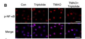
Application: IF/ICC Species: mouse Sample: BMDMs
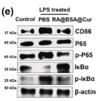
Application: WB Species: Mice Sample: spinal cord

Application: WB Species: Human Sample: ARPE‐19 cells
Restrictive clause
Affinity Biosciences tests all products strictly. Citations are provided as a resource for additional applications that have not been validated by Affinity Biosciences. Please choose the appropriate format for each application and consult Materials and Methods sections for additional details about the use of any product in these publications.
For Research Use Only.
Not for use in diagnostic or therapeutic procedures. Not for resale. Not for distribution without written consent. Affinity Biosciences will not be held responsible for patent infringement or other violations that may occur with the use of our products. Affinity Biosciences, Affinity Biosciences Logo and all other trademarks are the property of Affinity Biosciences LTD.