MMP2 Antibody - #AF0577


 Photographs of the PASMCs migration through the polycarbonate membrane stained by 0.2% crystal violet in hypoxia and treated with increasing concentrations of quercetin for 24 h. (B) Quantification of the number of cells migrating through the polycarbonate membrane of average of 3 independent experiments. (C) Full-length blots of MMP-2, MMP-9, CXCR4, Integrin α1, β1, and α5 and GAPDH are presented. (D) Results were quantified by densitometry analysis of the bands form (C) and then normalization to GAPDH protein. *Po0.05, **Po0.01 compared with control; #Po0.05, ##Po0.01 compared with hypoxia and quercetin treated PASMCs.")

 microscopy at x200 magnification was used to assess cell morphology. The A549 cells (parental cells) had an epithelioid, rounded cobblestone appearance and there was limited formation of pseudopodia. A549/PTX and A549/DDP cells exhibited a spindle-shaped morphology and an increased formation of pseudopodia, indicating a loss of cell polarity. (B) E-cadherin, β-catenin, vimentin, MMP-2 and MMP-9 which are EMT-related proteins, were assessed in terms of expression levels. EMT-related transcription factors (Snail, Slug, Twist and ZEB1) were measured in A549/PTX and A549/DDP cells using western blot analysis. (C) The expression changes were confirmed at the mRNA level by qRT-PCR. Expression was standardized to the expression of GAPDH and normalized to 1.0 in the parental cells (compared with the parental A549 cells, means ± SEM, n=3, * P<0.05)")

 Te expression of MMP2 and MMP9 at protein level was shown by Western blot. Band intensity is coming from densitometry, and data was shown as mean±SD")
 Representative immunohistochemical staining of MMP2 and MMP9 in AFG1-induced lung adenocarcinoma.")
 Twenty-four hours after transfection, cell morphology was observed by microscopy at x200 magnification for non-transfected A549 cells (blank), A549 cells transfected with mimic-NC or miR-181a-mimic. (B) E-cadherin, β-catenin, vimentin, MMP-9, MMP-2, Snail, Slug, Twist and ZEB1 expression levels after transfection were determined by western blot analysis.")
 The levels of cyclin A, cyclin B, cyclin D1, and CDK2 were reduced in A375 cells transfected with OVOS2-shRNA; (b) The downregulated expression of N-cadherin accompanied with the upregulated expression of E-cadherin and β-catenin were observed in A375 cells transfected with OVOS2-shRNA; (c) The expression of p-FAK, p-AKT, and p-ERK were reduced in A375 cells transfected with OVOS2-shRNA; (d) The increased production of MMP-2 was observed in A375 transfected with OVOS2-shRNA; (e) GAPDH was used as the reference.")

 The bands of protein
expression. (B)-(F) Correlation analysis between the depression-like behaviors and protein expression. Statistical analyses of single comparison were conducted
through the Student’s t-test. All data are presented as mean ± SD, *p < 0.05, **p < 0.01, n = 3 per group.")
 Expression of proliferation- and migration-associated genes (PCNA, MMP9 and TIMP-1) were evaluated using western blotting in HEY cells. (C and D) Western blotting of proteins involved in integrin-β1-FAK signaling pathway in the KRT7-overexpressing HEY cells. (E) Expression of MMPs after knockdown of KRT7 in OVCAR433 cells. (F and G) Expression of the TGF-β signaling pathway-related proteins was evaluated by western blotting in KRT7-overexpressing HEY cells and KRT7-knockdown OVCAR433 cells. All experiments were performed at least three times. Results are presented as the mean ± standard deviation. **P<0.01. FAK, focal adhesion kinase; PCNA, proliferating cell nuclear antigen; FN, fibronectin; TIMP-1, TIMP metallopeptidase inhibitor 1; p-, phosphorylated; MMP, matrix metalloproteinase; KRT7, keratin 7; sh, short hairpin RNA; NC, negative control.")
 Transwell migration assay and transwell invasion assay were performed in LoVo cells with increasing concentration of recombinant GSN. (C) The expression of MMP2 and MMP9 were examined by western blot analysis in HT29 cell with increasing concentration of recombinant GSN.")

. The change of cerebral edema rate in different groups (C). Representative protein (MMP-2and MMP-9) expression in different groups by
western blotting analysis (D–G). The date is expressed as the means ± S.E.M. (n = 3 rats in each group). *P < 0.05, **P < 0.01 and ***P < 0.001 vs MCAO
group.")
 Immunohistochemical analysis of lumican, MMP‑2, MMP‑14 and TIMP‑2 expression.")
 and statistically analyzed with Image J software (B, C).")
 Cell viability in the different transfection groups was eval‑uated by Cell Counting Kit‑8 assay. (B) Western blot analysis showing the protein expression levels of MMP2 and MMP9 in different groups.")
 mRNA and (B) protein levels, detected by reverse transcription-quantitative PCR and western blotting, respectively. (C) Immunohistochemical analysis of lumican, MMP-2, MMP-14 and TIMP-2 expression. (D) Quantification of results presented in panel C. *P<0.05 vs. control eyes (paired t-tests). TIMP, tissue inhibitor of metalloproteinases.")
. TIMP, tissue inhibitor of metalloproteinases.")
 Western blot assay was used to detect the expression of EMT markers, ZEB1, and MMP2 in ELTD1-interfered and control cells.")
 and statistically analyzed with Image J software (B, C). Data are presented as mean±SD of 3 independent experiments. * P<0.05, ** P<0.01, *** P<0.001, compared with the control group.")
 Cell viability in the different transfection groups was evaluated by Cell Counting Kit-8 assay. (B) Western blot analysis showing the protein expression levels of MMP2 and MMP9 in different groups. (C) Cell invasion and migration in different groups of cells after transfection. Scale bar, 100μm, ***P<0.001 vs. shRNA-NC; #P<0.05, ##P<0.01 and ###P<0.001 vs. shRNA-B3GALT5-AS1-1 + ov-NC. B3GALT5-AS1, β-1,3-galactosyltransferase 5-AS1; CSNK2A1, casein kinase 2 a1; MMP, matrix metalloproteinase; LncRNA, long non-coding RNA; Ov, expression; shRNA, short hairpin RNA; OD, optical density.")
 The Pearson correlation between ELTD1 and epithelial markers (CDH1, EPCAM), mesenchymal markers (VIM, CDH2) was analyzed using TCGA STAD data. (B) The Pearson correlation between ELTD1 and EMT transcription factors (ZEB1, ZEB2, SLUG, TWIST2) was analyzed using TCGA STAD data. (C) Cytoskeleton of indicated cells is visualized by staining F-actin with rhodamine-phalloidin. Scale bars, 5 μm. (D) qRT-PCR was used to detect the expression of EMT markers in ELTD1-interfered and control cells. (E) Western blot assay was used to detect the expression of EMT markers, ZEB1, and MMP2 in ELTD1-interfered and control cells.
Notes: **P< 0.01, ***P < 0.001.")

 The KEGG pathway enricherment analysis of DGEs. The length of the columns reflects the number of DGEs enrichment results are more significant. Different colours represent different P value. (B) Co-expression network map of differentially expressed genes KEGG enrichment. (C) Western blotting analysis of COL4A1, MMP2 and MMP9 expression in CNE1 and CNE2 cells. (D) Western blotting analysis of E-cadherin, N-cadherin, Vimentin and Snail expression in CNE1 and CNE2 cells. Data are shown as mean ± SD for three independent experiments, *p < 0.05, **p < 0.01 and ***p < 0.001.")
 After HSC-3 cells were treated with different concentrations (0, 0.625, 1.25, and 2.5 μM/L) of anlotinib for 24 h, expression of the p-Akt and Akt proteins was detected by immunofluorescence. (b) Quantitative immunofluorescence results of the p-Akt and Akt proteins. (c) HSC-3 cells were treated with anlotinib for 24 hours, and total MMP-2 and MMP-9, phosphorylated and total Akt and β-actin were detected by western blotting. (d) Ratio change of the migration markers MMP-2, MMP-9, phosphorylated, and total Akt. The data are expressed as the mean ± standard deviation of three repeats. ∗∗∗P < 0.001 compared with the control.")
 The apoptotic cells in pulmonary tissues were stained by tunel test kit and observed with a fluorescence microscope (magnification, 200×). (b) The quantitative result of TUNEL staining, n = 6. (c) The representative band of Western blot. (d)–(g) The quantitative result of Western blot, n = 3. Data were expressed as mean ± SD. ##P < 0.01 vs. control; ∗p < 0.05, ∗∗p < 0.01 vs. BLM.")
. C-F. The results of the Transwell invasion assay expressed as the number of invading cells in the experimental group compared to that in the respective control group (magnication: × 100, *P < 0.05, **P < 0.01, ***P < 1 0.001, Student's t test). G. circ-ITCH downregulated the mRNA expression of the cell migration-related genes MMP-2 and MMP-9 in PCa cells, as determined by qRT–PCR (*P < 0.05, **P < 0.01, Student’s t test). H. circ-ITCH downregulated the protein expression of MMP-2 and MMP-9 in PCa cells, as determined by Western blotting")
 Western blotting indicated that the upregulated protein expression levels of collagen I, collagen III, fibronectin, α-SMA and MMP2 in oe-PDCD4-HL-1 cells were attenuated by Pio and CBL0137. (B) Statistical analysis of western blotting. (C) Enzyme-linked immunosorbent assays showed that the upregulated protein concentrations of IL-6, IL-17A, IFN-γ and TNF-α, and downregulated protein concentration of IL-4, in oe-PDCD4-HL-1 cells were reversed by Pio and CBL0137. ***P")
 Cell proliferation was determined by CCK-8 assay after treatment with quercetin for 24 h, 48 h and 72 h. Cell invasion (B, C) and migration (D, E) was detected by transwell assay (scale bar = 100 μm). (F, G) Representative images and quantitative result of migration were detected by wound scratch assay (scale bar = 25 μm). (H) Expression of migration-related proteins E-cadherin, N-cadherin, MMP-9, MMP-2 and Snail in LOVO cells was determined by Western blot assay. ∗P < 0.05, ∗∗P < 0.01, ∗∗∗P < 0.001 vs. control. NC: Normoxia control; All other groups were treated in hypoxia.")
| Product: | MMP2 Antibody |
| Catalog: | AF0577 |
| Description: | Rabbit polyclonal antibody to MMP2 |
| Application: | WB IHC IF/ICC |
| Cited expt.: | WB, IHC, IF/ICC |
| Reactivity: | Human, Mouse, Rat |
| Prediction: | Pig, Bovine, Horse, Sheep, Rabbit, Dog |
| Mol.Wt.: | 74kDa; 74kD(Calculated). |
| Uniprot: | P08253 |
| RRID: | AB_2834154 |
Product Info
*The optimal dilutions should be determined by the end user. For optimal experimental results, antibody reuse is not recommended.
*Tips:
WB: For western blot detection of denatured protein samples. IHC: For immunohistochemical detection of paraffin sections (IHC-p) or frozen sections (IHC-f) of tissue samples. IF/ICC: For immunofluorescence detection of cell samples. ELISA(peptide): For ELISA detection of antigenic peptide.
Cite Format: Affinity Biosciences Cat# AF0577, RRID:AB_2834154.
Fold/Unfold
72 kDa gelatinase; 72kD type IV collagenase; CLG 4; CLG 4A; CLG4; CLG4A; Collagenase Type 4 alpha; Collagenase type IV A; Gelatinase A; Gelatinase alpha; Gelatinase neutrophil; Matrix metallopeptidase 2 gelatinase A 72kDa gelatinase 72kDa type IV collagenase; Matrix metalloproteinase 2 (gelatinase A, 72kDa gelatinase, 72kDa type IV collagenase); Matrix Metalloproteinase 2; Matrix metalloproteinase II; Matrix metalloproteinase-2; MMP 2; MMP II; MMP-2; MMP2; MMP2_HUMAN; MONA; Neutrophil gelatinase; PEX; TBE 1; TBE-1;
Immunogens
A synthesized peptide derived from human MMP2, corresponding to a region within C-terminal amino acids.
Produced by normal skin fibroblasts. PEX is expressed in a number of tumors including gliomas, breast and prostate.
- P08253 MMP2_HUMAN:
- Protein BLAST With
- NCBI/
- ExPASy/
- Uniprot
MEALMARGALTGPLRALCLLGCLLSHAAAAPSPIIKFPGDVAPKTDKELAVQYLNTFYGCPKESCNLFVLKDTLKKMQKFFGLPQTGDLDQNTIETMRKPRCGNPDVANYNFFPRKPKWDKNQITYRIIGYTPDLDPETVDDAFARAFQVWSDVTPLRFSRIHDGEADIMINFGRWEHGDGYPFDGKDGLLAHAFAPGTGVGGDSHFDDDELWTLGEGQVVRVKYGNADGEYCKFPFLFNGKEYNSCTDTGRSDGFLWCSTTYNFEKDGKYGFCPHEALFTMGGNAEGQPCKFPFRFQGTSYDSCTTEGRTDGYRWCGTTEDYDRDKKYGFCPETAMSTVGGNSEGAPCVFPFTFLGNKYESCTSAGRSDGKMWCATTANYDDDRKWGFCPDQGYSLFLVAAHEFGHAMGLEHSQDPGALMAPIYTYTKNFRLSQDDIKGIQELYGASPDIDLGTGPTPTLGPVTPEICKQDIVFDGIAQIRGEIFFFKDRFIWRTVTPRDKPMGPLLVATFWPELPEKIDAVYEAPQEEKAVFFAGNEYWIYSASTLERGYPKPLTSLGLPPDVQRVDAAFNWSKNKKTYIFAGDKFWRYNEVKKKMDPGFPKLIADAWNAIPDNLDAVVDLQGGGHSYFFKGAYYLKLENQSLKSVKFGSIKSDWLGC
Predictions
Score>80(red) has high confidence and is suggested to be used for WB detection. *The prediction model is mainly based on the alignment of immunogen sequences, the results are for reference only, not as the basis of quality assurance.
High(score>80) Medium(80>score>50) Low(score<50) No confidence
Research Backgrounds
Ubiquitinous metalloproteinase that is involved in diverse functions such as remodeling of the vasculature, angiogenesis, tissue repair, tumor invasion, inflammation, and atherosclerotic plaque rupture. As well as degrading extracellular matrix proteins, can also act on several nonmatrix proteins such as big endothelial 1 and beta-type CGRP promoting vasoconstriction. Also cleaves KISS at a Gly-|-Leu bond. Appears to have a role in myocardial cell death pathways. Contributes to myocardial oxidative stress by regulating the activity of GSK3beta. Cleaves GSK3beta in vitro. Involved in the formation of the fibrovascular tissues in association with MMP14.
PEX, the C-terminal non-catalytic fragment of MMP2, posseses anti-angiogenic and anti-tumor properties and inhibits cell migration and cell adhesion to FGF2 and vitronectin. Ligand for integrinv/beta3 on the surface of blood vessels.
Mediates the proteolysis of CHUK/IKKA and initiates a primary innate immune response by inducing mitochondrial-nuclear stress signaling with activation of the pro-inflammatory NF-kappaB, NFAT and IRF transcriptional pathways.
Phosphorylation on multiple sites modulates enzymatic activity. Phosphorylated by PKC in vitro.
The propeptide is processed by MMP14 (MT-MMP1) and MMP16 (MT-MMP3). Autocatalytic cleavage in the C-terminal produces the anti-angiogenic peptide, PEX. This processing appears to be facilitated by binding integrinv/beta3.
Secreted>Extracellular space>Extracellular matrix. Membrane. Nucleus.
Note: Colocalizes with integrin alphaV/beta3 at the membrane surface in angiogenic blood vessels and melanomas. Found in mitochondria, along microfibrils, and in nuclei of cardiomyocytes.
Cytoplasm. Mitochondrion.
Produced by normal skin fibroblasts. PEX is expressed in a number of tumors including gliomas, breast and prostate.
The conserved cysteine present in the cysteine-switch motif binds the catalytic zinc ion, thus inhibiting the enzyme. The dissociation of the cysteine from the zinc ion upon the activation-peptide release activates the enzyme.
Belongs to the peptidase M10A family.
Research Fields
· Human Diseases > Drug resistance: Antineoplastic > Endocrine resistance.
· Human Diseases > Cancers: Overview > Pathways in cancer. (View pathway)
· Human Diseases > Cancers: Overview > Proteoglycans in cancer.
· Human Diseases > Cancers: Specific types > Bladder cancer. (View pathway)
· Organismal Systems > Immune system > Leukocyte transendothelial migration. (View pathway)
· Organismal Systems > Endocrine system > Estrogen signaling pathway. (View pathway)
· Organismal Systems > Endocrine system > Relaxin signaling pathway.
References

Application: WB Species: human Sample:
Application: IHC Species: human Sample:
Application: WB Species: Human Sample: OS cells
Application: WB Species: mouse Sample: tumors

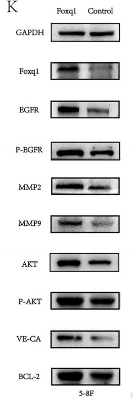
Application: IF/ICC Species: mouse Sample: 5–8F cells
Application: WB Species: human Sample: PLC-PRF-5 cells
Application: IHC Species: human Sample: tumor

Application: WB Species: Human Sample: breast cancer cells

Application: WB Species: Human Sample: VSMCs
Restrictive clause
Affinity Biosciences tests all products strictly. Citations are provided as a resource for additional applications that have not been validated by Affinity Biosciences. Please choose the appropriate format for each application and consult Materials and Methods sections for additional details about the use of any product in these publications.
For Research Use Only.
Not for use in diagnostic or therapeutic procedures. Not for resale. Not for distribution without written consent. Affinity Biosciences will not be held responsible for patent infringement or other violations that may occur with the use of our products. Affinity Biosciences, Affinity Biosciences Logo and all other trademarks are the property of Affinity Biosciences LTD.