Fibronectin Antibody - #AF0738

| Product: | Fibronectin Antibody |
| Catalog: | AF0738 |
| Description: | Rabbit polyclonal antibody to Fibronectin |
| Application: | WB IHC IF/ICC |
| Reactivity: | Human, Mouse, Rat |
| Prediction: | Pig, Bovine, Sheep, Rabbit |
| Mol.Wt.: | 260kDa; 272kD(Calculated). |
| Uniprot: | P02751 |
| RRID: | AB_2834137 |
Product Info
*The optimal dilutions should be determined by the end user.
*Tips:
WB: For western blot detection of denatured protein samples. IHC: For immunohistochemical detection of paraffin sections (IHC-p) or frozen sections (IHC-f) of tissue samples. IF/ICC: For immunofluorescence detection of cell samples. ELISA(peptide): For ELISA detection of antigenic peptide.
Cite Format: Affinity Biosciences Cat# AF0738, RRID:AB_2834137.
Fold/Unfold
CIG; Cold insoluble globulin; Cold-insoluble globulin; DKFZp686F10164; DKFZp686H0342; DKFZp686I1370; DKFZp686O13149; ED B; Fibronectin 1; FINC; FINC_HUMAN; FN; FN1; FNZ; GFND; GFND2; LETS; Migration stimulating factor; MSF; Ugl-Y3;
Immunogens
Expressed in the inner limiting membrane and around blood vessels in the retina (at protein level) (PubMed:29777959). Plasma FN (soluble dimeric form) is secreted by hepatocytes. Cellular FN (dimeric or cross-linked multimeric forms), made by fibroblasts, epithelial and other cell types, is deposited as fibrils in the extracellular matrix. Ugl-Y1, Ugl-Y2 and Ugl-Y3 are found in urine (PubMed:17614963).
- P02751 FINC_HUMAN:
- Protein BLAST With
- NCBI/
- ExPASy/
- Uniprot
MLRGPGPGLLLLAVQCLGTAVPSTGASKSKRQAQQMVQPQSPVAVSQSKPGCYDNGKHYQINQQWERTYLGNALVCTCYGGSRGFNCESKPEAEETCFDKYTGNTYRVGDTYERPKDSMIWDCTCIGAGRGRISCTIANRCHEGGQSYKIGDTWRRPHETGGYMLECVCLGNGKGEWTCKPIAEKCFDHAAGTSYVVGETWEKPYQGWMMVDCTCLGEGSGRITCTSRNRCNDQDTRTSYRIGDTWSKKDNRGNLLQCICTGNGRGEWKCERHTSVQTTSSGSGPFTDVRAAVYQPQPHPQPPPYGHCVTDSGVVYSVGMQWLKTQGNKQMLCTCLGNGVSCQETAVTQTYGGNSNGEPCVLPFTYNGRTFYSCTTEGRQDGHLWCSTTSNYEQDQKYSFCTDHTVLVQTRGGNSNGALCHFPFLYNNHNYTDCTSEGRRDNMKWCGTTQNYDADQKFGFCPMAAHEEICTTNEGVMYRIGDQWDKQHDMGHMMRCTCVGNGRGEWTCIAYSQLRDQCIVDDITYNVNDTFHKRHEEGHMLNCTCFGQGRGRWKCDPVDQCQDSETGTFYQIGDSWEKYVHGVRYQCYCYGRGIGEWHCQPLQTYPSSSGPVEVFITETPSQPNSHPIQWNAPQPSHISKYILRWRPKNSVGRWKEATIPGHLNSYTIKGLKPGVVYEGQLISIQQYGHQEVTRFDFTTTSTSTPVTSNTVTGETTPFSPLVATSESVTEITASSFVVSWVSASDTVSGFRVEYELSEEGDEPQYLDLPSTATSVNIPDLLPGRKYIVNVYQISEDGEQSLILSTSQTTAPDAPPDTTVDQVDDTSIVVRWSRPQAPITGYRIVYSPSVEGSSTELNLPETANSVTLSDLQPGVQYNITIYAVEENQESTPVVIQQETTGTPRSDTVPSPRDLQFVEVTDVKVTIMWTPPESAVTGYRVDVIPVNLPGEHGQRLPISRNTFAEVTGLSPGVTYYFKVFAVSHGRESKPLTAQQTTKLDAPTNLQFVNETDSTVLVRWTPPRAQITGYRLTVGLTRRGQPRQYNVGPSVSKYPLRNLQPASEYTVSLVAIKGNQESPKATGVFTTLQPGSSIPPYNTEVTETTIVITWTPAPRIGFKLGVRPSQGGEAPREVTSDSGSIVVSGLTPGVEYVYTIQVLRDGQERDAPIVNKVVTPLSPPTNLHLEANPDTGVLTVSWERSTTPDITGYRITTTPTNGQQGNSLEEVVHADQSSCTFDNLSPGLEYNVSVYTVKDDKESVPISDTIIPEVPQLTDLSFVDITDSSIGLRWTPLNSSTIIGYRITVVAAGEGIPIFEDFVDSSVGYYTVTGLEPGIDYDISVITLINGGESAPTTLTQQTAVPPPTDLRFTNIGPDTMRVTWAPPPSIDLTNFLVRYSPVKNEEDVAELSISPSDNAVVLTNLLPGTEYVVSVSSVYEQHESTPLRGRQKTGLDSPTGIDFSDITANSFTVHWIAPRATITGYRIRHHPEHFSGRPREDRVPHSRNSITLTNLTPGTEYVVSIVALNGREESPLLIGQQSTVSDVPRDLEVVAATPTSLLISWDAPAVTVRYYRITYGETGGNSPVQEFTVPGSKSTATISGLKPGVDYTITVYAVTGRGDSPASSKPISINYRTEIDKPSQMQVTDVQDNSISVKWLPSSSPVTGYRVTTTPKNGPGPTKTKTAGPDQTEMTIEGLQPTVEYVVSVYAQNPSGESQPLVQTAVTNIDRPKGLAFTDVDVDSIKIAWESPQGQVSRYRVTYSSPEDGIHELFPAPDGEEDTAELQGLRPGSEYTVSVVALHDDMESQPLIGTQSTAIPAPTDLKFTQVTPTSLSAQWTPPNVQLTGYRVRVTPKEKTGPMKEINLAPDSSSVVVSGLMVATKYEVSVYALKDTLTSRPAQGVVTTLENVSPPRRARVTDATETTITISWRTKTETITGFQVDAVPANGQTPIQRTIKPDVRSYTITGLQPGTDYKIYLYTLNDNARSSPVVIDASTAIDAPSNLRFLATTPNSLLVSWQPPRARITGYIIKYEKPGSPPREVVPRPRPGVTEATITGLEPGTEYTIYVIALKNNQKSEPLIGRKKTDELPQLVTLPHPNLHGPEILDVPSTVQKTPFVTHPGYDTGNGIQLPGTSGQQPSVGQQMIFEEHGFRRTTPPTTATPIRHRPRPYPPNVGEEIQIGHIPREDVDYHLYPHGPGLNPNASTGQEALSQTTISWAPFQDTSEYIISCHPVGTDEEPLQFRVPGTSTSATLTGLTRGATYNVIVEALKDQQRHKVREEVVTVGNSVNEGLNQPTDDSCFDPYTVSHYAVGDEWERMSESGFKLLCQCLGFGSGHFRCDSSRWCHDNGVNYKIGEKWDRQGENGQMMSCTCLGNGKGEFKCDPHEATCYDDGKTYHVGEQWQKEYLGAICSCTCFGGQRGWRCDNCRRPGGEPSPEGTTGQSYNQYSQRYHQRTNTNVNCPIECFMPLDVQADREDSRE
Predictions
Score>80(red) has high confidence and is suggested to be used for WB detection. *The prediction model is mainly based on the alignment of immunogen sequences, the results are for reference only, not as the basis of quality assurance.
High(score>80) Medium(80>score>50) Low(score<50) No confidence
PTMs - P02751 As Substrate
| Site | PTM Type | Enzyme | Source |
|---|---|---|---|
| S41 | Phosphorylation | Uniprot | |
| Y101 | Phosphorylation | Uniprot | |
| Y106 | Phosphorylation | Uniprot | |
| Y112 | Phosphorylation | Uniprot | |
| T136 | Phosphorylation | Uniprot | |
| T278 | O-Glycosylation | Uniprot | |
| T279 | O-Glycosylation | Uniprot | |
| S280 | O-Glycosylation | Uniprot | |
| S281 | O-Glycosylation | Uniprot | |
| S283 | O-Glycosylation | Uniprot | |
| T287 | O-Glycosylation | Uniprot | |
| Y372 | Phosphorylation | Uniprot | |
| N430 | N-Glycosylation | Uniprot | |
| K486 | Methylation | Uniprot | |
| N528 | N-Glycosylation | Uniprot | |
| N542 | N-Glycosylation | Uniprot | |
| Y588 | Phosphorylation | Uniprot | |
| Y641 | Phosphorylation | Uniprot | |
| T715 | Phosphorylation | Uniprot | |
| N877 | N-Glycosylation | Uniprot | |
| S904 | Phosphorylation | Uniprot | |
| S909 | Phosphorylation | Uniprot | |
| Y937 | Phosphorylation | Uniprot | |
| T960 | Phosphorylation | Uniprot | |
| S968 | Phosphorylation | Uniprot | |
| T972 | Phosphorylation | Uniprot | |
| K987 | Acetylation | Uniprot | |
| N1007 | N-Glycosylation | Uniprot | |
| Y1042 | Phosphorylation | Uniprot | |
| K1050 | Ubiquitination | Uniprot | |
| Y1206 | Phosphorylation | Uniprot | |
| N1244 | N-Glycosylation | Uniprot | |
| T1271 | Phosphorylation | Uniprot | |
| T1276 | O-Glycosylation | Uniprot | |
| S1565 | O-Glycosylation | Uniprot | |
| S1566 | O-Glycosylation | Uniprot | |
| S1567 | O-Glycosylation | Uniprot | |
| T1570 | O-Glycosylation | Uniprot | |
| T1641 | O-Glycosylation | Uniprot | |
| T1743 | Phosphorylation | Uniprot | |
| T1762 | Phosphorylation | Uniprot | |
| T1786 | Phosphorylation | Uniprot | |
| S1833 | Phosphorylation | Uniprot | |
| K1837 | Ubiquitination | Uniprot | |
| T1840 | Phosphorylation | Uniprot | |
| T1842 | Phosphorylation | Uniprot | |
| T1855 | Phosphorylation | Uniprot | |
| T1860 | Phosphorylation | Uniprot | |
| Y1879 | Phosphorylation | Uniprot | |
| K1880 | Acetylation | Uniprot | |
| Y1884 | Phosphorylation | Uniprot | |
| S1982 | Phosphorylation | Uniprot | |
| T1999 | O-Glycosylation | Uniprot | |
| T2060 | O-Glycosylation | Uniprot | |
| T2061 | O-Glycosylation | Uniprot | |
| T2064 | O-Glycosylation | Uniprot | |
| T2065 | O-Glycosylation | Uniprot | |
| T2067 | O-Glycosylation | Uniprot | |
| N2108 | N-Glycosylation | Uniprot | |
| Y2258 | Phosphorylation | Uniprot | |
| Y2312 | Phosphorylation | Uniprot | |
| S2318 | Phosphorylation | Uniprot | |
| S2341 | O-Glycosylation | Uniprot | |
| S2341 | Phosphorylation | Uniprot | |
| T2345 | O-Glycosylation | Uniprot | |
| T2346 | O-Glycosylation | Uniprot | |
| S2349 | O-Glycosylation | Uniprot | |
| S2349 | Phosphorylation | Uniprot | |
| Y2350 | Phosphorylation | Uniprot | |
| Y2353 | Phosphorylation | Uniprot | |
| S2354 | Phosphorylation | Uniprot | |
| T2363 | Phosphorylation | Uniprot | |
| S2384 | Phosphorylation | Uniprot |
Research Backgrounds
Fibronectins bind cell surfaces and various compounds including collagen, fibrin, heparin, DNA, and actin. Fibronectins are involved in cell adhesion, cell motility, opsonization, wound healing, and maintenance of cell shape. Involved in osteoblast compaction through the fibronectin fibrillogenesis cell-mediated matrix assembly process, essential for osteoblast mineralization. Participates in the regulation of type I collagen deposition by osteoblasts.
Anastellin binds fibronectin and induces fibril formation. This fibronectin polymer, named superfibronectin, exhibits enhanced adhesive properties. Both anastellin and superfibronectin inhibit tumor growth, angiogenesis and metastasis. Anastellin activates p38 MAPK and inhibits lysophospholipid signaling.
Sulfated.
It is not known whether both or only one of Thr-2155 and Thr-2156 are/is glycosylated.
Forms covalent cross-links mediated by a transglutaminase, such as F13A or TGM2, between a glutamine and the epsilon-amino group of a lysine residue, forming homopolymers and heteropolymers (e.g. fibrinogen-fibronectin, collagen-fibronectin heteropolymers).
Phosphorylated by FAM20C in the extracellular medium.
Proteolytic processing produces the C-terminal NC1 peptide, anastellin.
Some lysine residues are oxidized to allysine by LOXL3, promoting fibronectin activation and matrix formation.
Secreted>Extracellular space>Extracellular matrix.
Expressed in the inner limiting membrane and around blood vessels in the retina (at protein level). Plasma FN (soluble dimeric form) is secreted by hepatocytes. Cellular FN (dimeric or cross-linked multimeric forms), made by fibroblasts, epithelial and other cell types, is deposited as fibrils in the extracellular matrix. Ugl-Y1, Ugl-Y2 and Ugl-Y3 are found in urine.
Mostly heterodimers or multimers of alternatively spliced variants, connected by 2 disulfide bonds near the carboxyl ends; to a lesser extent homodimers. Interacts with FBLN1, AMBP, TNR, LGALS3BP and COL13A1. Interacts with FBLN7 (By similarity). Interacts with COMP. Interacts with TNR; the interaction inhibits cell adhesion and neurite outgrowth (By similarity). Interacts with FST3 and MYOC.
(Microbial infection) Interacts with S.aureus FnbA.
(Microbial infection) Interacts with M.bovis FbpB via the collagen-binding region.
(Microbial infection) Interacts with recombinant S.pneumoniae PavA (rqcH).
(Microbial infection) Interacts with recombinant S.suis FbpS (rqcH) via fibronectin's N-terminal 30 kDa region.
(Microbial infection) Interacts with fibronectin-binding proteins from other Mycobacteria.
Research Fields
· Cellular Processes > Cellular community - eukaryotes > Focal adhesion. (View pathway)
· Cellular Processes > Cell motility > Regulation of actin cytoskeleton. (View pathway)
· Environmental Information Processing > Signal transduction > PI3K-Akt signaling pathway. (View pathway)
· Environmental Information Processing > Signaling molecules and interaction > ECM-receptor interaction. (View pathway)
· Human Diseases > Infectious diseases: Bacterial > Bacterial invasion of epithelial cells.
· Human Diseases > Infectious diseases: Parasitic > Amoebiasis.
· Human Diseases > Infectious diseases: Viral > Human papillomavirus infection.
· Human Diseases > Cancers: Overview > Pathways in cancer. (View pathway)
· Human Diseases > Cancers: Overview > Proteoglycans in cancer.
· Human Diseases > Cancers: Specific types > Small cell lung cancer. (View pathway)
References

Application: IHC Species: Human Sample:
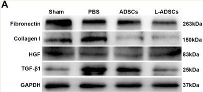
Application: WB Species: Rat Sample: penile tissues
Restrictive clause
Affinity Biosciences tests all products strictly. Citations are provided as a resource for additional applications that have not been validated by Affinity Biosciences. Please choose the appropriate format for each application and consult Materials and Methods sections for additional details about the use of any product in these publications.
For Research Use Only.
Not for use in diagnostic or therapeutic procedures. Not for resale. Not for distribution without written consent. Affinity Biosciences will not be held responsible for patent infringement or other violations that may occur with the use of our products. Affinity Biosciences, Affinity Biosciences Logo and all other trademarks are the property of Affinity Biosciences LTD.