Product: Collagen III Antibody
Catalog: AF0136
Description: Rabbit polyclonal antibody to Collagen III
Application: WB IHC IF/ICC
Reactivity: Human, Mouse, Rat
Prediction: Pig, Bovine, Horse, Sheep, Rabbit, Dog, Chicken, Xenopus
Mol.Wt.: 138kDa; 139kD(Calculated).
Uniprot: P02461
RRID: AB_2813770

View similar products>>

   Size Price Inventory
 100ul $280 In stock
 200ul $350 In stock

Lead Time: Same day delivery

For pricing and ordering contact:
Local distributors

Product Info

Source:
Rabbit
Application:
IHC 1:50-1:200, IF/ICC 1:100-1:500, WB 1:1000
*The optimal dilutions should be determined by the end user.
*Tips:

WB: For western blot detection of denatured protein samples. IHC: For immunohistochemical detection of paraffin sections (IHC-p) or frozen sections (IHC-f) of tissue samples. IF/ICC: For immunofluorescence detection of cell samples. ELISA(peptide): For ELISA detection of antigenic peptide.

Reactivity:
Human,Mouse,Rat
Prediction:
Pig(86%), Bovine(83%), Horse(83%), Sheep(100%), Rabbit(100%), Dog(100%), Chicken(100%), Xenopus(83%)
Clonality:
Polyclonal
Specificity:
Collagen III Antibody detects endogenous levels of total Collagen III.
RRID:
AB_2813770
Cite Format: Affinity Biosciences Cat# AF0136, RRID:AB_2813770.
Conjugate:
Unconjugated.
Purification:
The antiserum was purified by peptide affinity chromatography using SulfoLink™ Coupling Resin (Thermo Fisher Scientific).
Storage:
Rabbit IgG in phosphate buffered saline , pH 7.4, 150mM NaCl, 0.02% sodium azide and 50% glycerol. Store at -20 °C. Stable for 12 months from date of receipt.
Alias:

Fold/Unfold

Alpha 1 type III collagen; Alpha1 (III) collagen; CO3A1_HUMAN; COL 3A1; COL3A1; Collagen alpha 1(III) chain; Collagen alpha-1(III) chain; Collagen III alpha 1 chain precursor; Collagen III alpha 1 polypeptide; Collagen type III alpha 1 (Ehlers Danlos syndrome type IV autosomal dominant); Collagen type III alpha 1; Collagen type III alpha 1 chain; Collagen type III alpha; Collagen, fetal; EDS4A; Ehlers Danlos syndrome type IV, autosomal dominant; Fetal collagen; Type III collagen;

Immunogens

Immunogen:
Uniprot:
Gene(ID):
Description:
CO3A1 Collagen type III occurs in most soft connective tissues along with type I collagen. Belongs to the fibrillar collagen family. Trimers of identical alpha 1(III) chains. The chains are linked to each other by interchain disulfide bonds. Trimers are also cross-linked via hydroxylysines. 2 isoforms of the human protein are produced by alternative splicing. Note: This description may include information from UniProtKB.
Sequence:
MMSFVQKGSWLLLALLHPTIILAQQEAVEGGCSHLGQSYADRDVWKPEPCQICVCDSGSVLCDDIICDDQELDCPNPEIPFGECCAVCPQPPTAPTRPPNGQGPQGPKGDPGPPGIPGRNGDPGIPGQPGSPGSPGPPGICESCPTGPQNYSPQYDSYDVKSGVAVGGLAGYPGPAGPPGPPGPPGTSGHPGSPGSPGYQGPPGEPGQAGPSGPPGPPGAIGPSGPAGKDGESGRPGRPGERGLPGPPGIKGPAGIPGFPGMKGHRGFDGRNGEKGETGAPGLKGENGLPGENGAPGPMGPRGAPGERGRPGLPGAAGARGNDGARGSDGQPGPPGPPGTAGFPGSPGAKGEVGPAGSPGSNGAPGQRGEPGPQGHAGAQGPPGPPGINGSPGGKGEMGPAGIPGAPGLMGARGPPGPAGANGAPGLRGGAGEPGKNGAKGEPGPRGERGEAGIPGVPGAKGEDGKDGSPGEPGANGLPGAAGERGAPGFRGPAGPNGIPGEKGPAGERGAPGPAGPRGAAGEPGRDGVPGGPGMRGMPGSPGGPGSDGKPGPPGSQGESGRPGPPGPSGPRGQPGVMGFPGPKGNDGAPGKNGERGGPGGPGPQGPPGKNGETGPQGPPGPTGPGGDKGDTGPPGPQGLQGLPGTGGPPGENGKPGEPGPKGDAGAPGAPGGKGDAGAPGERGPPGLAGAPGLRGGAGPPGPEGGKGAAGPPGPPGAAGTPGLQGMPGERGGLGSPGPKGDKGEPGGPGADGVPGKDGPRGPTGPIGPPGPAGQPGDKGEGGAPGLPGIAGPRGSPGERGETGPPGPAGFPGAPGQNGEPGGKGERGAPGEKGEGGPPGVAGPPGGSGPAGPPGPQGVKGERGSPGGPGAAGFPGARGLPGPPGSNGNPGPPGPSGSPGKDGPPGPAGNTGAPGSPGVSGPKGDAGQPGEKGSPGAQGPPGAPGPLGIAGITGARGLAGPPGMPGPRGSPGPQGVKGESGKPGANGLSGERGPPGPQGLPGLAGTAGEPGRDGNPGSDGLPGRDGSPGGKGDRGENGSPGAPGAPGHPGPPGPVGPAGKSGDRGESGPAGPAGAPGPAGSRGAPGPQGPRGDKGETGERGAAGIKGHRGFPGNPGAPGSPGPAGQQGAIGSPGPAGPRGPVGPSGPPGKDGTSGHPGPIGPPGPRGNRGERGSEGSPGHPGQPGPPGPPGAPGPCCGGVGAAAIAGIGGEKAGGFAPYYGDEPMDFKINTDEIMTSLKSVNGQIESLISPDGSRKNPARNCRDLKFCHPELKSGEYWVDPNQGCKLDAIKVFCNMETGETCISANPLNVPRKHWWTDSSAEKKHVWFGESMDGGFQFSYGNPELPEDVLDVHLAFLRLLSSRASQNITYHCKNSIAYMDQASGNVKKALKLMGSNEGEFKAEGNSKFTYTVLEDGCTKHTGEWSKTVFEYRTRKAVRLPIVDIAPYDIGGPDQEFGVDVGPVCFL

Predictions

Predictions:

Score>80(red) has high confidence and is suggested to be used for WB detection. *The prediction model is mainly based on the alignment of immunogen sequences, the results are for reference only, not as the basis of quality assurance.

Species
Results
Score
Sheep
100
Dog
100
Chicken
100
Rabbit
100
Pig
86
Horse
83
Bovine
83
Xenopus
83
Zebrafish
0
Model Confidence:
High(score>80) Medium(80>score>50) Low(score<50) No confidence

PTMs - P02461 As Substrate

Site PTM Type Enzyme
S143 Phosphorylation
K757 Ubiquitination
S916 Phosphorylation
S1145 O-Glycosylation
Y1220 Phosphorylation
S1237 Phosphorylation
Y1370 Phosphorylation
Y1378 Phosphorylation
S1383 Phosphorylation
K1387 Acetylation

Research Backgrounds

Function:

Collagen type III occurs in most soft connective tissues along with type I collagen. Involved in regulation of cortical development. Is the major ligand of ADGRG1 in the developing brain and binding to ADGRG1 inhibits neuronal migration and activates the RhoA pathway by coupling ADGRG1 to GNA13 and possibly GNA12.

PTMs:

Proline residues at the third position of the tripeptide repeating unit (G-X-Y) are hydroxylated in some or all of the chains.

O-linked glycan consists of a Glc-Gal disaccharide bound to the oxygen atom of a post-translationally added hydroxyl group.

Subcellular Location:

Secreted>Extracellular space>Extracellular matrix.

Extracellular region or secreted Cytosol Plasma membrane Cytoskeleton Lysosome Endosome Peroxisome ER Golgi apparatus Nucleus Mitochondrion Manual annotation Automatic computational assertionSubcellular location
Subunit Structure:

Trimers of identical alpha 1(III) chains. The chains are linked to each other by interchain disulfide bonds. Trimers are also cross-linked via hydroxylysines. Interacts with ADGRG1.

Family&Domains:

The C-terminal propeptide, also known as COLFI domain, have crucial roles in tissue growth and repair by controlling both the intracellular assembly of procollagen molecules and the extracellular assembly of collagen fibrils. It binds a calcium ion which is essential for its function.

Belongs to the fibrillar collagen family.

Research Fields

· Human Diseases > Infectious diseases: Parasitic > Amoebiasis.

· Organismal Systems > Immune system > Platelet activation.   (View pathway)

· Organismal Systems > Endocrine system > Relaxin signaling pathway.

· Organismal Systems > Digestive system > Protein digestion and absorption.

References

1). An asymmetric chitosan scaffold for tendon tissue engineering: In vitro and in vivo evaluation with rat tendon stem/progenitor cells. Acta Biomaterialia, 2018 (PubMed: 29678676) [IF=9.7]

Application: IF/ICC    Species: rat    Sample: tendon

Fig. 6. | Analyses of tendon tissue by immunofluorescence. Fluorescence immunohistochemistry at the regenerated tendon in each group at 4 weeks. (A-D) The production of COL1; (E-H) The production of COL3; (I-L) The production of TNMD; (M-P) The production of CD31. Blue: DAPI (cell nuclei); Green:COL1/COL3/CD31; Red: TNMD. Scale bar = 100 μm.

Application: IHC    Species: rat    Sample: tendon

Fig. 5.| Detailed analyses of tendon tissue organization and immunohistochemistry. (A-E) DAPI staining and chart of calculated cell density. Data were mean ± S.D. * p < 0.05 vs. ED group. (F-Q)Representative images of regenerated tendon in each group at 4 weeks. Scale bar =100 μm.

2). Ethyl ferulate suppresses post-myocardial infarction myocardial fibrosis by inhibiting transforming growth factor receptor 1. Phytomedicine, 2023 (PubMed: 37801895) [IF=7.9]

3). Protective role of Gentianella acuta on isoprenaline induced myocardial fibrosis in rats via inhibition of NF-κB pathway. BIOMEDICINE & PHARMACOTHERAPY, 2019 (PubMed: 30554111) [IF=7.5]

4). Modified citrus pectin ameliorates myocardial fibrosis and inflammation via suppressing galectin-3 and TLR4/MyD88/NF-κB signaling pathway. Biomedicine & Pharmacotherapy, 2020 (PubMed: 32172066) [IF=7.5]

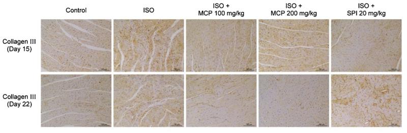
Application: IHC    Species: rat    Sample: heart

Fig. 5. |Effects of MCP on ISO-induced myocardial fibrosis in rats. (A and B) Immunohistochemical analysis of collagen I and collagen Ⅲ protein in heart cross sections of different groups on day 15 and day 22 (200× magnification). Scale bars 100 μm.

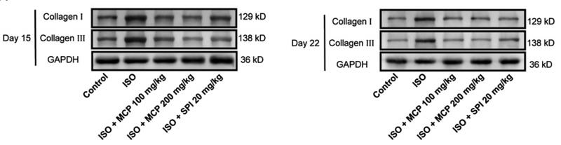
Application: WB    Species: rat    Sample: heart

Fig. 6. |Effects of MCP on the expression of collagen Ⅰ and collagen Ⅲ induced by ISO in rats. Protein and mRNA levels of collagen Ⅰ and collagen Ⅲ were analyzed by western blot and qRT-PCR in each group on day 15 and day 22. (A) Protein expression of collagen Ⅰ and collagen Ⅲ were measured by western blot.

5). Targeting the Akt/PI3K/mTOR signaling pathway for complete eradication of keloid disease by sunitinib. Apoptosis, 2022 (PubMed: 35802302) [IF=7.2]

6). Corilagin alleviates hypertrophic scars via inhibiting the transforming growth factor (TGF)-β/Smad signal pathway. LIFE SCIENCES, 2021 (PubMed: 33862115) [IF=6.1]

Application: WB    Species: Human    Sample: HSFs

Fig. 1. Corilagin downregulates mRNA and protein levels of collagen synthesis-related molecules in HSFs. A. Chemical structure of corilagin. B. Inhibitory effect of corilagin on COL1A1, COL1A2, COL3A1, and α-SMA mRNA expression level after HSFs were treated for 3 days. The expression levels were compared with that of GAPDH, n = 3. C. Protein levels of COL I, COL III, and α-SMA after corilagin application for 3 days by western blot analysis. The protein levels were compared with that of GAPDH. D. Quantification of protein levels detected by western blot analysis normalized to GAPDH level. n = 3. Data are shown as mean ± SD. *p < 0.05, **p < 0.01, ***p < 0.001. ns, no significance.

7). Bellidifolin Ameliorates Isoprenaline-Induced Myocardial Fibrosis by Regulating TGF-β1/Smads and p38 Signaling and Preventing NR4A1 Cytoplasmic Localization. Frontiers in Pharmacology, 2021 (PubMed: 33995055) [IF=5.6]

Application: WB    Species: mouse    Sample: heart

Fig. 2| (B–D) Western blotting detection of collagen Ⅰ, collagen Ⅲ, and α-smooth muscle actin (α-SMA). Bar graphs show fold changes for the collagen Ⅰ,collagen Ⅲ, and α-SMA expression as analyzed by western blotting. Glyceraldehyde 3-phosphate dehydrogenase (GAPDH) was used as a loading control (n 3). Datawere shown as mean ± SEM. **p < 0.01 vs. control, #p < 0.05 vs. ISO, ##p < 0.01 vs. ISO.

Application: IF/ICC    Species: mouse    Sample: CFs

FIGURE 4 | BEL decreases the expression of α-SMA, collagen Ⅰ, and collagen Ⅲ induced by TGF-β1 in CFs (A) The effects of different doses of BEL (15, 30, 60 μM)on α-SMA, collagen Ⅰ, and collagen Ⅲ expression induced by TGF-β1 using immunofluorescence staining.

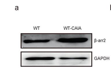
Application: WB    Species: Mouse    Sample: myocardial tissues

FIGURE 2 BEL ameliorates ISO-induced mouse myocardial fibrosis (A) HE staining to detect morphological changes; Masson trichrome staining to detect cardiac collagen deposition (B–D) Western blotting detection of collagen Ⅰ, collagen Ⅲ, and α-smooth muscle actin (α-SMA). Bar graphs show fold changes for the collagen Ⅰ, collagen Ⅲ, and α-SMA expression as analyzed by western blotting. Glyceraldehyde 3-phosphate dehydrogenase (GAPDH) was used as a loading control (n = 3). Data were shown as mean ± SEM. **p < 0.01 vs. control, #p < 0.05 vs. ISO, ##p < 0.01 vs. ISO.

8). Exosomes derived from induced pluripotent stem cells suppresses M2-type macrophages during pulmonary fibrosis via miR-302a-3p/TET1 axis. International Immunopharmacology, 2021 (PubMed: 34435585) [IF=5.6]

Application: WB    Species: Mice    Sample: lung tissues

Fig. 1. Exosomes derived from iPSCs mitigated pulmonary fibrosis. Mice models of pulmonary fibrosis were established by intratracheal instillation with bleomycin and then treated with iPSC-exosomes or MEF-exosomes. Then the histopathological changes in the lungs of mice from each group were detected by HE staining (A) and collagen deposition was detected by masson staining (B). Scale bar = 100 μm. The level of hydroxyproline in the lungs was examined with a hydroxyproline detection kit (C). The levels of TGF-β1, collagen I, and collagen III in the lungs were detected by western blot (D-E). The results are presented as mean ± SD. N = 6 for each group.

9). Oxygen–Glucose Deprivation Promoted Fibroblast Senescence and Collagen Expression via IL11. International Journal of Molecular Sciences, 2022 (PubMed: 36292942) [IF=5.6]

10). The Protective Effect of Evodiamine in Osteoarthritis: An In Vitro and In Vivo Study in Mice Model. Frontiers in Pharmacology, 2022 (PubMed: 35795554) [IF=5.6]

Load more

Restrictive clause


Affinity Biosciences tests all products strictly. Citations are provided as a resource for additional applications that have not been validated by Affinity Biosciences. Please choose the appropriate format for each application and consult Materials and Methods sections for additional details about the use of any product in these publications.

For Research Use Only.
Not for use in diagnostic or therapeutic procedures. Not for resale. Not for distribution without written consent. Affinity Biosciences will not be held responsible for patent infringement or other violations that may occur with the use of our products. Affinity Biosciences, Affinity Biosciences Logo and all other trademarks are the property of Affinity Biosciences LTD.