GHSR Antibody - #DF2794

Related Downloads
Protocols
Product Info
*The optimal dilutions should be determined by the end user.
*Tips:
WB: For western blot detection of denatured protein samples. IHC: For immunohistochemical detection of paraffin sections (IHC-p) or frozen sections (IHC-f) of tissue samples. IF/ICC: For immunofluorescence detection of cell samples. ELISA(peptide): For ELISA detection of antigenic peptide.
Cite Format: Affinity Biosciences Cat# DF2794, RRID:AB_2840000.
Fold/Unfold
GH releasing peptide receptor; GH-releasing peptide receptor; Ghrelin receptor; GHRP; GHRPR; GHS-R; GHSR; GHSR_HUMAN; Growth hormone secretagogue receptor 1; Growth hormone secretagogue receptor type 1; growth hormone secretatgogue receptor;
Immunogens
- Q92847 GHSR_HUMAN:
- Protein BLAST With
- NCBI/
- ExPASy/
- Uniprot
MWNATPSEEPGFNLTLADLDWDASPGNDSLGDELLQLFPAPLLAGVTATCVALFVVGIAGNLLTMLVVSRFRELRTTTNLYLSSMAFSDLLIFLCMPLDLVRLWQYRPWNFGDLLCKLFQFVSESCTYATVLTITALSVERYFAICFPLRAKVVVTKGRVKLVIFVIWAVAFCSAGPIFVLVGVEHENGTDPWDTNECRPTEFAVRSGLLTVMVWVSSIFFFLPVFCLTVLYSLIGRKLWRRRRGDAVVGASLRDQNHKQTVKMLAVVVFAFILCWLPFHVGRYLFSKSFEPGSLEIAQISQYCNLVSFVLFYLSAAINPILYNIMSKKYRVAVFRLLGFEPFSQRKLSTLKDESSRAWTESSINT
Predictions
Score>80(red) has high confidence and is suggested to be used for WB detection. *The prediction model is mainly based on the alignment of immunogen sequences, the results are for reference only, not as the basis of quality assurance.
High(score>80) Medium(80>score>50) Low(score<50) No confidence
PTMs - Q92847 As Substrate
| Site | PTM Type | Enzyme | Source |
|---|
Research Backgrounds
Receptor for ghrelin, coupled to G-alpha-11 proteins. Stimulates growth hormone secretion. Binds also other growth hormone releasing peptides (GHRP) (e.g. Met-enkephalin and GHRP-6) as well as non-peptide, low molecular weight secretagogues (e.g. L-692,429, MK-0677, adenosine).
Cell membrane>Multi-pass membrane protein.
Pituitary and hypothalamus.
Belongs to the G-protein coupled receptor 1 family.
Research Fields
· Environmental Information Processing > Signal transduction > cAMP signaling pathway. (View pathway)
· Environmental Information Processing > Signaling molecules and interaction > Neuroactive ligand-receptor interaction.
References
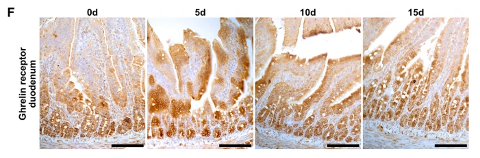
Application: IHC Species: Rat Sample:

Application: IHC Species: Rat Sample:
Restrictive clause
Affinity Biosciences tests all products strictly. Citations are provided as a resource for additional applications that have not been validated by Affinity Biosciences. Please choose the appropriate format for each application and consult Materials and Methods sections for additional details about the use of any product in these publications.
For Research Use Only.
Not for use in diagnostic or therapeutic procedures. Not for resale. Not for distribution without written consent. Affinity Biosciences will not be held responsible for patent infringement or other violations that may occur with the use of our products. Affinity Biosciences, Affinity Biosciences Logo and all other trademarks are the property of Affinity Biosciences LTD.