Product: VEGFA Antibody
Catalog: DF7470
Description: Rabbit polyclonal antibody to VEGFA
Application: WB IHC
Reactivity: Human, Mouse, Rat
Prediction: Pig, Bovine, Horse, Sheep, Rabbit, Dog
Mol.Wt.: 27kDa; 27kD(Calculated).
Uniprot: P15692
RRID: AB_2839407

View similar products>>

   Size Price Inventory
 100ul $280 In stock
 200ul $350 In stock

Lead Time: Same day delivery

For pricing and ordering contact:
Local distributors

Product Info

Source:
Rabbit
Application:
WB 1:500-1:2000, IHC 1:50-1:200
*The optimal dilutions should be determined by the end user.
*Tips:

WB: For western blot detection of denatured protein samples. IHC: For immunohistochemical detection of paraffin sections (IHC-p) or frozen sections (IHC-f) of tissue samples. IF/ICC: For immunofluorescence detection of cell samples. ELISA(peptide): For ELISA detection of antigenic peptide.

Reactivity:
Human,Mouse,Rat
Prediction:
Pig(100%), Bovine(88%), Horse(88%), Sheep(88%), Rabbit(100%), Dog(100%)
Clonality:
Polyclonal
Specificity:
VEGFA Antibody detects endogenous levels of total VEGFA.
RRID:
AB_2839407
Cite Format: Affinity Biosciences Cat# DF7470, RRID:AB_2839407.
Conjugate:
Unconjugated.
Purification:
The antiserum was purified by peptide affinity chromatography using SulfoLink™ Coupling Resin (Thermo Fisher Scientific).
Storage:
Rabbit IgG in phosphate buffered saline , pH 7.4, 150mM NaCl, 0.02% sodium azide and 50% glycerol. Store at -20 °C. Stable for 12 months from date of receipt.
Alias:

Fold/Unfold

Folliculostellate cell-derived growth factor; Glioma-derived endothelial cell mitogen; MGC70609; MVCD1; Vascular endothelial growth factor A; vascular endothelial growth factor A121; vascular endothelial growth factor A165; vascular endothelial growth factor; Vascular permeability factor; VEGF A; Vegf; VEGF-A; VEGF120; Vegfa; VEGFA_HUMAN; VPF;

Immunogens

Immunogen:
Uniprot:
Gene(ID):
Expression:
P15692 VEGFA_HUMAN:

Isoform VEGF189, isoform VEGF165 and isoform VEGF121 are widely expressed. Isoform VEGF206 and isoform VEGF145 are not widely expressed. A higher level expression seen in pituitary tumors as compared to the pituitary gland.

Description:
This gene is a member of the PDGF/VEGF growth factor family and encodes a protein that is often found as a disulfide linked homodimer. This protein is a glycosylated mitogen that specifically acts on endothelial cells and has various effects, including mediating increased vascular permeability, inducing angiogenesis, vasculogenesis and endothelial cell growth, promoting cell migration, and inhibiting apoptosis. Elevated levels of this protein is linked to POEMS syndrome, also known as Crow-Fukase syndrome. Mutations in this gene have been associated with proliferative and nonproliferative diabetic retinopathy. Alternatively spliced transcript variants, encoding either freely secreted or cell-associated isoforms, have been characterized. There is also evidence for the use of non-AUG (CUG) translation initiation sites upstream of, and in-frame with the first AUG, leading to additional isoforms.
Sequence:
MNFLLSWVHWSLALLLYLHHAKWSQAAPMAEGGGQNHHEVVKFMDVYQRSYCHPIETLVDIFQEYPDEIEYIFKPSCVPLMRCGGCCNDEGLECVPTEESNITMQIMRIKPHQGQHIGEMSFLQHNKCECRPKKDRARQEKKSVRGKGKGQKRKRKKSRYKSWSVYVGARCCLMPWSLPGPHPCGPCSERRKHLFVQDPQTCKCSCKNTDSRCKARQLELNERTCRCDKPRR

Predictions

Predictions:

Score>80(red) has high confidence and is suggested to be used for WB detection. *The prediction model is mainly based on the alignment of immunogen sequences, the results are for reference only, not as the basis of quality assurance.

Species
Results
Score
Pig
100
Dog
100
Rabbit
100
Horse
88
Bovine
88
Sheep
88
Xenopus
0
Zebrafish
0
Chicken
0
Model Confidence:
High(score>80) Medium(80>score>50) Low(score<50) No confidence

PTMs - P15692 As Substrate

Site PTM Type Enzyme

Research Backgrounds

Function:

Growth factor active in angiogenesis, vasculogenesis and endothelial cell growth. Induces endothelial cell proliferation, promotes cell migration, inhibits apoptosis and induces permeabilization of blood vessels. Binds to the FLT1/VEGFR1 and KDR/VEGFR2 receptors, heparan sulfate and heparin. NRP1/Neuropilin-1 binds isoforms VEGF-165 and VEGF-145. Isoform VEGF165B binds to KDR but does not activate downstream signaling pathways, does not activate angiogenesis and inhibits tumor growth. Binding to NRP1 receptor initiates a signaling pathway needed for motor neuron axon guidance and cell body migration, including for the caudal migration of facial motor neurons from rhombomere 4 to rhombomere 6 during embryonic development (By similarity).

Subcellular Location:

Secreted.
Note: VEGF121 is acidic and freely secreted. VEGF165 is more basic, has heparin-binding properties and, although a significant proportion remains cell-associated, most is freely secreted. VEGF189 is very basic, it is cell-associated after secretion and is bound avidly by heparin and the extracellular matrix, although it may be released as a soluble form by heparin, heparinase or plasmin.

Extracellular region or secreted Cytosol Plasma membrane Cytoskeleton Lysosome Endosome Peroxisome ER Golgi apparatus Nucleus Mitochondrion Manual annotation Automatic computational assertionSubcellular location
Tissue Specificity:

Isoform VEGF189, isoform VEGF165 and isoform VEGF121 are widely expressed. Isoform VEGF206 and isoform VEGF145 are not widely expressed. A higher level expression seen in pituitary tumors as compared to the pituitary gland.

Subunit Structure:

Homodimer; disulfide-linked. Also found as heterodimer with PGF (By similarity). Interacts with NRP1.

Family&Domains:

Belongs to the PDGF/VEGF growth factor family.

Research Fields

· Cellular Processes > Cellular community - eukaryotes > Focal adhesion.   (View pathway)

· Environmental Information Processing > Signal transduction > MAPK signaling pathway.   (View pathway)

· Environmental Information Processing > Signal transduction > Ras signaling pathway.   (View pathway)

· Environmental Information Processing > Signal transduction > Rap1 signaling pathway.   (View pathway)

· Environmental Information Processing > Signaling molecules and interaction > Cytokine-cytokine receptor interaction.   (View pathway)

· Environmental Information Processing > Signal transduction > HIF-1 signaling pathway.   (View pathway)

· Environmental Information Processing > Signal transduction > PI3K-Akt signaling pathway.   (View pathway)

· Human Diseases > Drug resistance: Antineoplastic > EGFR tyrosine kinase inhibitor resistance.

· Human Diseases > Infectious diseases: Viral > Human papillomavirus infection.

· Human Diseases > Cancers: Overview > Pathways in cancer.   (View pathway)

· Human Diseases > Cancers: Overview > Proteoglycans in cancer.

· Human Diseases > Cancers: Overview > MicroRNAs in cancer.

· Human Diseases > Cancers: Specific types > Renal cell carcinoma.   (View pathway)

· Human Diseases > Cancers: Specific types > Pancreatic cancer.   (View pathway)

· Human Diseases > Cancers: Specific types > Bladder cancer.   (View pathway)

· Human Diseases > Immune diseases > Rheumatoid arthritis.

· Organismal Systems > Endocrine system > Relaxin signaling pathway.

References

1). Injectable hydrogel with MSNs/microRNA-21-5p delivery enables both immunomodification and enhanced angiogenesis for myocardial infarction therapy in pigs. Science Advances (PubMed: 33627421) [IF=13.6]

Application: WB    Species:    Sample: endothelial cells

Fig. 3. |The mechanism underlying how the MSN/miR-21-5p complex promotes angiogenesis. (A) A heatmap of selected proteins representing strongly altered signaling pathways in three datasets of endothelial cells treated with MSN/miR-NC or MSN/miR-21-5p complexes. (B) KEGG pathway analysis of both up- and down-regulated pathways in endothelial cells after MSN/miR-21-5p complex treatment. (C) Western blot analysis of changes in SPRY1, P-ERK1/2, P-FAK, P-p38, P-AKT, VEGFA, and PDGF-BB protein content alteration in endothelial cells after treatment with the MSN/miR-21-5p complex.

2). MicroRNA-934 facilitates cell proliferation, migration, invasion and angiogenesis in colorectal cancer by targeting B-cell translocation gene 2. Bioengineered (PubMed: 34699325) [IF=4.9]

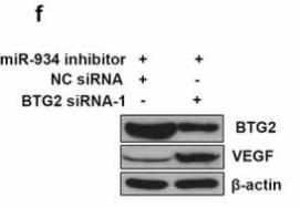
Application: WB    Species: Human    Sample: HCT116 cells

Figure 6. miR-934 mediated cell proliferation, migration, invasion and angiogenesis in dependent with BTG2. HCT116 cells were transfected with BTG2 siRNAs (50 pmol) and control siRNA with/without miR-934 inhibitor (50 pmol). (a) The mRNA levels of BTG2 in HCT116 cells were detected by qRT-PCR. (b) Cell proliferation was analyzed using MTT assay after the transfection. (c) The migratory ability of HCT116 cells was determined by wound healing assay. Scale bar: 200 μm. (d) The invasive ability of HCT116 cells was assessed by transwell assay. Scale bar: 100 μm. (e) The tube formation ratio of HUVECs were analyzed by tube formation assay. Scale bar: 200 μm. (f) The protein levels of BTG2 and VEGF were detected by western-blot. Bars and error bars represent mean values and the corresponding SD, n = 3. *P < 0.05, **P < 0.01, ***P < 0.001, ****P < 0.0001

3). Integrating Metabolomics and Network Pharmacology to Explore the Mechanism of Xiao-Yao-San in the Treatment of Inflammatory Response in CUMS Mice. Pharmaceuticals (Basel, Switzerland) (PubMed: 38004472) [IF=4.6]

4). Network Pharmacology Experiments Show That Emodin Can Exert a Protective Effect on MCAO Rats by Regulating Hif-1α/VEGF-A Signaling. ACS Omega (PubMed: 35811865) [IF=4.1]

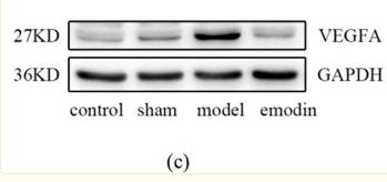
Application: WB    Species: Rat    Sample:

Figure 9 Effects of emodin on VEGF-A expression in MCAO rats. (a,b) are typical images and quantitative histograms of immunohistochemistry of VEGF-A, respectively. A, B, C, and D represent normal group, sham group, model group, and emodin group, respectively. (c,d) represent electrophoretic images and quantitative histogram of VEGF-A, respectively. Data are presented as mean ± SEM; *P < 0.05, **P < 0.01 vs sham group; #P < 0.05, ##P < 0.01 vs model group.

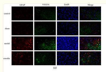
Application: IF/ICC    Species: Rat    Sample:

Figure 13 Immunofluorescence images and quantitative analysis of VEGF-A and Hif-1α. (a,c) VEGF-A and GFAP fluorescence double co-localization and the relevant quantitative analysis. VEGF-A (green), GFAP (red), and DAPI (nuclei marker, blue). (b,d) Hif-1α and NeuN fluorescence double co-localization and the relevant quantitative analysis (200×). Data are presented as mean ± SEM; *P < 0.05, **P < 0.01 vs sham group; #P < 0.05, ##P < 0.01 vs model group.

5). Molecular mechanism of Ginkgo biloba in treating type 2 diabetes mellitus combined with non-alcoholic fatty liver disease based on network pharmacology, molecular docking, and experimental evaluations. Journal of Food Biochemistry (PubMed: 36121703) [IF=4.0]

6). LINC00022 acts as an oncogene in colorectal cancer progression via sponging miR-375-3p to regulate FOXF1 expression. BMC CANCER (PubMed: 35468741) [IF=3.8]

Application: WB    Species: Human    Sample: HCT116, DLD1, and CaCo-2 cells

Fig. 5LINC00022 promoted angiogenesis in CRC. After HCT116, DLD1, and CaCo-2 cells were infected with LINC00022 low expression lentivirus or high expression lentivirus, the level of VEGFA in the cell supernatant was detected by the detection kit (a); The mRNA and protein levels of VEGFA in HCT116, DLD1, and CaCo-2 cells were measured using qRT-PCR and Western blot, respectively (b and c); The number of cavity formation was calculated (d). Scale bar = 200 μm. Data were presented as mean ± standard deviation (SD). N = 3. **P < 0.01 and ***P < 0.001 vs Lv-anti-NC group or Lv-NC group. VEGFA, vascular endothelial growth factor A; qRT-PCR, quantitative Real-time PCR

7). Screening of immunosuppressive factors for biomarkers of breast cancer malignancy phenotypes and subtype-specific targeted therapy. PeerJ (PubMed: 31293831) [IF=2.7]

Application: IHC    Species: human    Sample: breast

Figure 7 Immunohistochemical detection of the expression of MIF and VEGFA in a breast cancer tissue microarray. (A, D) Negative expression (-) in cancer-adjacent normal breast tissue.

8). Screening of Immunosuppressive Factors for Identifying Breast Cancer Malignant Phenotypes Using mRNA Microarray Datasets.

Application: IHC    Species: Human    Sample: breast cancer

Figure 3 Immunohistochemical detection of MIF (Row 1) and VEGFA (Row 2) in breast cancer tissue microarray.(a) and (D), Negative expression in Cancer adjacent normal breast tissue. (b) and (e), Negative expression in Invasive ductal carcinoma. (c) and (f), Positive expression in Invasive ductal carcinoma.

Restrictive clause


Affinity Biosciences tests all products strictly. Citations are provided as a resource for additional applications that have not been validated by Affinity Biosciences. Please choose the appropriate format for each application and consult Materials and Methods sections for additional details about the use of any product in these publications.

For Research Use Only.
Not for use in diagnostic or therapeutic procedures. Not for resale. Not for distribution without written consent. Affinity Biosciences will not be held responsible for patent infringement or other violations that may occur with the use of our products. Affinity Biosciences, Affinity Biosciences Logo and all other trademarks are the property of Affinity Biosciences LTD.