PARP1 Antibody - #DF7198

| Product: | PARP1 Antibody |
| Catalog: | DF7198 |
| Description: | Rabbit polyclonal antibody to PARP1 |
| Application: | WB IHC IF/ICC |
| Reactivity: | Human, Mouse, Rat |
| Prediction: | Pig, Zebrafish, Bovine, Horse, Sheep, Rabbit, Dog, Chicken, Xenopus |
| Mol.Wt.: | 89kDa(cleaved), 113kDa(precursor); 113kD(Calculated). |
| Uniprot: | P09874 |
| RRID: | AB_2839150 |
Related Downloads
Protocols
Product Info
*The optimal dilutions should be determined by the end user.
*Tips:
WB: For western blot detection of denatured protein samples. IHC: For immunohistochemical detection of paraffin sections (IHC-p) or frozen sections (IHC-f) of tissue samples. IF/ICC: For immunofluorescence detection of cell samples. ELISA(peptide): For ELISA detection of antigenic peptide.
Cite Format: Affinity Biosciences Cat# DF7198, RRID:AB_2839150.
Fold/Unfold
ADP-ribosyltransferase diphtheria toxin-like 1; ADPRT 1; ADPRT; ADPRT1; APOPAIN; ARTD1; NAD(+) ADP-ribosyltransferase 1; PARP; PARP-1; PARP1; PARP1_HUMAN; Poly [ADP-ribose] polymerase 1; Poly ADP ribose polymerase 1; Poly[ADP-ribose] synthase 1; PPOL; SCA1;
Immunogens
- P09874 PARP1_HUMAN:
- Protein BLAST With
- NCBI/
- ExPASy/
- Uniprot
MAESSDKLYRVEYAKSGRASCKKCSESIPKDSLRMAIMVQSPMFDGKVPHWYHFSCFWKVGHSIRHPDVEVDGFSELRWDDQQKVKKTAEAGGVTGKGQDGIGSKAEKTLGDFAAEYAKSNRSTCKGCMEKIEKGQVRLSKKMVDPEKPQLGMIDRWYHPGCFVKNREELGFRPEYSASQLKGFSLLATEDKEALKKQLPGVKSEGKRKGDEVDGVDEVAKKKSKKEKDKDSKLEKALKAQNDLIWNIKDELKKVCSTNDLKELLIFNKQQVPSGESAILDRVADGMVFGALLPCEECSGQLVFKSDAYYCTGDVTAWTKCMVKTQTPNRKEWVTPKEFREISYLKKLKVKKQDRIFPPETSASVAATPPPSTASAPAAVNSSASADKPLSNMKILTLGKLSRNKDEVKAMIEKLGGKLTGTANKASLCISTKKEVEKMNKKMEEVKEANIRVVSEDFLQDVSASTKSLQELFLAHILSPWGAEVKAEPVEVVAPRGKSGAALSKKSKGQVKEEGINKSEKRMKLTLKGGAAVDPDSGLEHSAHVLEKGGKVFSATLGLVDIVKGTNSYYKLQLLEDDKENRYWIFRSWGRVGTVIGSNKLEQMPSKEDAIEHFMKLYEEKTGNAWHSKNFTKYPKKFYPLEIDYGQDEEAVKKLTVNPGTKSKLPKPVQDLIKMIFDVESMKKAMVEYEIDLQKMPLGKLSKRQIQAAYSILSEVQQAVSQGSSDSQILDLSNRFYTLIPHDFGMKKPPLLNNADSVQAKVEMLDNLLDIEVAYSLLRGGSDDSSKDPIDVNYEKLKTDIKVVDRDSEEAEIIRKYVKNTHATTHNAYDLEVIDIFKIEREGECQRYKPFKQLHNRRLLWHGSRTTNFAGILSQGLRIAPPEAPVTGYMFGKGIYFADMVSKSANYCHTSQGDPIGLILLGEVALGNMYELKHASHISKLPKGKHSVKGLGKTTPDPSANISLDGVDVPLGTGISSGVNDTSLLYNEYIVYDIAQVNLKYLLKLKFNFKTSLW
Predictions
Score>80(red) has high confidence and is suggested to be used for WB detection. *The prediction model is mainly based on the alignment of immunogen sequences, the results are for reference only, not as the basis of quality assurance.
High(score>80) Medium(80>score>50) Low(score<50) No confidence
PTMs - P09874 As Substrate
| Site | PTM Type | Enzyme | Source |
|---|---|---|---|
| A2 | Acetylation | Uniprot | |
| S5 | Phosphorylation | Uniprot | |
| K7 | Ubiquitination | Uniprot | |
| K15 | Ubiquitination | Uniprot | |
| S16 | Phosphorylation | Uniprot | |
| C24 | S-Nitrosylation | Uniprot | |
| S25 | Phosphorylation | Uniprot | |
| S27 | Phosphorylation | Uniprot | |
| S32 | Phosphorylation | Uniprot | |
| S41 | Phosphorylation | Uniprot | |
| R65 | Methylation | Uniprot | |
| S75 | Phosphorylation | Uniprot | |
| R78 | Methylation | Uniprot | |
| K84 | Acetylation | Uniprot | |
| K84 | Ubiquitination | Uniprot | |
| K87 | Ubiquitination | Uniprot | |
| T88 | Phosphorylation | Uniprot | |
| K97 | Acetylation | Uniprot | |
| K97 | Sumoylation | Uniprot | |
| K97 | Ubiquitination | Uniprot | |
| K105 | Acetylation | Uniprot | |
| K105 | Sumoylation | Uniprot | |
| K108 | Acetylation | Uniprot | |
| K108 | Ubiquitination | Uniprot | |
| Y117 | Phosphorylation | Uniprot | |
| K119 | Ubiquitination | Uniprot | |
| K131 | Acetylation | Uniprot | |
| K131 | Ubiquitination | Uniprot | |
| K148 | Acetylation | Uniprot | |
| K148 | Sumoylation | Uniprot | |
| K148 | Ubiquitination | Uniprot | |
| R156 | Methylation | Uniprot | |
| K165 | Ubiquitination | Uniprot | |
| S177 | Phosphorylation | P54646 (PRKAA2) | Uniprot |
| S179 | Phosphorylation | Uniprot | |
| K182 | Ubiquitination | Uniprot | |
| S185 | Phosphorylation | Uniprot | |
| T189 | Phosphorylation | Uniprot | |
| K192 | Sumoylation | Uniprot | |
| K192 | Ubiquitination | Uniprot | |
| K197 | Ubiquitination | Uniprot | |
| K203 | Sumoylation | Uniprot | |
| S204 | Phosphorylation | Uniprot | |
| K209 | Ubiquitination | Uniprot | |
| K221 | Ubiquitination | Uniprot | |
| S224 | Phosphorylation | Uniprot | |
| K239 | Ubiquitination | Uniprot | |
| K249 | Acetylation | Uniprot | |
| K249 | Sumoylation | Uniprot | |
| K249 | Ubiquitination | Uniprot | |
| K253 | Acetylation | Uniprot | |
| K254 | Acetylation | Uniprot | |
| K254 | Ubiquitination | Uniprot | |
| S257 | Phosphorylation | Uniprot | |
| K262 | Acetylation | Uniprot | |
| K262 | Sumoylation | Uniprot | |
| K262 | Ubiquitination | Uniprot | |
| K269 | Ubiquitination | Uniprot | |
| S274 | Phosphorylation | Uniprot | |
| S277 | Phosphorylation | Uniprot | |
| R282 | Methylation | Uniprot | |
| K320 | Ubiquitination | Uniprot | |
| K331 | Ubiquitination | Uniprot | |
| K337 | Sumoylation | Uniprot | |
| K337 | Ubiquitination | Uniprot | |
| S343 | Phosphorylation | Uniprot | |
| S362 | Phosphorylation | Uniprot | |
| S364 | Phosphorylation | Uniprot | |
| T368 | Phosphorylation | Uniprot | |
| S372 | Phosphorylation | P28482 (MAPK1) | Uniprot |
| T373 | Phosphorylation | P28482 (MAPK1) | Uniprot |
| S375 | Phosphorylation | Uniprot | |
| S391 | Phosphorylation | Uniprot | |
| K394 | Ubiquitination | Uniprot | |
| K400 | Acetylation | Uniprot | |
| K400 | Ubiquitination | Uniprot | |
| K409 | Sumoylation | Uniprot | |
| K414 | Ubiquitination | Uniprot | |
| K418 | Acetylation | Uniprot | |
| K418 | Ubiquitination | Uniprot | |
| T420 | Phosphorylation | Uniprot | |
| K425 | Ubiquitination | Uniprot | |
| K433 | Acetylation | Uniprot | |
| K433 | Sumoylation | Uniprot | |
| K434 | Ubiquitination | Uniprot | |
| K447 | Ubiquitination | Uniprot | |
| S455 | Phosphorylation | Uniprot | |
| S465 | Phosphorylation | Uniprot | |
| K467 | Sumoylation | Uniprot | |
| S479 | Phosphorylation | Uniprot | |
| K486 | Sumoylation | Uniprot | |
| K486 | Ubiquitination | Uniprot | |
| K498 | Acetylation | Uniprot | |
| S499 | Phosphorylation | Uniprot | |
| S504 | Phosphorylation | Uniprot | |
| K505 | Acetylation | Uniprot | |
| K508 | Acetylation | Uniprot | |
| K508 | Methylation | Uniprot | |
| K512 | Sumoylation | Uniprot | |
| K518 | Acetylation | Uniprot | |
| K518 | Sumoylation | Uniprot | |
| S519 | Phosphorylation | Uniprot | |
| K521 | Acetylation | Uniprot | |
| K524 | Acetylation | Uniprot | |
| K528 | Sumoylation | Uniprot | |
| K528 | Ubiquitination | Uniprot | |
| S537 | Phosphorylation | Uniprot | |
| S542 | Phosphorylation | Uniprot | |
| K548 | Acetylation | Uniprot | |
| K548 | Ubiquitination | Uniprot | |
| K551 | Ubiquitination | Uniprot | |
| K564 | Ubiquitination | Uniprot | |
| K571 | Ubiquitination | Uniprot | |
| K579 | Acetylation | Uniprot | |
| K579 | Ubiquitination | Uniprot | |
| R582 | Methylation | Uniprot | |
| K600 | Acetylation | Uniprot | |
| K600 | Sumoylation | Uniprot | |
| K600 | Ubiquitination | Uniprot | |
| S606 | Phosphorylation | Uniprot | |
| K607 | Ubiquitination | Uniprot | |
| K621 | Acetylation | Uniprot | |
| K621 | Ubiquitination | Uniprot | |
| K629 | Acetylation | Uniprot | |
| K629 | Ubiquitination | Uniprot | |
| K633 | Acetylation | Uniprot | |
| K633 | Ubiquitination | Uniprot | |
| K637 | Sumoylation | Uniprot | |
| K637 | Ubiquitination | Uniprot | |
| Y645 | Phosphorylation | Uniprot | |
| K653 | Ubiquitination | Uniprot | |
| K654 | Sumoylation | Uniprot | |
| K654 | Ubiquitination | Uniprot | |
| T661 | Phosphorylation | Uniprot | |
| K662 | Ubiquitination | Uniprot | |
| K664 | Ubiquitination | Uniprot | |
| K667 | Ubiquitination | Uniprot | |
| K674 | Methylation | Uniprot | |
| S681 | Phosphorylation | Uniprot | |
| K683 | Acetylation | Uniprot | |
| Y689 | Phosphorylation | Uniprot | |
| K695 | Ubiquitination | Uniprot | |
| K700 | Ubiquitination | Uniprot | |
| S702 | Phosphorylation | Uniprot | |
| S733 | Phosphorylation | Uniprot | |
| Y737 | Phosphorylation | Uniprot | |
| T738 | Phosphorylation | Uniprot | |
| K748 | Sumoylation | Uniprot | |
| K748 | Ubiquitination | Uniprot | |
| Y775 | Phosphorylation | Uniprot | |
| R779 | Methylation | Uniprot | |
| S782 | Phosphorylation | Uniprot | |
| S785 | Phosphorylation | Uniprot | |
| S786 | Phosphorylation | Uniprot | |
| K787 | Ubiquitination | Uniprot | |
| Y794 | Phosphorylation | Uniprot | |
| K796 | Sumoylation | Uniprot | |
| K796 | Ubiquitination | Uniprot | |
| K798 | Ubiquitination | Uniprot | |
| R806 | Methylation | Uniprot | |
| S808 | Phosphorylation | Uniprot | |
| R815 | Methylation | Uniprot | |
| K819 | Ubiquitination | Uniprot | |
| T824 | Phosphorylation | Uniprot | |
| K838 | Ubiquitination | Uniprot | |
| K852 | Acetylation | Uniprot | |
| K852 | Ubiquitination | Uniprot | |
| S864 | Phosphorylation | Uniprot | |
| S874 | Phosphorylation | Uniprot | |
| R878 | Methylation | Uniprot | |
| Y896 | Phosphorylation | Uniprot | |
| Y907 | Phosphorylation | P08581 (MET) | Uniprot |
| K940 | Ubiquitination | Uniprot | |
| K949 | Ubiquitination | Uniprot | |
| K1010 | Methylation | Uniprot | |
| K1010 | Ubiquitination | Uniprot |
Research Backgrounds
Poly-ADP-ribosyltransferase that mediates poly-ADP-ribosylation of proteins and plays a key role in DNA repair. Mainly mediates glutamate and aspartate ADP-ribosylation of target proteins: the ADP-D-ribosyl group of NAD(+) is transferred to the acceptor carboxyl group of glutamate and aspartate residues and further ADP-ribosyl groups are transferred to the 2'-position of the terminal adenosine moiety, building up a polymer with an average chain length of 20-30 units. Mediates the poly(ADP-ribosyl)ation of a number of proteins, including itself, APLF and CHFR. Also mediates serine ADP-ribosylation of target proteins following interaction with HPF1; HPF1 conferring serine specificity. Probably also catalyzes tyrosine ADP-ribosylation of target proteins following interaction with HPF1. Catalyzes the poly-ADP-ribosylation of histones in a HPF1-dependent manner. Involved in the base excision repair (BER) pathway by catalyzing the poly-ADP-ribosylation of a limited number of acceptor proteins involved in chromatin architecture and in DNA metabolism. ADP-ribosylation follows DNA damage and appears as an obligatory step in a detection/signaling pathway leading to the reparation of DNA strand breaks. In addition to base excision repair (BER) pathway, also involved in double-strand breaks (DSBs) repair: together with TIMELESS, accumulates at DNA damage sites and promotes homologous recombination repair by mediating poly-ADP-ribosylation. In addition to proteins, also able to ADP-ribosylate DNA: catalyzes ADP-ribosylation of DNA strand break termini containing terminal phosphates and a 2'-OH group in single- and double-stranded DNA, respectively. Required for PARP9 and DTX3L recruitment to DNA damage sites. PARP1-dependent PARP9-DTX3L-mediated ubiquitination promotes the rapid and specific recruitment of 53BP1/TP53BP1, UIMC1/RAP80, and BRCA1 to DNA damage sites. Acts as a regulator of transcription: positively regulates the transcription of MTUS1 and negatively regulates the transcription of MTUS2/TIP150. With EEF1A1 and TXK, forms a complex that acts as a T-helper 1 (Th1) cell-specific transcription factor and binds the promoter of IFN-gamma to directly regulate its transcription, and is thus involved importantly in Th1 cytokine production. Involved in the synthesis of ATP in the nucleus, together with NMNAT1, PARG and NUDT5. Nuclear ATP generation is required for extensive chromatin remodeling events that are energy-consuming.
Phosphorylated by PRKDC and TXK.
Poly-ADP-ribosylated on glutamate and aspartate residues by autocatalysis. Poly-ADP-ribosylated by PARP2; poly-ADP-ribosylation mediates the recruitment of CHD1L to DNA damage sites. ADP-ribosylated on serine by autocatalysis; serine ADP-ribosylation takes place following interaction with HPF1.
S-nitrosylated, leading to inhibit transcription regulation activity.
Nucleus. Nucleus>Nucleolus. Chromosome.
Note: Localizes to sites of DNA damage.
Homo- and heterodimer with PARP2. Interacts with APTX. Component of a base excision repair (BER) complex, containing at least XRCC1, PARP1, PARP2, POLB and LRIG3 (By similarity). Interacts with SRY. The SWAP complex consists of NPM1, NCL, PARP1 and SWAP70 (By similarity). Interacts with TIAM2 (By similarity). Interacts with PARP3; leading to activate PARP1 in absence of DNA. Interacts (when poly-ADP-ribosylated) with CHD1L. Interacts with the DNA polymerase alpha catalytic subunit POLA1; this interaction functions as part of the control of replication fork progression. Interacts with EEF1A1 and TXK. Interacts with RNF4. Interacts with RNF146. Interacts with ZNF423. Interacts with APLF. Interacts with SNAI1 (via zinc fingers); the interaction requires SNAI1 to be poly-ADP-ribosylated and non-phosphorylated (active) by GSK3B. Interacts (when poly-ADP-ribosylated) with PARP9. Interacts with NR4A3; activates PARP1 by improving acetylation of PARP1 and suppressing the interaction between PARP1 and SIRT1 (By similarity). Interacts (via catalytic domain) with PUM3; the interaction inhibits the poly-ADP-ribosylation activity of PARP1 and the degradation of PARP1 by CASP3 following genotoxic stress. Interacts (via the PARP catalytic domain) with HPF1. Interacts with ZNF365. Interacts with RRP1B. Interacts with TIMELESS; the interaction is direct. Interacts with CGAS; leading to impede the formation of the PARP1-TIMELESS complex.
Research Fields
· Cellular Processes > Cell growth and death > Apoptosis. (View pathway)
· Cellular Processes > Cell growth and death > Necroptosis. (View pathway)
· Environmental Information Processing > Signal transduction > NF-kappa B signaling pathway. (View pathway)
· Genetic Information Processing > Replication and repair > Base excision repair.
References
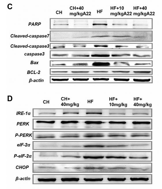
Application: WB Species: mouse Sample: liver

Application: WB Species: rat Sample:
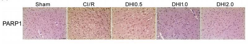
Application: IHC Species: rat Sample: brain
Application: WB Species: Rat Sample: NRK-52E cells
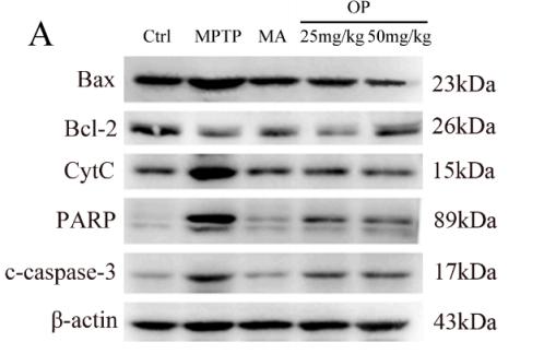
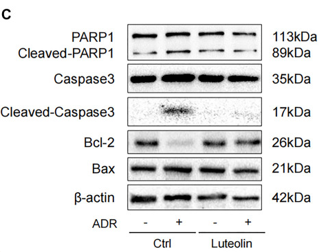
Application: WB Species: mouse Sample:

Application: WB Species: Human Sample: SCLC cells
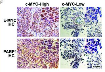
Application: IHC Species: Human Sample: tumor tissues
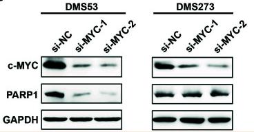
Application: WB Species: Human Sample: tumor tissues
Restrictive clause
Affinity Biosciences tests all products strictly. Citations are provided as a resource for additional applications that have not been validated by Affinity Biosciences. Please choose the appropriate format for each application and consult Materials and Methods sections for additional details about the use of any product in these publications.
For Research Use Only.
Not for use in diagnostic or therapeutic procedures. Not for resale. Not for distribution without written consent. Affinity Biosciences will not be held responsible for patent infringement or other violations that may occur with the use of our products. Affinity Biosciences, Affinity Biosciences Logo and all other trademarks are the property of Affinity Biosciences LTD.