IRF3 Antibody - #DF6895

| Product: | IRF3 Antibody |
| Catalog: | DF6895 |
| Description: | Rabbit polyclonal antibody to IRF3 |
| Application: | WB IHC IF/ICC |
| Reactivity: | Human, Mouse, Rat |
| Prediction: | Pig, Bovine, Horse, Sheep, Dog |
| Mol.Wt.: | 49kDa; 47kD(Calculated). |
| Uniprot: | Q14653 |
| RRID: | AB_2838854 |
Product Info
*The optimal dilutions should be determined by the end user.
*Tips:
WB: For western blot detection of denatured protein samples. IHC: For immunohistochemical detection of paraffin sections (IHC-p) or frozen sections (IHC-f) of tissue samples. IF/ICC: For immunofluorescence detection of cell samples. ELISA(peptide): For ELISA detection of antigenic peptide.
Cite Format: Affinity Biosciences Cat# DF6895, RRID:AB_2838854.
Fold/Unfold
IIAE7; Interferon regulatory factor 3; IRF 3; IRF-3; IRF3; IRF3_HUMAN; MGC94729;
Immunogens
- Q14653 IRF3_HUMAN:
- Protein BLAST With
- NCBI/
- ExPASy/
- Uniprot
MGTPKPRILPWLVSQLDLGQLEGVAWVNKSRTRFRIPWKHGLRQDAQQEDFGIFQAWAEATGAYVPGRDKPDLPTWKRNFRSALNRKEGLRLAEDRSKDPHDPHKIYEFVNSGVGDFSQPDTSPDTNGGGSTSDTQEDILDELLGNMVLAPLPDPGPPSLAVAPEPCPQPLRSPSLDNPTPFPNLGPSENPLKRLLVPGEEWEFEVTAFYRGRQVFQQTISCPEGLRLVGSEVGDRTLPGWPVTLPDPGMSLTDRGVMSYVRHVLSCLGGGLALWRAGQWLWAQRLGHCHTYWAVSEELLPNSGHGPDGEVPKDKEGGVFDLGPFIVDLITFTEGSGRSPRYALWFCVGESWPQDQPWTKRLVMVKVVPTCLRALVEMARVGGASSLENTVDLHISNSHPLSLTSDQYKAYLQDLVEGMDFQGPGES
Predictions
Score>80(red) has high confidence and is suggested to be used for WB detection. *The prediction model is mainly based on the alignment of immunogen sequences, the results are for reference only, not as the basis of quality assurance.
High(score>80) Medium(80>score>50) Low(score<50) No confidence
PTMs - Q14653 As Substrate
| Site | PTM Type | Enzyme | Source |
|---|---|---|---|
| T3 | Phosphorylation | Uniprot | |
| S14 | Phosphorylation | Uniprot | |
| K29 | Ubiquitination | Uniprot | |
| K70 | Sumoylation | Uniprot | |
| K70 | Ubiquitination | Uniprot | |
| T75 | Phosphorylation | Uniprot | |
| K77 | Ubiquitination | Uniprot | |
| K87 | Sumoylation | Uniprot | |
| K87 | Ubiquitination | Uniprot | |
| S97 | Phosphorylation | Uniprot | |
| S123 | Phosphorylation | Uniprot | |
| T135 | Phosphorylation | P78527 (PRKDC) | Uniprot |
| S173 | Phosphorylation | P45984 (MAPK9) , P45983 (MAPK8) , Q9UHD2 (TBK1) | Uniprot |
| S175 | Phosphorylation | Q9UHD2 (TBK1) | Uniprot |
| T180 | Phosphorylation | Uniprot | |
| S188 | Phosphorylation | Uniprot | |
| K193 | Ubiquitination | Uniprot | |
| T237 | Phosphorylation | Uniprot | |
| T244 | Phosphorylation | Uniprot | |
| T253 | Phosphorylation | Uniprot | |
| S259 | Phosphorylation | Uniprot | |
| K313 | Ubiquitination | Uniprot | |
| K315 | Ubiquitination | Uniprot | |
| S339 | Phosphorylation | Uniprot | |
| T370 | Phosphorylation | Uniprot | |
| S385 | Phosphorylation | Q9UHD2 (TBK1) | Uniprot |
| S386 | Phosphorylation | Q14164 (IKBKE) , Q9UQM7 (CAMK2A) , Q9UHD2 (TBK1) | Uniprot |
| T390 | Phosphorylation | Uniprot | |
| S396 | Phosphorylation | O14920 (IKBKB) , Q14164 (IKBKE) , Q9UHD2 (TBK1) | Uniprot |
| S398 | Phosphorylation | O14920 (IKBKB) , Q14164 (IKBKE) , Q9UHD2 (TBK1) | Uniprot |
| S402 | Phosphorylation | O14920 (IKBKB) , Q14164 (IKBKE) , Q9UHD2 (TBK1) | Uniprot |
| T404 | Phosphorylation | O14920 (IKBKB) , Q9UHD2 (TBK1) | Uniprot |
| S405 | Phosphorylation | O14920 (IKBKB) , Q9UHD2 (TBK1) | Uniprot |
| S427 | Phosphorylation | Uniprot |
Research Backgrounds
Key transcriptional regulator of type I interferon (IFN)-dependent immune responses which plays a critical role in the innate immune response against DNA and RNA viruses. Regulates the transcription of type I IFN genes (IFN-alpha and IFN-beta) and IFN-stimulated genes (ISG) by binding to an interferon-stimulated response element (ISRE) in their promoters. Acts as a more potent activator of the IFN-beta (IFNB) gene than the IFN-alpha (IFNA) gene and plays a critical role in both the early and late phases of the IFNA/B gene induction. Found in an inactive form in the cytoplasm of uninfected cells and following viral infection, double-stranded RNA (dsRNA), or toll-like receptor (TLR) signaling, is phosphorylated by IKBKE and TBK1 kinases. This induces a conformational change, leading to its dimerization and nuclear localization and association with CREB binding protein (CREBBP) to form dsRNA-activated factor 1 (DRAF1), a complex which activates the transcription of the type I IFN and ISG genes. Can activate distinct gene expression programs in macrophages and can induce significant apoptosis in primary macrophages.
Constitutively phosphorylated on many Ser/Thr residues. Activated following phosphorylation by TBK1 and IKBKE. Innate adapter protein MAVS, STING1 or TICAM1 are first activated by viral RNA, cytosolic DNA, and bacterial lipopolysaccharide (LPS), respectively, leading to activation of the kinases TBK1 and IKBKE. These kinases then phosphorylate the adapter proteins on the pLxIS motif, leading to recruitment of IRF3, thereby licensing IRF3 for phosphorylation by TBK1. Phosphorylated IRF3 dissociates from the adapter proteins, dimerizes, and then enters the nucleus to induce IFNs.
(Microbial infection) Phosphorylation and subsequent activation of IRF3 is inhibited by vaccinia virus protein E3.
Ubiquitinated; ubiquitination involves RBCK1 leading to proteasomal degradation. Polyubiquitinated; ubiquitination involves TRIM21 leading to proteasomal degradation.
ISGylated by HERC5 resulting in sustained IRF3 activation and in the inhibition of IRF3 ubiquitination by disrupting PIN1 binding. The phosphorylation state of IRF3 does not alter ISGylation.
Cytoplasm. Nucleus.
Note: Shuttles between cytoplasmic and nuclear compartments, with export being the prevailing effect (PubMed:10805757). When activated, IRF3 interaction with CREBBP prevents its export to the cytoplasm (PubMed:10805757).
Expressed constitutively in a variety of tissues.
Monomer. Homodimer; phosphorylation-induced. Interacts (when phosphorylated) with CREBBP. Interacts with MAVS (via phosphorylated pLxIS motif). Interacts with TICAM1 (via phosphorylated pLxIS motif). Interacts with STING1 (via phosphorylated pLxIS motif). Interacts with IKBKE and TBK1. Interacts with TICAM2. Interacts with RBCK1. Interacts with HERC5. Interacts with DDX3X (phosphorylated at 'Ser-102'); the interaction allows the phosphorylation and activation of IRF3 by IKBKE. Interacts with TRIM21 and ULK1, in the presence of TRIM21; this interaction leads to IRF3 degradation by autophagy. Interacts with RIOK3; RIOK3 probably mediates the interaction of TBK1 with IRF3. Interacts with ILRUN; the interaction inhibits IRF3 binding to its DNA consensus sequence. Interacts with LYAR; this interaction impairs IRF3 DNA-binding activity.
(Microbial infection) Interacts with rotavirus A NSP1 (via pLxIS motif); this interaction leads to the proteasome-dependent degradation of IRF3.
(Microbial infection) Interacts with herpes virus 8/HHV-8 protein VIRF1.
(Microbial infection) Interacts with Seneca Valley virus protease 3C; this interaction is involved in the suppression of IRF3 expression and phosphorylation by the virus.
Belongs to the IRF family.
Research Fields
· Human Diseases > Infectious diseases: Bacterial > Pertussis.
· Human Diseases > Infectious diseases: Viral > Hepatitis C.
· Human Diseases > Infectious diseases: Viral > Hepatitis B.
· Human Diseases > Infectious diseases: Viral > Measles.
· Human Diseases > Infectious diseases: Viral > Influenza A.
· Human Diseases > Infectious diseases: Viral > Human papillomavirus infection.
· Human Diseases > Infectious diseases: Viral > Herpes simplex infection.
· Human Diseases > Infectious diseases: Viral > Epstein-Barr virus infection.
· Human Diseases > Cancers: Overview > Viral carcinogenesis.
· Organismal Systems > Immune system > Toll-like receptor signaling pathway. (View pathway)
· Organismal Systems > Immune system > NOD-like receptor signaling pathway. (View pathway)
· Organismal Systems > Immune system > RIG-I-like receptor signaling pathway. (View pathway)
· Organismal Systems > Immune system > Cytosolic DNA-sensing pathway. (View pathway)
References
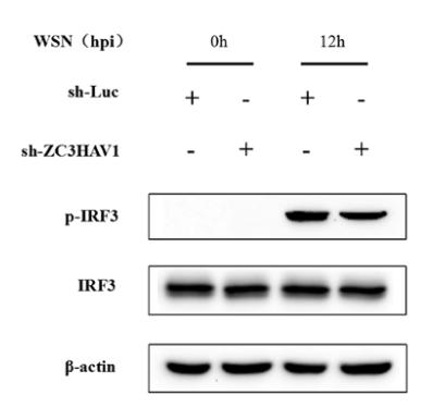
Application: WB Species: Sample: ZC3HAV1 knockdown cells
Application: WB Species: mouse Sample: BV-2 cells
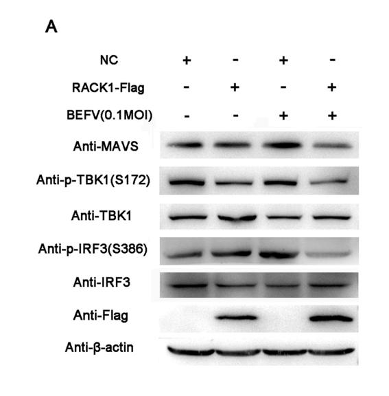
Application: WB Species: Mouse Sample: BHK-21 cell
Application: WB Species: Mice Sample: brain tissues
Restrictive clause
Affinity Biosciences tests all products strictly. Citations are provided as a resource for additional applications that have not been validated by Affinity Biosciences. Please choose the appropriate format for each application and consult Materials and Methods sections for additional details about the use of any product in these publications.
For Research Use Only.
Not for use in diagnostic or therapeutic procedures. Not for resale. Not for distribution without written consent. Affinity Biosciences will not be held responsible for patent infringement or other violations that may occur with the use of our products. Affinity Biosciences, Affinity Biosciences Logo and all other trademarks are the property of Affinity Biosciences LTD.