ARG1 Antibody - #DF6657

 and mouse anti-beta tubulin Ab(T0023 1:200) for 1 hour at 37°C. An AlexaFluor594 conjugated goat anti-rabbit IgG(H+L) Ab(Red) and an AlexaFluor488 conjugated goat anti-mouse IgG(H+L) Ab(Green) were used as the secondary antibody.
The nuclear counter stain is DAPI(blue).")
. The green signal represents the staining of activated ornithine decarboxylase, and the blue signal represents the DAPI-stained nuclei. *P < .05; **P < .01; ***P < .001. Bars indicate mean and SEM of triplicate experiments and show a representative experiment of at least 3 independent experiments performed for each panel")
 activation.E, iNOS and ARG1 protein levels were determined by Western blot in IL-33–induced M2 macrophage with or without knocking down ODC. *P < .05; **P < .01; ***P < .001. Bars indicate mean and SEM of triplicate experiments and show a representative experiment of at least 3 independent experiments performed for each panel")
-,macrophage (Mp)-related molecules, proinflammatory cytokines, and anti-inflammatory cytokines. A: immunohistochemical distribution of the expression of PPAR-, proinflammatory cytokines, and Mp-related molecules in the kidneys as well as the ratio of areas with positive expression. PPAR- expression in pioglitazone (PGZ)-treated groups was significantly higher than in other groups. Expression of monocyte chemotactic protein (MCP)-1, F4/80, inducible nitric oxide synthase (iNOS), arginine 1 (ARG1), interferon regulatory factor 1 (IRF1), and PKNOX1 was significantly higher in 3-day (d) glyoxylic acid (GA)- and 6-day GA-treated groups than in the control group and was significantly lower in PGZ-treated groups than in the 6-day GA group. Original magnification: 200.")
 and Pknox1 are two miR-23 target genes, and miR-23 is important in regulating macrophage polarization and inflammation.B: Western blots were performed in calcium oxalate monohydrate (COM)-stimulated bone marrow-derived macrophages(BMDMs) with miR-23 mimic (MI23) or miR-23 inhibitor (IN23) for 48 h. All Western blot data were quantified. Data are presented as means SE.")

, caspase-1 (a and c), and cleaved
caspase-1 (a and d) are normalized by BV2 cells (n = 4 in each group).
One-way ANOVA: F(2,9) = 16.679, p = 0.02 for NLRP3; F(2,9) = 0.669,
p = 0.527 for caspase-1; F(2,9) = 9.466, p = 0.003 for cleaved caspase-1.e–h IL-1β, TNF-α, Il-4, and IL-13 mRNA were determined by real-time.
The relative protein levels of iNOS (i and j) and Arg-1 (i and k) are
normalized by BV2 cells. Data are expressed as mean ± SEM,
*p < 0.05, **p < 0.01 e–h IL-1β, TNF-α, Il-4, and IL-13 mRNA were determined by real-time.
The relative protein levels of iNOS (i and j) and Arg-1 (i and k) are
normalized by BV2 cells. Data are expressed as mean ± SEM,
*p < 0.05, **p < 0.01")


 iNOS, CD86,
CD206, and Arg1 protein levels in tibialis anterior muscles were determined by western
blot at day 3 (n=4). Data are expressed as the means ± standard error of the mean. *
P<0.05, ** P<0.01. (G) Immunofluorescence localization and relative expression of CD206,
maker of M2, in tibialis anterior muscles between contusion group and BMSC-Exos group
(contusion + BMSC-Exos injection) at day 3. All photographs were taken at 200x
magnification. Red arrows pointed to CD206 positive cells.")


 and elevated body swing test
(B) were used to evaluate the neurological
deficits of the Sham, SOL, EMO and DCQ rats (n
= 15/group). Prussian blue staining was used to
evaluate the iron deposition of rats (C, Bar = 50
μm). The intensities of prussian blue positive
were analysed by ImageJ (D). Network showed
the DEPs related to the lysosome and proteasome in DCQ/SOL (E). The DEPs involved in
glial cell activation in CPu of DCQ rats were
showed in (F). Prussian blue positive ameboid
microglia were shown by immunohistochemical
staining with Iba1 (a marker of microglia) (G,
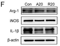
Bar = 50 μm). Levels of pro-inflammatory cytokines including IL-1β, IL-10, TNF-α and Arg1
in the CPu (H) and Hp (J) were measured by
Western blotting (n = 6) and quantitatively
analysed (I, K). Data were presented as means ±
SEM. * p < 0.05, ** p < 0.01, *** p < 0.001 vs.
Sham. # p < 0.05 vs. SOL. ▴ p < 0.05 vs. EMO.")
 Relative mRNA level of IL10, Arg-1, and TGF-β1in the colon was measured through qPCR. Expression of IL-10, Arg-1, and TGF-β1 in colon tissue lysates were determined by Western blot from DSS-challenged mice. n = 3,.means ± SEM.*p < 0.05, **p < 0.01, ***p < 0.001.")
 Fluorescence immunohistochemical distribution of Arg1 (green) and iNOS (red) in THP-1 cells with the stimulus of COM or ROSI (1 μM) or GW9662 (10 μM) (arrows). (b) Evaluation of mean fluorescence intensity (MFI) of Arg1 and iNOS. (c) ELISA. The expression levels of IL-4, IL-10, IL-6, and TNF-α. (d) Crystal adhesion assay. COM crystal adhesion to HK-2 cells was observed (arrows). COM: calcium oxalate monohydrate; ROSI: rosiglitazone; iNOS: induced nitric oxide synthase; TNF-α: tumor necrosis factor α. ∗p < 0.05; ∗∗p < 0.01.")
 mRNA levels in THP-1 cells stimulated with COM or ROSI (1 μM) or GW9662 (10 μM) by qRT-PCR. (b) Genetic expression determined by Western blot. COM: calcium oxalate monohydrate; ROSI: rosiglitazone. ∗p < 0.05; ∗∗p < 0.01.")
. (n = 4). Data are expressed as the mean ± SD. aP < 0.05, bP < 0.01, cP < 0.001, dP < 0.0001; E: Immunofluorescence localization and relative expression of iNOS, a marker of inflammatory level, in macrophage medium after culture with different concentrations of C2C12-Exos for 24 h. Scale bar = 90 μm; F: The concentration of cytokine interleukin (IL)-6, transforming growth factor-β, tumor necrosis factor-α, and IL-1β in supernatants of macrophage cells after culture with IF-C2C12-Exos or phosphate-buffered saline for 24 h measured by ELISA (n = 3). aP < 0.05, bP < 0.01, cP < 0.001. PBS: Phosphate-buffered saline. IL: Interleukin; TGF-β: Transforming growth factor-β; TNF-α: Tumor necrosis factor-α; NS: Not significant.")
 The relative mRNA expression of MCP-1, CD86, and CD206 in left ventricular tissues was detected by qRT-PCR. (D and F) The contents of M1 (CD68+, iNOS+) and M2 (CD68+, Arg-1+) macrophages were examined by double-immunofluorescence staining. Scale bars, 50 μm. Arrows point to double-immunofluorescence staining cells. (E and G) The percentage of iNOS-positive CD68 macrophages and the percentage of Arg-1-positive CD68 macrophages (six mice/group, three sections/mouse). The data are expressed as means ± SD (n = 6/group). ***, P < 0.001; **, P < 0.01; *, P < 0.05 versus control mice. †††, P < 0.001; ††, P < 0.01; †, P < 0.05 versus DM mice. ###, P < 0.001 versus GVDM + shNC mice. Arg-1, arginase 1; DAPI, 4',6-diamidino-2-phenylindole; iNOS, inducible nitric-oxide synthase.")
 PWT in rats after intrathecal BMP4 application showed a significant effect on time: F (4.143, 66.29) = 16.09, P<0.01, left; F (3.960, 63.35) = 12.67, P<0.01, right, intrathecal application: F (1, 16) = 161.3, P<0.01, left; F (1, 16) = 160.5, P<0.01, right and interaction: F (7, 112) = 11.76, P<0.01, left; F (7, 112) = 5.653, P<0.01, right. Compared with the Sham group, rats in the BMP4 group developed a significant decrease in bilateral PWT for the whole first week, P<0.01, n=9 at each time point for both groups. (B) Representative Western blotting showed a sustained increase of CD11b (P<0.01) and CD16 (P<0.01) expressions for the whole 1st week in the BMP4 group compared with the Sham group. Meanwhile, ARG-1 levels increased at day 1 (P<0.01) and day 4 (P<0.01), then fell below the levels of the Sham group (P<0.01). n=3 for each column. (C and D) Double-immunofluorescence further detected that compared with the Sham group (P7), CD11b expression was elevated in the dorsal horn of spinal cord after BMP4 treatment. Moreover, expressions of CD16 and ARG-1 showed similar pattern with Western blotting results, which were both mainly accumulated with CD11b+ cells. n=3 for each column. Two-way ANOVA, followed by a Bonferroni test (A) and one-way ANOVA, followed by Sidak’s multiple comparisons test (B–D) were performed to analyze the statistical differences. *Represented P<0.05 and **Represented P<0.01 compared with the Sham group.")
 PWT in rats after intrathecal BMP4 application showed a significant effect on time: F (4.143, 66.29) = 16.09, P<0.01, left; F (3.960, 63.35) = 12.67, P<0.01, right, intrathecal application: F (1, 16) = 161.3, P<0.01, left; F (1, 16) = 160.5, P<0.01, right and interaction: F (7, 112) = 11.76, P<0.01, left; F (7, 112) = 5.653, P<0.01, right. Compared with the Sham group, rats in the BMP4 group developed a significant decrease in bilateral PWT for the whole first week, P<0.01, n=9 at each time point for both groups. (B) Representative Western blotting showed a sustained increase of CD11b (P<0.01) and CD16 (P<0.01) expressions for the whole 1st week in the BMP4 group compared with the Sham group. Meanwhile, ARG-1 levels increased at day 1 (P<0.01) and day 4 (P<0.01), then fell below the levels of the Sham group (P<0.01). n=3 for each column. (C and D) Double-immunofluorescence further detected that compared with the Sham group (P7), CD11b expression was elevated in the dorsal horn of spinal cord after BMP4 treatment. Moreover, expressions of CD16 and ARG-1 showed similar pattern with Western blotting results, which were both mainly accumulated with CD11b+ cells. n=3 for each column. Two-way ANOVA, followed by a Bonferroni test (A) and one-way ANOVA, followed by Sidak’s multiple comparisons test (B–D) were performed to analyze the statistical differences. *Represented P<0.05 and **Represented P<0.01 compared with the Sham group.")
. Representative blots are shown. *P")
 Observation of cell morphology, (b) Cytotoxicity assay, (c) Screening the effective concentration (p")
: Protein expression levels of CD16, iNOS, TNF-α, Arg-1 and CD206 in BV2 microglia after exposure to OGD/R. (b-f): Protein expression levels of M1 markers (TNF-α, iNOS, and CD16) and M2 markers (Arg-1 and CD206) in BV2 microglia 24 h after exposure to OGD/R. (g-h): Representative images of immunofluorescence staining of CD16 and CD206. q-PCR analysis of the mRNA expression levels of CD32 (i), iNOS (j), Arg-1 (k) and CD206 (l) in OGD/R-exposed BV2 microglia (n = 3). #P < 0.05, ##P < 0.01, ###P < 0.001 vs. the control group; *P < 0.05, **P < 0.01, ***P < 0.001 vs. the OGD/R group. The data are the mean ± S.E.M. of three independent experiments.")
 and hypoxia/reoxygenation (H/R) groups. C, Protein concentrations of TNF-α and IL-10 in supernatants of the cell culture experiment with RAW264.7 macrophages of different groups. D and E, Representative Western blots and summarized data showing the effects of ERK inhibitor PD98059 on the protein expression of hypoxia/reoxygenation-induced pro-inflammatory factor TNF-α and anti-inflammatory factor ARG-1. **P < .01 vs normoxia (N); #P < .05, ##P < .01 vs H/R. N: normoxia; H/R: Hypoxia/reoxygenation")
 Representative images and quantitative analysis showing iNOS+/Iba1+ immunofluorescence staining. Lv-shPARP14 injection enhanced the SCI-induced increase in iNOS expression (pro-inflammatory phenotype). White arrows indicate iNOS+ (green, FITC-labeled)/Iba1+ (red, Cy3-labeled, microglia marker) cells. (B) Representative images and quantitative analysis showing Arg-1+/Iba1+ immunofluorescence staining. Lv-shPARP14 injection reversed the SCI-induced increase in Arg-1 expression (anti-inflammatory phenotype). White arrows indicate Arg-1+ (green, FITC-labeled)/Iba1+ (red, Cy3-labeled, microglia marker) cells. Scale bars: 50 µm in A and B. (C) Relative protein expression of CD16 and CD206 in each group. Lv-shPARP14 injection enhanced the SCI-induced increase in CD16 (M1-type marker) expression and decreased the SCI-induced increase in CD206 (M2-type marker) expression. (D) The concentration of pro-inflammatory cytokines (TNF-α, IL-1β, and IL-6) and anti-inflammatory cytokines (IL-10, TGF-β1, and IL-4) in each group was measured by enzyme-linked immunosorbent assay. Lv-shPARP14 injection increased the concentration of pro-inflammatory cytokines but decreased anti-inflammatory cytokine accumulation. Values are shown as mean ± SD (n = 6). *P < 0.05, **P < 0.01 (one-way analysis of variance followed by Tukey’s post hoc test). Images were taken of the gray matter ventral horn at the injury site. Spinal cord tissues from the injury site were used for western blot and enzyme-linked immunosorbent assay detection. Arg-1: Arginase-1; DAPI: 4′,6-diamidino-2-phenylindole; FITC: fluorescein isothiocyanate; Iba1: ionized calcium-binding adaptor molecule 1; IL: interleukin; iNOS: inducible nitric oxide synthase; PARP14: poly(ADP-ribose)polymerase, member 14; SCI: spinal cord injury; TGF-β1: transforming growth factor-beta1; TNF-α: tumor necrosis factor alpha.")
 Representative images of double immunofluorescence of iNOS antibody (green, M1 macrophage), Arg-1 (red, M2 macrophage), and nuclei (blue). Scale bar is 20 μm. (B) Illustration of macrophages displaying different degrees of polarization cultured on the AMRs. (C) Real-time PCR analysis of the RAW 264.7 cells cultured on the AMRs for 1 day, housekeeping gene β-actin was used as an internal control. (D) Flow cytometric analysis of CD206 and CD86 after treatment with AMRs. (E) Quantitative data of M1 CD86 and M2 CD206 population (%). Error bars are ±SEM. p values were calculated by one-way ANOVA, ∗p < 0.05.")
 immunofluorescence staining (original magnification, × 40, bar = 50 μm) and quantitative analysis of macrophage accumulation and macrophage polarization by detection of (a) the macrophage marker cluster of differentiation (CD)68 and M1 marker inducible nitric oxide synthase (iNOS), and (b) the M2 markers CD206 and arginase (Arg)-1, in aortic sections from MiR-22-3p agomir or normal control (NC) mice; and (c) western blot and quantitative analysis of relative protein levels of the M1 markers (iNOS, interleukin [IL]-6, and tumour necrosis factor [TNF]-α) and M2 markers (Arg1 and CD206) in aortic sections from MiR-22-3p agomir or NC mice.")
 immunofluorescence staining (original magnification, × 40, bar = 50 μm) and quantitative analysis of macrophage accumulation and macrophage polarization by detection of (a) the macrophage marker cluster of differentiation (CD)68 and M1 marker inducible nitric oxide synthase (iNOS), and (b) the M2 markers CD206 and arginase (Arg)-1, in aortic sections from MiR-22-3p agomir or normal control (NC) mice; and (c) western blot and quantitative analysis of relative protein levels of the M1 markers (iNOS, interleukin [IL]-6, and tumour necrosis factor [TNF]-α) and M2 markers (Arg1 and CD206) in aortic sections from MiR-22-3p agomir or NC mice.")
 The expression levels of iNOS and Arg1 proteins were assessed using representative blots and quantification. (D and E) Immunofluorescence staining was performed to visualize and quantify the number of iNOS-positive cells in the anterior horn of the spinal cord at the lesion epicenter. The scale bar represents 200 µm. (F and G) Immunofluorescence staining was also conducted to assess the number of Arg1-positive cells in the anterior horn of the spinal cord at the lesion epicenter. The scale bar represents 200 µm. *P≤0.05. iNOS, inducible nitric oxide synthase; Arg1, arginase 1; Mel, Melatonin; Veh, Vehicle; 4PP, 4-phenyl-2-propionamidotetralin.")
 Protein Levels of Arg-1 (M2 marker) were measured by Western blot analysis, and quantitative analysis was performed on the corresponding bands (n = 8–10). (b) The mRNA levels of interleukin-4 (IL-4), (c) IL-10, and (d) TGF-β (M2 markers) in aortas (n = 5–6) were measured by qRT-PCR. Serum cytokine levels of (e) IL-4, (f) IL-10 and (g) TGF-β were measured by Enzyme-linked immunosorbent assay kits (n = 5–6). Means ± standard error of mean. *P < 0.05, **P < 0.01 versus Con; #P < 0.05, ##P < 0.01 versus Mod.")
Related Downloads
Protocols
Product Info
*The optimal dilutions should be determined by the end user.
*Tips:
WB: For western blot detection of denatured protein samples. IHC: For immunohistochemical detection of paraffin sections (IHC-p) or frozen sections (IHC-f) of tissue samples. IF/ICC: For immunofluorescence detection of cell samples. ELISA(peptide): For ELISA detection of antigenic peptide.
Cite Format: Affinity Biosciences Cat# DF6657, RRID:AB_2838619.
Fold/Unfold
A I; Al; ARG 1; arg1; ARGI1_HUMAN; Arginase 1; Arginase liver; Arginase type I; Arginase, liver; Arginase-1; Arginase1; Liver type arginase; Liver-type arginase; Type I arginase;
Immunogens
Within the immune system initially reported to be selectively expressed in granulocytes (polymorphonuclear leukocytes [PMNs]) (PubMed:15546957). Also detected in macrophages mycobacterial granulomas (PubMed:23749634). Expressed in group2 innate lymphoid cells (ILC2s) during lung disease (PubMed:27043409).
- P05089 ARGI1_HUMAN:
- Protein BLAST With
- NCBI/
- ExPASy/
- Uniprot
MSAKSRTIGIIGAPFSKGQPRGGVEEGPTVLRKAGLLEKLKEQECDVKDYGDLPFADIPNDSPFQIVKNPRSVGKASEQLAGKVAEVKKNGRISLVLGGDHSLAIGSISGHARVHPDLGVIWVDAHTDINTPLTTTSGNLHGQPVSFLLKELKGKIPDVPGFSWVTPCISAKDIVYIGLRDVDPGEHYILKTLGIKYFSMTEVDRLGIGKVMEETLSYLLGRKKRPIHLSFDVDGLDPSFTPATGTPVVGGLTYREGLYITEEIYKTGLLSGLDIMEVNPSLGKTPEEVTRTVNTAVAITLACFGLAREGNHKPIDYLNPPK
Predictions
Score>80(red) has high confidence and is suggested to be used for WB detection. *The prediction model is mainly based on the alignment of immunogen sequences, the results are for reference only, not as the basis of quality assurance.
High(score>80) Medium(80>score>50) Low(score<50) No confidence
PTMs - P05089 As Substrate
| Site | PTM Type | Enzyme | Source |
|---|---|---|---|
| S16 | Phosphorylation | Uniprot | |
| T29 | Phosphorylation | Uniprot | |
| K33 | Ubiquitination | Uniprot | |
| Y50 | Phosphorylation | Uniprot | |
| S62 | Phosphorylation | Uniprot | |
| S72 | Phosphorylation | Uniprot | |
| S94 | Phosphorylation | Uniprot | |
| S102 | Phosphorylation | Uniprot | |
| S107 | Phosphorylation | Uniprot | |
| S109 | Phosphorylation | Uniprot | |
| S163 | Phosphorylation | Uniprot | |
| T166 | Phosphorylation | Uniprot | |
| C168 | S-Nitrosylation | Uniprot | |
| S170 | Phosphorylation | Uniprot | |
| K191 | Acetylation | Uniprot | |
| S217 | Phosphorylation | Uniprot | |
| Y265 | Phosphorylation | Uniprot | |
| C303 | S-Nitrosylation | Uniprot |
Research Backgrounds
Key element of the urea cycle converting L-arginine to urea and L-ornithine, which is further metabolized into metabolites proline and polyamides that drive collagen synthesis and bioenergetic pathways critical for cell proliferation, respectively; the urea cycle takes place primarily in the liver and, to a lesser extent, in the kidneys.
Functions in L-arginine homeostasis in nonhepatic tissues characterized by the competition between nitric oxide synthase (NOS) and arginase for the available intracellular substrate arginine. Arginine metabolism is a critical regulator of innate and adaptive immune responses. Involved in an antimicrobial effector pathway in polymorphonuclear granulocytes (PMN). Upon PMN cell death is liberated from the phagolysosome and depletes arginine in the microenvironment leading to suppressed T cell and natural killer (NK) cell proliferation and cytokine secretion. In group 2 innate lymphoid cells (ILC2s) promotes acute type 2 inflammation in the lung and is involved in optimal ILC2 proliferation but not survival (By similarity). In humans, the immunological role in the monocytic/macrophage/dendritic cell (DC) lineage is unsure.
Cytoplasm. Cytoplasmic granule.
Note: Localized in azurophil granules of neutrophils (PubMed:15546957).
Within the immune system initially reported to be selectively expressed in granulocytes (polymorphonuclear leukocytes [PMNs]). Also detected in macrophages mycobacterial granulomas. Expressed in group2 innate lymphoid cells (ILC2s) during lung disease.
Homotrimer. Interacts with CMTM6.
Belongs to the arginase family.
Research Fields
· Human Diseases > Infectious diseases: Parasitic > Amoebiasis.
· Metabolism > Amino acid metabolism > Arginine biosynthesis.
· Metabolism > Amino acid metabolism > Arginine and proline metabolism.
· Metabolism > Global and overview maps > Metabolic pathways.
· Metabolism > Global and overview maps > Biosynthesis of amino acids.
References
Application: WB Species: Mice Sample:
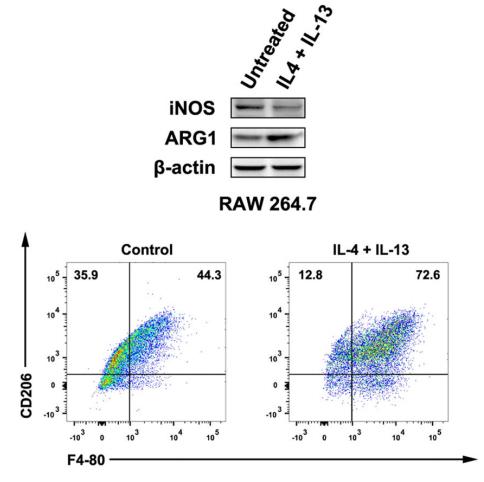
Application: WB Species: mouse Sample: RAW264.7 cells
Application: WB Species: human Sample: prostate cancer cells

Application: IHC Species: mouse Sample: tumor
Restrictive clause
Affinity Biosciences tests all products strictly. Citations are provided as a resource for additional applications that have not been validated by Affinity Biosciences. Please choose the appropriate format for each application and consult Materials and Methods sections for additional details about the use of any product in these publications.
For Research Use Only.
Not for use in diagnostic or therapeutic procedures. Not for resale. Not for distribution without written consent. Affinity Biosciences will not be held responsible for patent infringement or other violations that may occur with the use of our products. Affinity Biosciences, Affinity Biosciences Logo and all other trademarks are the property of Affinity Biosciences LTD.