Product: DCN Antibody
Catalog: DF6543
Description: Rabbit polyclonal antibody to DCN
Application: WB IHC IF/ICC
Reactivity: Human, Mouse, Rat
Prediction: Pig, Bovine, Sheep
Mol.Wt.: 30kDa; 40kD(Calculated).
Uniprot: P07585
RRID: AB_2838505

View similar products>>

   Size Price Inventory
 100ul $280 In stock
 200ul $350 In stock

Lead Time: Same day delivery

For pricing and ordering contact:
Local distributors

Product Info

Source:
Rabbit
Application:
WB 1:500-1:2000, IHC 1:50-1:200, IF/ICC 1:100-1:500
*The optimal dilutions should be determined by the end user.
*Tips:

WB: For western blot detection of denatured protein samples. IHC: For immunohistochemical detection of paraffin sections (IHC-p) or frozen sections (IHC-f) of tissue samples. IF/ICC: For immunofluorescence detection of cell samples. ELISA(peptide): For ELISA detection of antigenic peptide.

Reactivity:
Human,Mouse,Rat
Prediction:
Pig(92%), Bovine(83%), Sheep(83%)
Clonality:
Polyclonal
Specificity:
DCN Antibody detects endogenous levels of total DCN.
RRID:
AB_2838505
Cite Format: Affinity Biosciences Cat# DF6543, RRID:AB_2838505.
Conjugate:
Unconjugated.
Purification:
The antiserum was purified by peptide affinity chromatography using SulfoLink™ Coupling Resin (Thermo Fisher Scientific).
Storage:
Rabbit IgG in phosphate buffered saline , pH 7.4, 150mM NaCl, 0.02% sodium azide and 50% glycerol. Store at -20 °C. Stable for 12 months from date of receipt.
Alias:

Fold/Unfold

Bone proteoglycan II; CSCD; DCN; DCN protein; Decorin; Decorin proteoglycan; Dermatan sulphate proteoglycans II; DKFZp686J19238; DSPG 2; DSPG2; PG 40; PG II; PG S2; PG-S2; PG40; PGII; PGS 2; PGS2; PGS2_HUMAN; Proteoglycan core protein; SLRR1B; Small leucine rich protein 1B;

Immunogens

Immunogen:
Uniprot:
Gene(ID):
Description:
The protein encoded by this gene is a small cellular or pericellular matrix proteoglycan that is closely related in structure to biglycan protein. The encoded protein and biglycan are thought to be the result of a gene duplication. This protein is a component of connective tissue, binds to type I collagen fibrils, and plays a role in matrix assembly. It contains one attached glycosaminoglycan chain. This protein is capable of suppressing the growth of various tumor cell lines. There are multiple alternatively spliced transcript variants known for this gene. This gene is a candidate gene for Marfan syndrome. [provided by RefSeq]
Sequence:
MKATIILLLLAQVSWAGPFQQRGLFDFMLEDEASGIGPEVPDDRDFEPSLGPVCPFRCQCHLRVVQCSDLGLDKVPKDLPPDTTLLDLQNNKITEIKDGDFKNLKNLHALILVNNKISKVSPGAFTPLVKLERLYLSKNQLKELPEKMPKTLQELRAHENEITKVRKVTFNGLNQMIVIELGTNPLKSSGIENGAFQGMKKLSYIRIADTNITSIPQGLPPSLTELHLDGNKISRVDAASLKGLNNLAKLGLSFNSISAVDNGSLANTPHLRELHLDNNKLTRVPGGLAEHKYIQVVYLHNNNISVVGSSDFCPPGHNTKKASYSGVSLFSNPVQYWEIQPSTFRCVYVRSAIQLGNYK

Predictions

Predictions:

Score>80(red) has high confidence and is suggested to be used for WB detection. *The prediction model is mainly based on the alignment of immunogen sequences, the results are for reference only, not as the basis of quality assurance.

Species
Results
Score
Pig
92
Bovine
83
Sheep
83
Horse
75
Dog
75
Rabbit
67
Xenopus
0
Zebrafish
0
Chicken
0
Model Confidence:
High(score>80) Medium(80>score>50) Low(score<50) No confidence

PTMs - P07585 As Substrate

Site PTM Type Enzyme
S34 O-Glycosylation
C67 S-Nitrosylation
K92 Ubiquitination
K102 Acetylation
T126 Phosphorylation
K138 Acetylation
K147 Acetylation
K200 Acetylation
K201 Acetylation
N211 N-Glycosylation
N262 N-Glycosylation
K280 Ubiquitination
S328 Phosphorylation
S331 Phosphorylation

Research Backgrounds

Function:

May affect the rate of fibrils formation.

PTMs:

The attached glycosaminoglycan chain can be either chondroitin sulfate or dermatan sulfate depending upon the tissue of origin.

Subcellular Location:

Secreted>Extracellular space>Extracellular matrix.

Extracellular region or secreted Cytosol Plasma membrane Cytoskeleton Lysosome Endosome Peroxisome ER Golgi apparatus Nucleus Mitochondrion Manual annotation Automatic computational assertionSubcellular location
Subunit Structure:

Binds to type I and type II collagen, fibronectin and TGF-beta. Forms a ternary complex with MFAP2 and ELN. Interacts with DPT (By similarity).

Family&Domains:

Belongs to the small leucine-rich proteoglycan (SLRP) family. SLRP class I subfamily.

Research Fields

· Environmental Information Processing > Signal transduction > TGF-beta signaling pathway.   (View pathway)

· Human Diseases > Cancers: Overview > Proteoglycans in cancer.

References

1). Biomimetic multilayer polycaprolactone/sodium alginate hydrogel scaffolds loaded with melatonin facilitate tendon regeneration. Carbohydrate Polymers (PubMed: 34893270) [IF=11.2]

2). MicroRNA engineered umbilical cord stem cell-derived exosomes direct tendon regeneration by mTOR signaling. Journal of Nanobiotechnology (PubMed: 34090456) [IF=10.2]

3). Promotion of collagen deposition during skin healing through Smad3/mTOR pathway by parathyroid hormone-loaded microneedle. Materials Science and Engineering: C (PubMed: 33321586) [IF=7.9]

4). Role of intestinal extracellular matrix-related signaling in porcine epidemic diarrhea virus infection. Virulence (PubMed: 34515624) [IF=5.2]

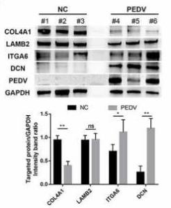
Application: WB    Species: Pig    Sample: Porcine intestinal epithelial cells

Figure 1. Enrichment analysis and expression profiles of differentially expressed genes (DEGs). (a) Functional map for downregulated DEGs enriched for GO terms. (b) Statistical analysis of the top 30 enriched KEGG pathways during PEDV infection. The x-axis indicates the factor of enrichment, whereas the y-axis indicates the KEGG channel. The dot color indicates the q value. The dot size corresponds to the number of DEGs mapped to the reference category. (c and d) Hierarchical clustering of extracellular matrix-constituent (c) and -regulated (d) DEGs. Rows indicate hierarchical clustering in accordance with average linkage based on Pearson correlation coefficients with the measured distance. Expression was visualized using a gradient color scheme; the scale from the minimal to maximal abundance fell in the range of −3.0 to 3.0. Blue represents low expression, whereas red represents high expression. (e) The total protein collected from the small intestine of PEDV-infected piglets (48 h post-infection) was prepared, and the expression levels of ECM-associated proteins (i.e., COL4A1, LAMB2, ITG6, and DCN) and PEDV N protein were determined by western blotting. The data shown are means ± SDs from triplicate experiments. *P < 0.05, **P < 0.01

5). Parathyroid Hormone-Loaded Microneedle Promotes Tendon Healing Through Activation of mTOR. Advanced Therapeutics [IF=4.6]

Restrictive clause


Affinity Biosciences tests all products strictly. Citations are provided as a resource for additional applications that have not been validated by Affinity Biosciences. Please choose the appropriate format for each application and consult Materials and Methods sections for additional details about the use of any product in these publications.

For Research Use Only.
Not for use in diagnostic or therapeutic procedures. Not for resale. Not for distribution without written consent. Affinity Biosciences will not be held responsible for patent infringement or other violations that may occur with the use of our products. Affinity Biosciences, Affinity Biosciences Logo and all other trademarks are the property of Affinity Biosciences LTD.