NOX2 Antibody - #DF6520

| Product: | NOX2 Antibody |
| Catalog: | DF6520 |
| Description: | Rabbit polyclonal antibody to NOX2 |
| Application: | WB IHC |
| Reactivity: | Human, Mouse, Rat |
| Prediction: | Dog |
| Mol.Wt.: | 65kDa; 65kD(Calculated). |
| Uniprot: | P04839 |
| RRID: | AB_2838482 |
Related Downloads
Protocols
Product Info
*The optimal dilutions should be determined by the end user.
*Tips:
WB: For western blot detection of denatured protein samples. IHC: For immunohistochemical detection of paraffin sections (IHC-p) or frozen sections (IHC-f) of tissue samples. IF/ICC: For immunofluorescence detection of cell samples. ELISA(peptide): For ELISA detection of antigenic peptide.
Cite Format: Affinity Biosciences Cat# DF6520, RRID:AB_2838482.
Fold/Unfold
AMCBX2; CGD; CGD91-phox; CY24B_HUMAN; CYBB; Cytochrome b 245, beta polypeptide; Cytochrome b(558) beta chain; Cytochrome b(558) subunit beta; Cytochrome b-245 heavy chain; Cytochrome b558 subunit beta; GP91 PHOX; gp91-1; gp91-phox; GP91PHOX; Heme-binding membrane glycoprotein gp91phox; NADPH oxidase 2; Neutrophil cytochrome b 91 kDa polypeptide; NOX2; p22 phagocyte B-cytochrome; P91 PHOX; p91-PHOX; Superoxide-generating NADPH oxidase heavy chain subunit;
Immunogens
- P04839 CY24B_HUMAN:
- Protein BLAST With
- NCBI/
- ExPASy/
- Uniprot
MGNWAVNEGLSIFVILVWLGLNVFLFVWYYRVYDIPPKFFYTRKLLGSALALARAPAACLNFNCMLILLPVCRNLLSFLRGSSACCSTRVRRQLDRNLTFHKMVAWMIALHSAIHTIAHLFNVEWCVNARVNNSDPYSVALSELGDRQNESYLNFARKRIKNPEGGLYLAVTLLAGITGVVITLCLILIITSSTKTIRRSYFEVFWYTHHLFVIFFIGLAIHGAERIVRGQTAESLAVHNITVCEQKISEWGKIKECPIPQFAGNPPMTWKWIVGPMFLYLCERLVRFWRSQQKVVITKVVTHPFKTIELQMKKKGFKMEVGQYIFVKCPKVSKLEWHPFTLTSAPEEDFFSIHIRIVGDWTEGLFNACGCDKQEFQDAWKLPKIAVDGPFGTASEDVFSYEVVMLVGAGIGVTPFASILKSVWYKYCNNATNLKLKKIYFYWLCRDTHAFEWFADLLQLLESQMQERNNAGFLSYNIYLTGWDESQANHFAVHHDEEKDVITGLKQKTLYGRPNWDNEFKTIASQHPNTRIGVFLCGPEALAETLSKQSISNSESGPRGVHFIFNKENF
Predictions
Score>80(red) has high confidence and is suggested to be used for WB detection. *The prediction model is mainly based on the alignment of immunogen sequences, the results are for reference only, not as the basis of quality assurance.
High(score>80) Medium(80>score>50) Low(score<50) No confidence
PTMs - P04839 As Substrate
| Site | PTM Type | Enzyme | Source |
|---|---|---|---|
| Y41 | Phosphorylation | Uniprot | |
| T42 | Phosphorylation | Uniprot | |
| K44 | Ubiquitination | Uniprot | |
| N132 | N-Glycosylation | Uniprot | |
| N149 | N-Glycosylation | Uniprot | |
| Y168 | Phosphorylation | Uniprot | |
| N240 | N-Glycosylation | Uniprot | |
| K253 | Ubiquitination | Uniprot | |
| K294 | Ubiquitination | Uniprot | |
| T298 | Phosphorylation | Uniprot | |
| Y324 | Phosphorylation | Uniprot | |
| K334 | Ubiquitination | Uniprot | |
| S486 | Phosphorylation | Uniprot | |
| K548 | Ubiquitination | Uniprot |
Research Backgrounds
Critical component of the membrane-bound oxidase of phagocytes that generates superoxide. It is the terminal component of a respiratory chain that transfers single electrons from cytoplasmic NADPH across the plasma membrane to molecular oxygen on the exterior. Also functions as a voltage-gated proton channel that mediates the H(+) currents of resting phagocytes. It participates in the regulation of cellular pH and is blocked by zinc.
Glycosylated.
Phosphorylated on Ser and Thr residues.
Undergoes 'Lys-48'-linked polyubiquitination, likely by RNF145, triggering endoplasmic reticulum-associated degradation.
Cell membrane>Multi-pass membrane protein.
Note: As unassembled monomer may localize to the endoplasmic reticulum.
Detected in neutrophils (at protein level).
Composed of a heavy chain (beta) and a light chain (alpha). Component of an NADPH oxidase complex composed of a heterodimer formed by the membrane proteins CYBA and CYBB and the cytosolic subunits NCF1, NCF2 and NCF4. Interacts with NCF1. Interacts with calprotectin (S100A8/9). Interacts with NRROS; the interaction is direct and impairs formation of a stable NADPH oxidase complex. Interacts with CYBC1; CYBC1 may act as a chaperone stabilizing Cytochrome b-245 heterodimer.
Research Fields
· Cellular Processes > Transport and catabolism > Phagosome. (View pathway)
· Cellular Processes > Cell growth and death > Ferroptosis. (View pathway)
· Cellular Processes > Cell growth and death > Necroptosis. (View pathway)
· Environmental Information Processing > Signal transduction > HIF-1 signaling pathway. (View pathway)
· Human Diseases > Infectious diseases: Parasitic > Leishmaniasis.
· Organismal Systems > Immune system > NOD-like receptor signaling pathway. (View pathway)
· Organismal Systems > Immune system > Leukocyte transendothelial migration. (View pathway)
References
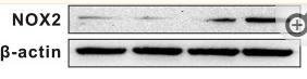
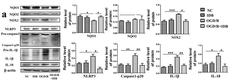
Application: WB Species: Rat Sample: DRGs
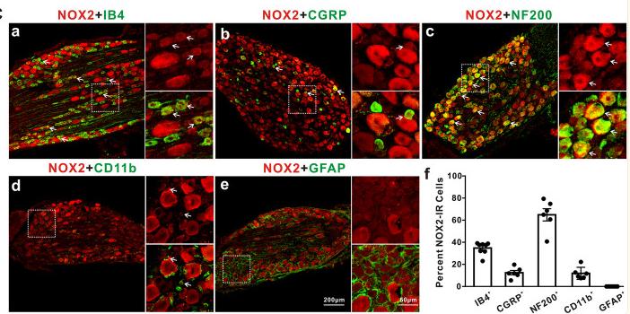
Application: IF/ICC Species: Rat Sample: DRGs

Application: IHC Species: Human Sample: HK-2 cells
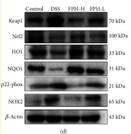
Application: WB Species: Mouse Sample:
Application: WB Species: human Sample: HK-2 cells
Application: WB Species: mouse Sample: BV3 cells
Application: WB Species: Mouse Sample:
Restrictive clause
Affinity Biosciences tests all products strictly. Citations are provided as a resource for additional applications that have not been validated by Affinity Biosciences. Please choose the appropriate format for each application and consult Materials and Methods sections for additional details about the use of any product in these publications.
For Research Use Only.
Not for use in diagnostic or therapeutic procedures. Not for resale. Not for distribution without written consent. Affinity Biosciences will not be held responsible for patent infringement or other violations that may occur with the use of our products. Affinity Biosciences, Affinity Biosciences Logo and all other trademarks are the property of Affinity Biosciences LTD.