Product: AIF1/IBA1 Antibody
Catalog: DF6442
Description: Rabbit polyclonal antibody to AIF1/IBA1
Application: WB IHC IF/ICC
Reactivity: Human, Mouse, Rat
Prediction: Pig, Bovine, Horse, Sheep, Rabbit, Dog
Mol.Wt.: 17kDa; 17kD(Calculated).
Uniprot: P55008
RRID: AB_2838405

View similar products>>

   Size Price Inventory
 100ul $280 In stock
 200ul $350 In stock

Lead Time: Same day delivery

For pricing and ordering contact:
Local distributors

Product Info

Source:
Rabbit
Application:
WB 1:500-1:2000, IHC 1:50-1:200, IF/ICC 1:100-1:500
*The optimal dilutions should be determined by the end user.
*Tips:

WB: For western blot detection of denatured protein samples. IHC: For immunohistochemical detection of paraffin sections (IHC-p) or frozen sections (IHC-f) of tissue samples. IF/ICC: For immunofluorescence detection of cell samples. ELISA(peptide): For ELISA detection of antigenic peptide.

Reactivity:
Human,Mouse,Rat
Prediction:
Pig(100%), Bovine(91%), Horse(91%), Sheep(91%), Rabbit(91%), Dog(90%)
Clonality:
Polyclonal
Specificity:
AIF1/IBA1 Antibody detects endogenous levels of total AIF1/IBA1.
RRID:
AB_2838405
Cite Format: Affinity Biosciences Cat# DF6442, RRID:AB_2838405.
Conjugate:
Unconjugated.
Purification:
The antiserum was purified by peptide affinity chromatography using SulfoLink™ Coupling Resin (Thermo Fisher Scientific).
Storage:
Rabbit IgG in phosphate buffered saline , pH 7.4, 150mM NaCl, 0.02% sodium azide and 50% glycerol. Store at -20 °C. Stable for 12 months from date of receipt.
Alias:

Fold/Unfold

AIF 1; AIF-1; Aif1; AIF1 protein; AIF1_HUMAN; Allograft inflammatory factor 1; Allograft inflammatory factor 1 splice variant G; allograft inflammatory factor-1 splice variant Hara-1; balloon angioplasty responsive transcription; BART 1; G1; G1 putative splice variant of allograft inflamatory factor 1; IBA 1; IBA1; interferon gamma responsive transcript; Interferon responsive transcript 1; interferon responsive transcript factor 1; Ionized calcium binding adapter molecule 1; Ionized calcium-binding adapter molecule 1; ionized calcium-binding adapter molecule; IRT 1; IRT1; Microglia response factor; MRF1; Protein g1;

Immunogens

Immunogen:
Uniprot:
Gene(ID):
Expression:
P55008 AIF1_HUMAN:

Detected in T-lymphocytes and peripheral blood mononuclear cells.

Description:
This gene is induced by cytokines and interferon. Its protein product is thought to be involved in negative regulation of growth of vascular smooth muscle cells, which contributes to the anti-inflammatory response to vessel wall trauma. Three transcript variants encoding different isoforms have been found for this gene. [provided by RefSeq, Sep 2008]
Sequence:
MSQTRDLQGGKAFGLLKAQQEERLDEINKQFLDDPKYSSDEDLPSKLEGFKEKYMEFDLNGNGDIDIMSLKRMLEKLGVPKTHLELKKLIGEVSSGSGETFSYPDFLRMMLGKRSAILKMILMYEEKAREKEKPTGPPAKKAISELP

Predictions

Predictions:

Score>80(red) has high confidence and is suggested to be used for WB detection. *The prediction model is mainly based on the alignment of immunogen sequences, the results are for reference only, not as the basis of quality assurance.

Species
Results
Score
Pig
100
Horse
91
Bovine
91
Sheep
91
Rabbit
91
Dog
90
Xenopus
0
Zebrafish
0
Chicken
0
Model Confidence:
High(score>80) Medium(80>score>50) Low(score<50) No confidence

PTMs - P55008 As Substrate

Site PTM Type Enzyme
S2 Phosphorylation
K11 Acetylation
K11 Ubiquitination
Y37 Phosphorylation
S38 Phosphorylation
S39 Phosphorylation
Y54 Phosphorylation
S69 Phosphorylation
K76 Ubiquitination
K81 Ubiquitination
K113 Methylation
Y124 Phosphorylation

Research Backgrounds

Function:

Actin-binding protein that enhances membrane ruffling and RAC activation. Enhances the actin-bundling activity of LCP1. Binds calcium. Plays a role in RAC signaling and in phagocytosis. May play a role in macrophage activation and function. Promotes the proliferation of vascular smooth muscle cells and of T-lymphocytes. Enhances lymphocyte migration. Plays a role in vascular inflammation.

PTMs:

Phosphorylated on serine residues.

Subcellular Location:

Cytoplasm>Cytoskeleton. Cell projection>Ruffle membrane>Peripheral membrane protein>Cytoplasmic side. Cell projection>Phagocytic cup.
Note: Associated with the actin cytoskeleton at membrane ruffles and at sites of phagocytosis.

Extracellular region or secreted Cytosol Plasma membrane Cytoskeleton Lysosome Endosome Peroxisome ER Golgi apparatus Nucleus Mitochondrion Manual annotation Automatic computational assertionSubcellular location
Tissue Specificity:

Detected in T-lymphocytes and peripheral blood mononuclear cells.

Subunit Structure:

Homodimer (Potential). Monomer. Interacts with LCP1.

References

1). Huoluo Xiaoling Pellet promotes microglia M2 polarization through increasing MCPIP1 expression for ischemia stroke alleviation. Biomedicine & Pharmacotherapy (PubMed: 37236023) [IF=7.5]

Application: IF/ICC    Species: Rat    Sample:

Fig. 3. HXP reduced neuroinflammation in MCAO rats by promoting MCPIP1-mediated microglia M2 polarization. A. After reperfusion for 7 d, brain water contents were examined. B. Brain infract volume was detected by 2,3,5-Triphentltetrazolium chloride staining. C. Changes in IL-1β, IL-6, iNOS, and TNF-α were measured via ELISA assy. D. Representative images of histopathological analysis. Scale bar, 20 µm. E. Representative immunofluorescence staining for CD206 (red) and Iba1 (green) in brain tissues. Scale bar, 20 µm. F. The expression of MCPIP1, CD16, iNOS, CD206, Arg1, and PPARγ was detected using Western blotting assay. Data are represented as mean ± standard deviation (N = 5). * * P 

2). Effects of hydrogen-rich saline in neuroinflammation and mitochondrial dysfunction in rat model of sepsis-associated encephalopathy. Journal of Translational Medicine (PubMed: 36435787) [IF=7.4]

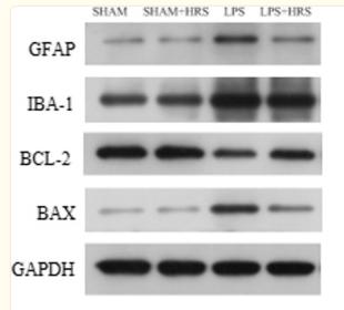
Application: WB    Species: Rat    Sample:

Fig. 8 Effects of HRS treatment on protein expressions in septic rats 48 h after LPS challenge. A Representative images of protein expression levels by western blot analysis in each group. B–E Statistical representation of relative protein expressions of GFAP, IBA-1, BCL-2 and BAX; respectively. All data are presented as mean ± SD ****p < 0.0001, ***p < 0.001, **p < 0.005, *p < 0.05. HRS Hydrogen-rich saline, LPS Lipopolysaccharide, GFAP Glial fibrillary acidic protein, IBA-1 Ionised calcium binding adaptor molecule 1, BCL-2 B-cell lymphoma 2

3). Chitosan oligosaccharides exert neuroprotective effects via modulating the PI3K/Akt/Bcl-2 pathway in a Parkinsonian model. Food & Function (PubMed: 35545086) [IF=6.1]

4). Inhibition of NLRP3 inflammasome activation and pyroptosis with the ethyl acetate fraction of Bungeanum ameliorated cognitive dysfunction in aged mice. Food & Function (PubMed: 34231604) [IF=6.1]

5). Aucubin improves chronic unpredictable mild stress-induced depressive behavior in mice via the GR/NF-κB/NLRP3 axis. International Immunopharmacology (PubMed: 37523973) [IF=5.6]

6). MKP-1 regulates the inflammatory activation of microglia against Alzheimer's disease. CNS Neuroscience & Therapeutics (PubMed: 37602891) [IF=5.5]

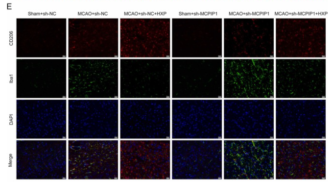
Application: IF/ICC    Species: Mouse    Sample: brains

FIGURE 1 The expression of microglia and amyloid‐β (Aβ) plaques in wild‐type (WT) and Alzheimer's disease (AD) mice brains. (A) The distribution of microglia and Aβ plaques in mice brains. (a1, b1) Visual cortex, (a2, b2) auditory cortex, (a3, b3) pear‐shaped cortex, (a4, b4) DG, (a5, b5) CA1, (a6, b6) CA2, (a7, b7) CA3, (a8, b8) amygdala, and (a9, b9) hypothalamus. Iba1 (red) for microglia, 4G8 (green) for Aβ, DAPI (blue) for nuclei. Scale bar: a–b, 500 μm; a1–a9, 100 μm; b1–b9, 100 μm. (B–E) The number and area ratio of microglia and Aβ plaques were detected. The results are presented as the mean values ± SEM, n = 4 per group.

7). CTRP1 attenuates cerebral ischemia/reperfusion injury via the PERK signaling pathway. Frontiers in Cell and Developmental Biology (PubMed: 34422821) [IF=5.5]

Application: IF/ICC    Species: Rat    Sample: Brain

FIGURE 2 The location and expression of CTRP1 in the brain analyzed by immunofluorescence (n = 3). (A) The expression of CTRP1 in neuron in the cortex, NeuN was used to label neuron. CTRP1 expression was observed by fluorescence microscope and is shown by green fluorescence. NeuN expression is shown by red fluorescence. The nuclei were stained with DAPI and is shown by blue fluorescence. (B) The expression of CTRP1 in astroglia in the cortex. GFAP was used to label astroglia. CTRP1 expression is shown by red fluorescence. GFAP expression is shown by green fluorescence. (C) The expression of CTRP1 in microglia in the cortex. IBA1 was used to label microglia. CTRP1 expression is shown by green fluorescence. IBA1 expression is shown by red fluorescence. (D) The intensity of CTRP1 and NeuN in the cortex. The representative images were acquired under × 400 magnification, scale bars = 50 μm. ****p < 0.0001 vs. sham group.

8). Phosphorylation at Ser 727 Increases STAT3 Interaction with PKCε Regulating Neuron–Glia Crosstalk via IL-6-Mediated Hyperalgesia In Vivo and In Vitro. MEDIATORS OF INFLAMMATION (PubMed: 35125963) [IF=4.6]

9). NaF-induced neurotoxicity via activation of the IL-1β/JNK signaling pathway. TOXICOLOGY (PubMed: 35172196) [IF=4.5]

10). Enriched environment mitigates depressive behavior by changing the inflammatory activation phenotype of microglia in the hippocampus of depression model rats. Brain Research Bulletin (PubMed: 34653561) [IF=3.8]

Load more

Restrictive clause


Affinity Biosciences tests all products strictly. Citations are provided as a resource for additional applications that have not been validated by Affinity Biosciences. Please choose the appropriate format for each application and consult Materials and Methods sections for additional details about the use of any product in these publications.

For Research Use Only.
Not for use in diagnostic or therapeutic procedures. Not for resale. Not for distribution without written consent. Affinity Biosciences will not be held responsible for patent infringement or other violations that may occur with the use of our products. Affinity Biosciences, Affinity Biosciences Logo and all other trademarks are the property of Affinity Biosciences LTD.