Product: CD86 Antibody
Catalog: DF6332
Description: Rabbit polyclonal antibody to CD86
Application: WB IHC IF/ICC
Reactivity: Human, Mouse
Mol.Wt.: 38kDa,60-85kD; 38kD(Calculated).
Uniprot: P42081
RRID: AB_2838296

View similar products>>

   Size Price Inventory
 100ul $280 In stock
 200ul $350 In stock

Lead Time: Same day delivery

For pricing and ordering contact:
Local distributors

Product Info

Source:
Rabbit
Application:
WB 1:500-1:2000, IHC 1:50-1:200, IF/ICC 1:100-1:500
*The optimal dilutions should be determined by the end user.
*Tips:

WB: For western blot detection of denatured protein samples. IHC: For immunohistochemical detection of paraffin sections (IHC-p) or frozen sections (IHC-f) of tissue samples. IF/ICC: For immunofluorescence detection of cell samples. ELISA(peptide): For ELISA detection of antigenic peptide.

Reactivity:
Human,Mouse
Clonality:
Polyclonal
Specificity:
CD86 Antibody detects endogenous levels of total CD86.
RRID:
AB_2838296
Cite Format: Affinity Biosciences Cat# DF6332, RRID:AB_2838296.
Conjugate:
Unconjugated.
Purification:
The antiserum was purified by peptide affinity chromatography using SulfoLink™ Coupling Resin (Thermo Fisher Scientific).
Storage:
Rabbit IgG in phosphate buffered saline , pH 7.4, 150mM NaCl, 0.02% sodium azide and 50% glycerol. Store at -20 °C. Stable for 12 months from date of receipt.
Alias:

Fold/Unfold

Activation B7-2 antigen 3; Activation B7-2 antigen; B-lymphocyte activation antigen B7-2 2; B-lymphocyte activation antigen B7-2; B7 2; B7; B7-2; B7.2; B70; B72 antigen; BU63; CD28 antigen ligand 2 2; CD28 antigen ligand 2; Cd28l2; CD28LG2; CD86; CD86 antigen (CD28 antigen ligand 2, B7-2 antigen) 1, 2; CD86 antigen (CD28 antigen ligand 2, B7-2 antigen); CD86 antigen; CD86 molecule; CD86_HUMAN; CLS1; CTLA-4 counter-receptor B7.2 2, 3; CTLA-4 counter-receptor B7.2; Early T-cell costimulatory molecule 1; ETC-1; FUN 1; FUN-1; LAB72; Ly-58; Ly58; MB7; MB7-2; Membrane glycoprotein; MGC34413; T-lymphocyte activation antigen CD86; TS/A-2;

Immunogens

Immunogen:
Uniprot:
Gene(ID):
Expression:
P42081 CD86_HUMAN:

Expressed by activated B-lymphocytes and monocytes.

Description:
This gene encodes a type I membrane protein that is a member of the immunoglobulin superfamily. This protein is expressed by antigen-presenting cells, and it is the ligand for two proteins at the cell surface of T cells, CD28 antigen and cytotoxic T-lymphocyte-associated protein 4. Binding of this protein with CD28 antigen is a costimulatory signal for activation of the T-cell. Binding of this protein with cytotoxic T-lymphocyte-associated protein 4 negatively regulates T-cell activation and diminishes the immune response. Alternative splicing results in several transcript variants encoding different isoforms.[provided by RefSeq, May 2011]
Sequence:
MDPQCTMGLSNILFVMAFLLSGAAPLKIQAYFNETADLPCQFANSQNQSLSELVVFWQDQENLVLNEVYLGKEKFDSVHSKYMGRTSFDSDSWTLRLHNLQIKDKGLYQCIIHHKKPTGMIRIHQMNSELSVLANFSQPEIVPISNITENVYINLTCSSIHGYPEPKKMSVLLRTKNSTIEYDGVMQKSQDNVTELYDVSISLSVSFPDVTSNMTIFCILETDKTRLLSSPFSIELEDPQPPPDHIPWITAVLPTVIICVMVFCLILWKWKKKKRPRNSYKCGTNTMEREESEQTKKREKIHIPERSDEAQRVFKSSKTSSCDKSDTCF

PTMs - P42081 As Substrate

Site PTM Type Enzyme
S77 Phosphorylation
S80 Phosphorylation
Y108 Phosphorylation
T175 Phosphorylation
K315 Acetylation

Research Backgrounds

Function:

Receptor involved in the costimulatory signal essential for T-lymphocyte proliferation and interleukin-2 production, by binding CD28 or CTLA-4. May play a critical role in the early events of T-cell activation and costimulation of naive T-cells, such as deciding between immunity and anergy that is made by T-cells within 24 hours after activation. Isoform 2 interferes with the formation of CD86 clusters, and thus acts as a negative regulator of T-cell activation.

(Microbial infection) Acts as a receptor for adenovirus subgroup B.

PTMs:

Polyubiquitinated; which is promoted by MARCH8 and results in endocytosis and lysosomal degradation.

Subcellular Location:

Cell membrane>Single-pass type I membrane protein.

Extracellular region or secreted Cytosol Plasma membrane Cytoskeleton Lysosome Endosome Peroxisome ER Golgi apparatus Nucleus Mitochondrion Manual annotation Automatic computational assertionSubcellular location
Tissue Specificity:

Expressed by activated B-lymphocytes and monocytes.

Subunit Structure:

Homodimer. Interacts with MARCH8.

(Microbial infection) Interacts with adenovirus subgroup b fiber protein.

Research Fields

· Environmental Information Processing > Signaling molecules and interaction > Cell adhesion molecules (CAMs).   (View pathway)

· Human Diseases > Endocrine and metabolic diseases > Type I diabetes mellitus.

· Human Diseases > Cancers: Overview > Transcriptional misregulation in cancer.

· Human Diseases > Immune diseases > Autoimmune thyroid disease.

· Human Diseases > Immune diseases > Systemic lupus erythematosus.

· Human Diseases > Immune diseases > Rheumatoid arthritis.

· Human Diseases > Immune diseases > Allograft rejection.

· Human Diseases > Immune diseases > Graft-versus-host disease.

· Human Diseases > Cardiovascular diseases > Viral myocarditis.

· Organismal Systems > Immune system > Toll-like receptor signaling pathway.   (View pathway)

· Organismal Systems > Immune system > Intestinal immune network for IgA production.   (View pathway)

References

1). Toxin-Enabled “On-Demand” Liposomes for Enhanced Phototherapy to Treat and Protect against Methicillin-Resistant Staphylococcus aureus Infection. Small (PubMed: 35859534) [IF=13.3]

2). Bacterial Toxin‐Responsive Biomimetic Nanobubbles for Precision Photodynamic Therapy against Bacterial Infections. Advanced Healthcare Materials (PubMed: 35836329) [IF=10.0]

3). Ganoderma lucidum polysaccharide hydrogel accelerates diabetic wound healing by regulating macrophage polarization. International journal of biological macromolecules (PubMed: 38266851) [IF=8.2]

4). Multimodal imaging and photothermal synergistic immunotherapy of retinoblastoma with tuftsin-loaded carbonized MOF nanoparticles. DRUG DELIVERY (PubMed: 35642917) [IF=6.0]

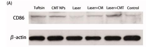
Application: WB    Species: Mice    Sample: Y79 tumors

Figure 11. (A, B) The contents of CD86 in Y79 tumors after different treatments. (C) TNF-α levels in Y79 tumors after different treatments (**p<.01).

5). Isosteviol reduces the acute inflammatory response after burns by upregulating MMP9 in macrophages leading to M2 polarization. International Immunopharmacology (PubMed: 35176589) [IF=5.6]

6). Zanubrutinib ameliorates lipopolysaccharide-induced acute lung injury via regulating macrophage polarization. International Immunopharmacology (PubMed: 35973369) [IF=5.6]

7). miR-146a-5p enhances embryo survival in unexplained recurrent spontaneous abortion by promoting M2 polarization of decidual macrophages. International Immunopharmacology (PubMed: 35764020) [IF=5.6]

8). Vascular endothelial growth factor A is a potential prognostic biomarker and correlates with immune cell infiltration in hepatocellular carcinoma. Journal of Cellular and Molecular Medicine (PubMed: 36729917) [IF=5.3]

9). Contribution of Oxidative Stress Induced by Sonodynamic Therapy to the Calcium Homeostasis Imbalance Enhances Macrophage Infiltration in Glioma Cells. Cancers (PubMed: 35454942) [IF=5.2]

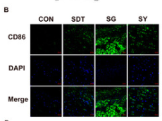
Application: IF/ICC    Species: Mouse    Sample: brain

Figure 8. (A) GL261 glioma-bearing C57BL/6 to undergo xenograft tumor and brain treatment and MRI tests on days 0 and 24. (B,D) At the end of the experiment (30 days), the mouse brains were made into sections for immunofluorescence staining (CD86 and CD206); (C,E) analysis of the immunofluorescence intensity showed that the SG group induced more CD86 expression in the gliomas than CD206 expression.

10). Erythropoietin-Induced Autophagy Protects Against Spinal Cord Injury and Improves Neurological Function via the Extracellular-Regulated Protein Kinase Signaling Pathway. MOLECULAR NEUROBIOLOGY (PubMed: 32647973) [IF=5.1]

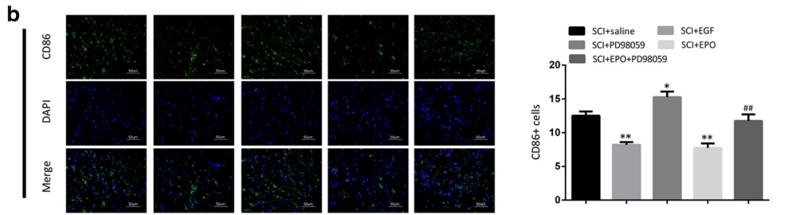
Application: IF/ICC    Species: rat    Sample:

Fig. 6 |EPO benefit antiinflammation and axonal regeneration can be neutralized by ERK inhibition, and activator can mimic the effect of EPO. a The axonal special protein GAP43 immunofluorescence staining and histogram at 14 days (magnification × 400). b–e The inflammatory marker proteins immunofluorescence staining and histograms at 14 days, including CD86, GFAP, TNF-α, iNOS(magnification × 400). All data presented as mean ± SEM in each group. *p < 0.05 vs. SCI + saline group, **p < 0.01 vs. SCI + saline group. #p < 0.05 comparison between SCI + EPO group and SCI + EPO + PD98059, ##p < 0.01 comparison between SCI + EPO group and SCI + EPO + PD98059

Load more

Restrictive clause


Affinity Biosciences tests all products strictly. Citations are provided as a resource for additional applications that have not been validated by Affinity Biosciences. Please choose the appropriate format for each application and consult Materials and Methods sections for additional details about the use of any product in these publications.

For Research Use Only.
Not for use in diagnostic or therapeutic procedures. Not for resale. Not for distribution without written consent. Affinity Biosciences will not be held responsible for patent infringement or other violations that may occur with the use of our products. Affinity Biosciences, Affinity Biosciences Logo and all other trademarks are the property of Affinity Biosciences LTD.