EPHA4 Antibody - #AF5496

| Product: | EPHA4 Antibody |
| Catalog: | AF5496 |
| Description: | Rabbit polyclonal antibody to EPHA4 |
| Application: | WB IHC |
| Reactivity: | Human, Mouse, Rat |
| Prediction: | Pig, Bovine, Horse, Sheep, Rabbit, Dog, Chicken, Xenopus |
| Mol.Wt.: | 110 kd; 110kD(Calculated). |
| Uniprot: | P54764 |
| RRID: | AB_2837975 |
Product Info
*The optimal dilutions should be determined by the end user.
*Tips:
WB: For western blot detection of denatured protein samples. IHC: For immunohistochemical detection of paraffin sections (IHC-p) or frozen sections (IHC-f) of tissue samples. IF/ICC: For immunofluorescence detection of cell samples. ELISA(peptide): For ELISA detection of antigenic peptide.
Cite Format: Affinity Biosciences Cat# AF5496, RRID:AB_2837975.
Fold/Unfold
Cek 8; CEK8; EK8; eph receptor a4; EPH-like kinase 8; EPHA4; EPHA4_HUMAN; Ephrin type-A receptor 4; HEK 8; hEK8; Receptor protein-tyrosine kinase HEK8; Sek 1; SEK; TYRO 1 protein tyrosine kinase; TYRO1; Tyrosine-protein kinase receptor SEK; Tyrosine-protein kinase TYRO1;
Immunogens
- P54764 EPHA4_HUMAN:
- Protein BLAST With
- NCBI/
- ExPASy/
- Uniprot
MAGIFYFALFSCLFGICDAVTGSRVYPANEVTLLDSRSVQGELGWIASPLEGGWEEVSIMDEKNTPIRTYQVCNVMEPSQNNWLRTDWITREGAQRVYIEIKFTLRDCNSLPGVMGTCKETFNLYYYESDNDKERFIRENQFVKIDTIAADESFTQVDIGDRIMKLNTEIRDVGPLSKKGFYLAFQDVGACIALVSVRVFYKKCPLTVRNLAQFPDTITGADTSSLVEVRGSCVNNSEEKDVPKMYCGADGEWLVPIGNCLCNAGHEERSGECQACKIGYYKALSTDATCAKCPPHSYSVWEGATSCTCDRGFFRADNDAASMPCTRPPSAPLNLISNVNETSVNLEWSSPQNTGGRQDISYNVVCKKCGAGDPSKCRPCGSGVHYTPQQNGLKTTKVSITDLLAHTNYTFEIWAVNGVSKYNPNPDQSVSVTVTTNQAAPSSIALVQAKEVTRYSVALAWLEPDRPNGVILEYEVKYYEKDQNERSYRIVRTAARNTDIKGLNPLTSYVFHVRARTAAGYGDFSEPLEVTTNTVPSRIIGDGANSTVLLVSVSGSVVLVVILIAAFVISRRRSKYSKAKQEADEEKHLNQGVRTYVDPFTYEDPNQAVREFAKEIDASCIKIEKVIGVGEFGEVCSGRLKVPGKREICVAIKTLKAGYTDKQRRDFLSEASIMGQFDHPNIIHLEGVVTKCKPVMIITEYMENGSLDAFLRKNDGRFTVIQLVGMLRGIGSGMKYLSDMSYVHRDLAARNILVNSNLVCKVSDFGMSRVLEDDPEAAYTTRGGKIPIRWTAPEAIAYRKFTSASDVWSYGIVMWEVMSYGERPYWDMSNQDVIKAIEEGYRLPPPMDCPIALHQLMLDCWQKERSDRPKFGQIVNMLDKLIRNPNSLKRTGTESSRPNTALLDPSSPEFSAVVSVGDWLQAIKMDRYKDNFTAAGYTTLEAVVHVNQEDLARIGITAITHQNKILSSVQAMRTQMQQMHGRMVPV
Predictions
Score>80(red) has high confidence and is suggested to be used for WB detection. *The prediction model is mainly based on the alignment of immunogen sequences, the results are for reference only, not as the basis of quality assurance.
High(score>80) Medium(80>score>50) Low(score<50) No confidence
PTMs - P54764 As Substrate
| Site | PTM Type | Enzyme | Source |
|---|---|---|---|
| T117 | Phosphorylation | Uniprot | |
| T168 | Phosphorylation | Uniprot | |
| S349 | Phosphorylation | Uniprot | |
| S350 | Phosphorylation | Uniprot | |
| T354 | Phosphorylation | Uniprot | |
| T595 | Phosphorylation | Uniprot | |
| Y596 | Phosphorylation | P54764 (EPHA4) | Uniprot |
| T601 | Phosphorylation | Uniprot | |
| Y602 | Phosphorylation | P54764 (EPHA4) | Uniprot |
| S637 | Phosphorylation | Uniprot | |
| K693 | Acetylation | Uniprot | |
| K735 | Ubiquitination | Uniprot | |
| S741 | Phosphorylation | Uniprot | |
| Y779 | Phosphorylation | Uniprot | |
| T781 | Phosphorylation | Uniprot | |
| Y798 | Phosphorylation | Uniprot | |
| S887 | Phosphorylation | Uniprot | |
| T957 | Phosphorylation | Uniprot |
PTMs - P54764 As Enzyme
Research Backgrounds
Receptor tyrosine kinase which binds membrane-bound ephrin family ligands residing on adjacent cells, leading to contact-dependent bidirectional signaling into neighboring cells. The signaling pathway downstream of the receptor is referred to as forward signaling while the signaling pathway downstream of the ephrin ligand is referred to as reverse signaling. Highly promiscuous, it has the unique property among Eph receptors to bind and to be physiologically activated by both GPI-anchored ephrin-A and transmembrane ephrin-B ligands including EFNA1 and EFNB3. Upon activation by ephrin ligands, modulates cell morphology and integrin-dependent cell adhesion through regulation of the Rac, Rap and Rho GTPases activity. Plays an important role in the development of the nervous system controlling different steps of axonal guidance including the establishment of the corticospinal projections. May also control the segregation of motor and sensory axons during neuromuscular circuit development. In addition to its role in axonal guidance plays a role in synaptic plasticity. Activated by EFNA1 phosphorylates CDK5 at 'Tyr-15' which in turn phosphorylates NGEF regulating RHOA and dendritic spine morphogenesis. In the nervous system, plays also a role in repair after injury preventing axonal regeneration and in angiogenesis playing a role in central nervous system vascular formation. Additionally, its promiscuity makes it available to participate in a variety of cell-cell signaling regulating for instance the development of the thymic epithelium. During development of the cochlear organ of Corti, regulates pillar cell separation by forming a ternary complex with ADAM10 and CADH1 which facilitates the cleavage of CADH1 by ADAM10 and disruption of adherens junctions (By similarity).
Cell membrane>Single-pass type I membrane protein. Cell projection>Axon. Cell projection>Dendrite. Cell junction>Synapse>Postsynaptic density membrane. Early endosome. Cell junction>Adherens junction.
Note: Clustered upon activation and targeted to early endosome.
Ubiquitous.
Heterotetramer upon binding of the ligand. The heterotetramer is composed of an ephrin dimer and a receptor dimer. Oligomerization is probably required to induce biological responses. Interacts (phosphorylated at position Tyr-602) with FYN. Interacts with CDK5, CDK5R1 and NGEF; upon activation by EFNA1 induces NGEF phosphorylation by the kinase CDK5. Interacts with CHN1; effector of EPHA4 in axon guidance linking EPHA4 activation to RAC1 regulation (By similarity). Interacts (via PDZ motif) with SIPA1L1 (via PDZ domain); controls neuronal morphology through regulation of the RAP1 (RAP1A or RAP1B) and RAP2 (RAP2A, RAP2B or RAP2C) GTPases. Forms a ternary complex composed of ADAM10, CADH1 and EPHA4; within the complex, CADH1 is cleaved by ADAM10 which disrupts adherens junctions (By similarity).
The protein kinase domain mediates interaction with NGEF.
Belongs to the protein kinase superfamily. Tyr protein kinase family. Ephrin receptor subfamily.
Research Fields
· Organismal Systems > Development > Axon guidance. (View pathway)
References
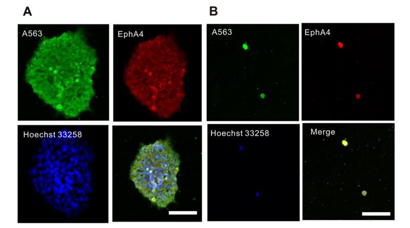
Application: IF/ICC Species: rat Sample: neural stem cells
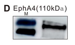
Application: WB Species: rat Sample: neural stem cells
Restrictive clause
Affinity Biosciences tests all products strictly. Citations are provided as a resource for additional applications that have not been validated by Affinity Biosciences. Please choose the appropriate format for each application and consult Materials and Methods sections for additional details about the use of any product in these publications.
For Research Use Only.
Not for use in diagnostic or therapeutic procedures. Not for resale. Not for distribution without written consent. Affinity Biosciences will not be held responsible for patent infringement or other violations that may occur with the use of our products. Affinity Biosciences, Affinity Biosciences Logo and all other trademarks are the property of Affinity Biosciences LTD.