BMPR2 Antibody - #AF5383

| Product: | BMPR2 Antibody |
| Catalog: | AF5383 |
| Description: | Rabbit polyclonal antibody to BMPR2 |
| Application: | WB IHC IF/ICC |
| Reactivity: | Human, Mouse, Rat, Monkey |
| Prediction: | Bovine, Horse, Sheep, Rabbit, Dog, Chicken, Xenopus |
| Mol.Wt.: | 115 kDa; 115kD(Calculated). |
| Uniprot: | Q13873 |
| RRID: | AB_2837868 |
Product Info
*The optimal dilutions should be determined by the end user.
*Tips:
WB: For western blot detection of denatured protein samples. IHC: For immunohistochemical detection of paraffin sections (IHC-p) or frozen sections (IHC-f) of tissue samples. IF/ICC: For immunofluorescence detection of cell samples. ELISA(peptide): For ELISA detection of antigenic peptide.
Cite Format: Affinity Biosciences Cat# AF5383, RRID:AB_2837868.
Fold/Unfold
BMP type II receptor; BMP type-2 receptor; BMPR 2; BMPR 3; BMPR II; BMPR-2; BMPR-II; Bmpr2; BMPR2_HUMAN; BMPR3; BMPRII; BMR 2; BMR2; Bone morphogenetic protein receptor type 2; Bone morphogenetic protein receptor type II; Bone morphogenetic protein receptor type-2; Bone morphogenic protein receptor type II serine threonine kinase; BRK 3; BRK3; PPH 1; PPH1; Serine threonine kinase type II activin receptor like kinase; T ALK; TALK; Type II activin receptor like kinase;
Immunogens
- Q13873 BMPR2_HUMAN:
- Protein BLAST With
- NCBI/
- ExPASy/
- Uniprot
MTSSLQRPWRVPWLPWTILLVSTAAASQNQERLCAFKDPYQQDLGIGESRISHENGTILCSKGSTCYGLWEKSKGDINLVKQGCWSHIGDPQECHYEECVVTTTPPSIQNGTYRFCCCSTDLCNVNFTENFPPPDTTPLSPPHSFNRDETIIIALASVSVLAVLIVALCFGYRMLTGDRKQGLHSMNMMEAAASEPSLDLDNLKLLELIGRGRYGAVYKGSLDERPVAVKVFSFANRQNFINEKNIYRVPLMEHDNIARFIVGDERVTADGRMEYLLVMEYYPNGSLCKYLSLHTSDWVSSCRLAHSVTRGLAYLHTELPRGDHYKPAISHRDLNSRNVLVKNDGTCVISDFGLSMRLTGNRLVRPGEEDNAAISEVGTIRYMAPEVLEGAVNLRDCESALKQVDMYALGLIYWEIFMRCTDLFPGESVPEYQMAFQTEVGNHPTFEDMQVLVSREKQRPKFPEAWKENSLAVRSLKETIEDCWDQDAEARLTAQCAEERMAELMMIWERNKSVSPTVNPMSTAMQNERNLSHNRRVPKIGPYPDYSSSSYIEDSIHHTDSIVKNISSEHSMSSTPLTIGEKNRNSINYERQQAQARIPSPETSVTSLSTNTTTTNTTGLTPSTGMTTISEMPYPDETNLHTTNVAQSIGPTPVCLQLTEEDLETNKLDPKEVDKNLKESSDENLMEHSLKQFSGPDPLSSTSSSLLYPLIKLAVEATGQQDFTQTANGQACLIPDVLPTQIYPLPKQQNLPKRPTSLPLNTKNSTKEPRLKFGSKHKSNLKQVETGVAKMNTINAAEPHVVTVTMNGVAGRNHSVNSHAATTQYANGTVLSGQTTNIVTHRAQEMLQNQFIGEDTRLNINSSPDEHEPLLRREQQAGHDEGVLDRLVDRRERPLEGGRTNSNNNNSNPCSEQDVLAQGVPSTAADPGPSKPRRAQRPNSLDLSATNVLDGSSIQIGESTQDGKSGSGEKIKKRVKTPYSLKRWRPSTWVISTESLDCEVNNNGSNRAVHSKSSTAVYLAEGGTATTMVSKDIGMNCL
Predictions
Score>80(red) has high confidence and is suggested to be used for WB detection. *The prediction model is mainly based on the alignment of immunogen sequences, the results are for reference only, not as the basis of quality assurance.
High(score>80) Medium(80>score>50) Low(score<50) No confidence
PTMs - Q13873 As Substrate
| Site | PTM Type | Enzyme | Source |
|---|---|---|---|
| K219 | Acetylation | Uniprot | |
| Y247 | Phosphorylation | Uniprot | |
| Y314 | Phosphorylation | Uniprot | |
| S336 | Phosphorylation | Uniprot | |
| S355 | Phosphorylation | Uniprot | |
| T359 | Phosphorylation | Uniprot | |
| S375 | Phosphorylation | Uniprot | |
| T379 | Phosphorylation | Uniprot | |
| S513 | Phosphorylation | Uniprot | |
| S515 | Phosphorylation | Uniprot | |
| K539 | Ubiquitination | Uniprot | |
| Y546 | Phosphorylation | Uniprot | |
| S547 | Phosphorylation | Uniprot | |
| S550 | Phosphorylation | Uniprot | |
| Y551 | Phosphorylation | Uniprot | |
| K564 | Ubiquitination | Uniprot | |
| S574 | Phosphorylation | Uniprot | |
| T575 | Phosphorylation | Uniprot | |
| S586 | Phosphorylation | Uniprot | |
| Y589 | Phosphorylation | Uniprot | |
| S680 | Phosphorylation | Uniprot | |
| S681 | Phosphorylation | Uniprot | |
| Y708 | Phosphorylation | Uniprot | |
| S757 | Phosphorylation | Uniprot | |
| K763 | Ubiquitination | Uniprot | |
| T803 | Phosphorylation | Uniprot | |
| T805 | Phosphorylation | Uniprot | |
| Y825 | Phosphorylation | Uniprot | |
| S862 | Phosphorylation | Uniprot | |
| S863 | Phosphorylation | Uniprot | |
| T977 | Phosphorylation | Uniprot | |
| Y979 | Phosphorylation | Uniprot | |
| S980 | Phosphorylation | Uniprot |
Research Backgrounds
On ligand binding, forms a receptor complex consisting of two type II and two type I transmembrane serine/threonine kinases. Type II receptors phosphorylate and activate type I receptors which autophosphorylate, then bind and activate SMAD transcriptional regulators. Binds to BMP7, BMP2 and, less efficiently, BMP4. Binding is weak but enhanced by the presence of type I receptors for BMPs. Mediates induction of adipogenesis by GDF6.
Cell membrane>Single-pass type I membrane protein.
Highly expressed in heart and liver.
Interacts with GDF5.
Belongs to the protein kinase superfamily. TKL Ser/Thr protein kinase family. TGFB receptor subfamily.
Research Fields
· Cellular Processes > Cellular community - eukaryotes > Signaling pathways regulating pluripotency of stem cells. (View pathway)
· Environmental Information Processing > Signaling molecules and interaction > Cytokine-cytokine receptor interaction. (View pathway)
· Environmental Information Processing > Signal transduction > TGF-beta signaling pathway. (View pathway)
· Environmental Information Processing > Signal transduction > Hippo signaling pathway. (View pathway)
· Human Diseases > Cancers: Overview > MicroRNAs in cancer.
· Organismal Systems > Development > Axon guidance. (View pathway)
References
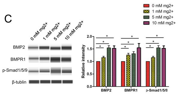
Application: WB Species: Human Sample: DPSCs
Application: WB Species: Human Sample: HUVECs
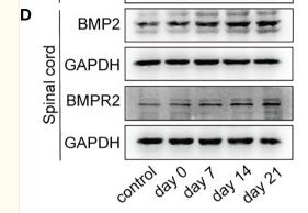
Application: WB Species: Mice Sample: brain and spinal cord
Restrictive clause
Affinity Biosciences tests all products strictly. Citations are provided as a resource for additional applications that have not been validated by Affinity Biosciences. Please choose the appropriate format for each application and consult Materials and Methods sections for additional details about the use of any product in these publications.
For Research Use Only.
Not for use in diagnostic or therapeutic procedures. Not for resale. Not for distribution without written consent. Affinity Biosciences will not be held responsible for patent infringement or other violations that may occur with the use of our products. Affinity Biosciences, Affinity Biosciences Logo and all other trademarks are the property of Affinity Biosciences LTD.