Product: MMP2 Antibody
Catalog: AF5330
Description: Rabbit polyclonal antibody to MMP2
Application: WB IHC IF/ICC
Reactivity: Human, Mouse, Rat, Monkey
Prediction: Pig, Bovine, Horse, Sheep, Rabbit, Dog
Mol.Wt.: 74 kDa; 74kD(Calculated).
Uniprot: P08253
RRID: AB_2837815

View similar products>>

   Size Price Inventory
 100ul $280 In stock
 200ul $350 In stock

Lead Time: Same day delivery

For pricing and ordering contact:
Local distributors

Product Info

Source:
Rabbit
Application:
WB 1:500-1:2000, IF/ICC 1:100-1:500, IHC 1:50-1:200
*The optimal dilutions should be determined by the end user.
*Tips:

WB: For western blot detection of denatured protein samples. IHC: For immunohistochemical detection of paraffin sections (IHC-p) or frozen sections (IHC-f) of tissue samples. IF/ICC: For immunofluorescence detection of cell samples. ELISA(peptide): For ELISA detection of antigenic peptide.

Reactivity:
Human,Mouse,Rat,Monkey
Prediction:
Pig(100%), Bovine(100%), Horse(94%), Sheep(100%), Rabbit(89%), Dog(100%)
Clonality:
Polyclonal
Specificity:
MMP2 Antibody detects endogenous levels of total MMP2.
RRID:
AB_2837815
Cite Format: Affinity Biosciences Cat# AF5330, RRID:AB_2837815.
Conjugate:
Unconjugated.
Purification:
The antiserum was purified by peptide affinity chromatography using SulfoLink™ Coupling Resin (Thermo Fisher Scientific).
Storage:
Rabbit IgG in phosphate buffered saline , pH 7.4, 150mM NaCl, 0.02% sodium azide and 50% glycerol. Store at -20 °C. Stable for 12 months from date of receipt.
Alias:

Fold/Unfold

72 kDa gelatinase; 72kD type IV collagenase; CLG 4; CLG 4A; CLG4; CLG4A; Collagenase Type 4 alpha; Collagenase type IV A; Gelatinase A; Gelatinase alpha; Gelatinase neutrophil; Matrix metallopeptidase 2 gelatinase A 72kDa gelatinase 72kDa type IV collagenase; Matrix metalloproteinase 2 (gelatinase A, 72kDa gelatinase, 72kDa type IV collagenase); Matrix Metalloproteinase 2; Matrix metalloproteinase II; Matrix metalloproteinase-2; MMP 2; MMP II; MMP-2; MMP2; MMP2_HUMAN; MONA; Neutrophil gelatinase; PEX; TBE 1; TBE-1;

Immunogens

Immunogen:
Uniprot:
Gene(ID):
Expression:
P08253 MMP2_HUMAN:

Produced by normal skin fibroblasts. PEX is expressed in a number of tumors including gliomas, breast and prostate.

Description:
Ubiquitinous metalloproteinase that is involved in diverse functions such as remodeling of the vasculature, angiogenesis, tissue repair, tumor invasion, inflammation, and atherosclerotic plaque rupture.
Sequence:
MEALMARGALTGPLRALCLLGCLLSHAAAAPSPIIKFPGDVAPKTDKELAVQYLNTFYGCPKESCNLFVLKDTLKKMQKFFGLPQTGDLDQNTIETMRKPRCGNPDVANYNFFPRKPKWDKNQITYRIIGYTPDLDPETVDDAFARAFQVWSDVTPLRFSRIHDGEADIMINFGRWEHGDGYPFDGKDGLLAHAFAPGTGVGGDSHFDDDELWTLGEGQVVRVKYGNADGEYCKFPFLFNGKEYNSCTDTGRSDGFLWCSTTYNFEKDGKYGFCPHEALFTMGGNAEGQPCKFPFRFQGTSYDSCTTEGRTDGYRWCGTTEDYDRDKKYGFCPETAMSTVGGNSEGAPCVFPFTFLGNKYESCTSAGRSDGKMWCATTANYDDDRKWGFCPDQGYSLFLVAAHEFGHAMGLEHSQDPGALMAPIYTYTKNFRLSQDDIKGIQELYGASPDIDLGTGPTPTLGPVTPEICKQDIVFDGIAQIRGEIFFFKDRFIWRTVTPRDKPMGPLLVATFWPELPEKIDAVYEAPQEEKAVFFAGNEYWIYSASTLERGYPKPLTSLGLPPDVQRVDAAFNWSKNKKTYIFAGDKFWRYNEVKKKMDPGFPKLIADAWNAIPDNLDAVVDLQGGGHSYFFKGAYYLKLENQSLKSVKFGSIKSDWLGC

Predictions

Predictions:

Score>80(red) has high confidence and is suggested to be used for WB detection. *The prediction model is mainly based on the alignment of immunogen sequences, the results are for reference only, not as the basis of quality assurance.

Species
Results
Score
Pig
100
Bovine
100
Sheep
100
Dog
100
Horse
94
Rabbit
89
Chicken
56
Xenopus
0
Zebrafish
0
Model Confidence:
High(score>80) Medium(80>score>50) Low(score<50) No confidence

PTMs - P08253 As Substrate

Site PTM Type Enzyme
T11 Phosphorylation
S160 Phosphorylation P17252 (PRKCA)
S246 Phosphorylation
T250 Phosphorylation P17252 (PRKCA)
T261 Phosphorylation
Y360 Phosphorylation
S362 Phosphorylation
T364 Phosphorylation
S365 Phosphorylation P17252 (PRKCA)
S369 Phosphorylation
T377 Phosphorylation P17252 (PRKCA)
T378 Phosphorylation P17252 (PRKCA)

Research Backgrounds

Function:

Ubiquitinous metalloproteinase that is involved in diverse functions such as remodeling of the vasculature, angiogenesis, tissue repair, tumor invasion, inflammation, and atherosclerotic plaque rupture. As well as degrading extracellular matrix proteins, can also act on several nonmatrix proteins such as big endothelial 1 and beta-type CGRP promoting vasoconstriction. Also cleaves KISS at a Gly-|-Leu bond. Appears to have a role in myocardial cell death pathways. Contributes to myocardial oxidative stress by regulating the activity of GSK3beta. Cleaves GSK3beta in vitro. Involved in the formation of the fibrovascular tissues in association with MMP14.

PEX, the C-terminal non-catalytic fragment of MMP2, posseses anti-angiogenic and anti-tumor properties and inhibits cell migration and cell adhesion to FGF2 and vitronectin. Ligand for integrinv/beta3 on the surface of blood vessels.

Mediates the proteolysis of CHUK/IKKA and initiates a primary innate immune response by inducing mitochondrial-nuclear stress signaling with activation of the pro-inflammatory NF-kappaB, NFAT and IRF transcriptional pathways.

PTMs:

Phosphorylation on multiple sites modulates enzymatic activity. Phosphorylated by PKC in vitro.

The propeptide is processed by MMP14 (MT-MMP1) and MMP16 (MT-MMP3). Autocatalytic cleavage in the C-terminal produces the anti-angiogenic peptide, PEX. This processing appears to be facilitated by binding integrinv/beta3.

Subcellular Location:

Secreted>Extracellular space>Extracellular matrix. Membrane. Nucleus.
Note: Colocalizes with integrin alphaV/beta3 at the membrane surface in angiogenic blood vessels and melanomas. Found in mitochondria, along microfibrils, and in nuclei of cardiomyocytes.

Cytoplasm. Mitochondrion.

Extracellular region or secreted Cytosol Plasma membrane Cytoskeleton Lysosome Endosome Peroxisome ER Golgi apparatus Nucleus Mitochondrion Manual annotation Automatic computational assertionSubcellular location
Tissue Specificity:

Produced by normal skin fibroblasts. PEX is expressed in a number of tumors including gliomas, breast and prostate.

Subunit Structure:

Interacts (via the C-terminal hemopexin-like domains-containing region) with the integrin alpha-V/beta-3; the interaction promotes vascular invasion in angiogenic vessels and melamoma cells. Interacts (via the C-terminal PEX domain) with TIMP2 (via the C-terminal); the interaction inhibits the degradation activity. Interacts with GSK3B.

Family&Domains:

The conserved cysteine present in the cysteine-switch motif binds the catalytic zinc ion, thus inhibiting the enzyme. The dissociation of the cysteine from the zinc ion upon the activation-peptide release activates the enzyme.

Belongs to the peptidase M10A family.

Research Fields

· Human Diseases > Drug resistance: Antineoplastic > Endocrine resistance.

· Human Diseases > Cancers: Overview > Pathways in cancer.   (View pathway)

· Human Diseases > Cancers: Overview > Proteoglycans in cancer.

· Human Diseases > Cancers: Specific types > Bladder cancer.   (View pathway)

· Organismal Systems > Immune system > Leukocyte transendothelial migration.   (View pathway)

· Organismal Systems > Endocrine system > Estrogen signaling pathway.   (View pathway)

· Organismal Systems > Endocrine system > Relaxin signaling pathway.

References

1). Phenytoin silver: a new nanocompound for promoting dermal wound healing via comprehensive pharmacological action. Theranostics (PubMed: 28255340) [IF=12.4]

Application: WB    Species: human    Sample:

Figure 6. PnAg regulates gp130/Jak/Stat3 signaling pathway (A) and (B) NIH-3T3 and HaCat Cells were treated with PnAg at different concentrations and cell viability was tested using MTT analysis. (C) Wound healing assay reflected the effect of PnAg on cell migration. (D) Binding mode of PnAg in the active pocket of gp130. (E) and (F) MMPs activity and expression levels of Stat3, VEGF, TGFB-1, and TGFB1 detected using zymographic and Western blot assays. (G) Diagram of the proposed function of PnAg in wound inflammation and re-epithelialization controls.

Application: IHC    Species: human    Sample:

Figure 2. PnAg promotes wound healing in SD rats. (A) Photographs of rat skin full-thickness excision wounds on different post-excision days. (B) Change in wound areas of SD rats after treatment; (C) and (D) Expression levels of collagen I, NF-κB, TGF-ß, MMP-2, and MMP-9 in tissues on day 7 and 17 detected by immunohistochemistry. (E) Histogram of protein expression levels in these tissues. (F) and (G) Histomorphological changes in wound tissues stained by Masson trichrome and HE on day 17.

2). Downregulation of castor zinc finger 1 predicts poor prognosis and facilitates hepatocellular carcinoma progression via MAPK/ERK signaling. Journal of Experimental & Clinical Cancer Research (PubMed: 29506567) [IF=11.3]

3). The NQO1/PKLR axis promotes lymph node metastasis and breast cancer progression by modulating glycolytic reprogramming. CANCER LETTERS (PubMed: 30954648) [IF=9.7]

Application: WB    Species: mouse    Sample: NQO1 cells

Figure 3| NQO1 promotes cellular invasion, metastasis and the EMT in vitro and in vivo.

Application: WB    Species: mouse    Sample: NQO1 cells

Figure 3| NQO1 promotes cellular invasion, metastasis and the EMT in vitro and in vivo.F: The expression of epithelial markers (E-cadherin and ZO-1) and mesenchymal markers (Vimentin, Snail, Slug, Twist and MMP-2) was determined by western blot analysis. β-Actin was used as a loading control.

4). Foxq1 promotes metastasis of nasopharyngeal carcinoma by inducing vasculogenic mimicry via the EGFR signaling pathway. Cell Death & Disease (PubMed: 33875643) [IF=9.0]

Application: WB    Species: mouse    Sample: tumors

Fig. 5 |Foxq1 promotes VM formation, and NPC growth and metastasis through regulating EGFR in vivo.H Metastatic nodules by tail vein injection of each group and normal lung (left). Image of H&E staining of lung sections from each group (right); Scale bars represent 500 μm. I Statistical results of the metastatic nodules in each group; p < 0.001. The expression of related genes in xenograft tumors from each group were monitored by qRT-PCR (J)and western blot (K). Data are presented as mean ± SD of three independent experiments.

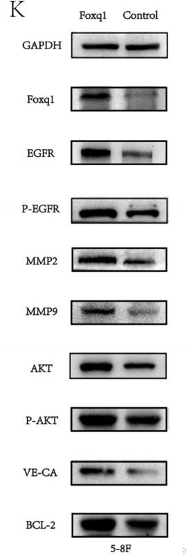
Application: IF/ICC    Species: mouse    Sample: 5–8F cells

Fig. 7| MiR-124 inhibits the EGFR signaling pathway and VM formation, that could be rescued by Foxq1 expression. E Immunofluorescence staining of Foxq1, EGFR, VE-cadherin, MMP2 and MMP9 in 5–8F cells that overexpressed miR-124, control or simultaneously Foxq1 and miR-124, respectively; scale bars represent 50μm. qRT-PCR (F) and western blot (G) were used to monitor the expression of EGFR signaling pathway and VM-related genes in 5–8F cells that overexpressed miR-124, control or simultaneously Foxq1 and miR-124, respectively.Data are presented as mean ± SD of three independent experiments.

5). Hsp90β promotes aggressive vasculogenic mimicry via epithelial-mesenchymal transition in hepatocellular carcinoma. ONCOGENE (PubMed: 30087438) [IF=8.0]

Application: WB    Species: human    Sample: PLC-PRF-5 cells

Fig. 5| Hsp90β promotes Twist1 nuclear translocation and binding to VE-cadherin promoter to increase VM-related gene networks. eWestern blot analysis of VM and EMT-related markers, including VE-cadherin, VEGFR1, VEGFR2, E-cadherin, Vimentin, MMP2, and MMP9 in PLC-PRF-5 cells overexpressed Hsp90β or under lack of Twist1.

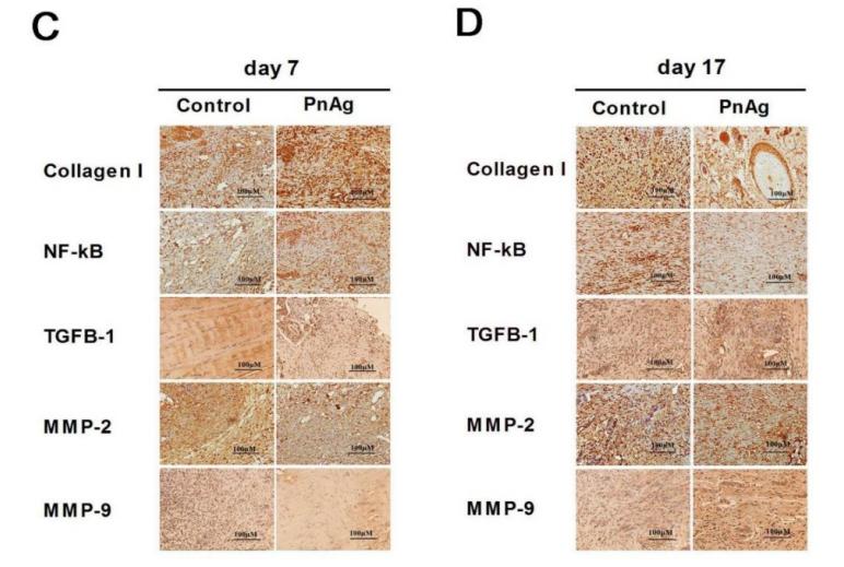
Application: IHC    Species: human    Sample: tumor

Fig. 6 | Hsp90β promotes tumor growth and VM formation relied on Twist1 in vivo and Hsp90 inhibitor depresses the promotion effect.e, f Hsp90β, Twist1, VE-cadherin, E-cadherin, Vimentin,MMP2, and MMP9 expression levels were measured in tumor tissue of the control, Hsp90β overexpression, Hsp90β+ Twist1 siRNA,control shRNA, and Hsp90β knockdown groups by IHC.

6). Myricetin suppresses the proliferation and migration of vascular smooth muscle cells and inhibits neointimal hyperplasia via suppressing TGFBR1 signaling pathways. PHYTOMEDICINE (PubMed: 34500301) [IF=7.9]

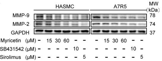
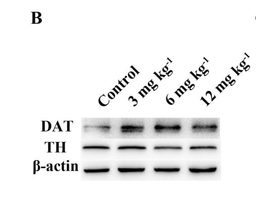
Application: WB    Species:    Sample: VSMCs

Fig. 2.| Myricetin suppresses migration of VSMCs.(H) Western blotting was used to assess MMP-9 and MMP-2 expression in cells treated with different doses of myricetin for 24 h.

7). Aiduqing formula suppresses breast cancer metastasis via inhibiting CXCL1-mediated autophagy. PHYTOMEDICINE (PubMed: 34247114) [IF=7.9]

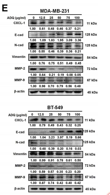
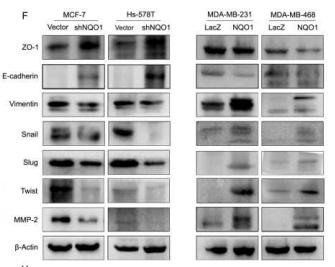
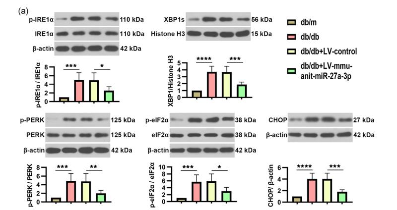
Application: WB    Species: human    Sample: MDA-MB-231 and BT-549 cells

Fig. 1. |ADQ formula suppresses autophagy and induces apoptosis of high-metastatic breast cancer cells.(E) The expression levels of metastasis-related proteins in MDA-MB-231 and BT-549 cells using western blotting analysis after treating with ADQ formula in a concentration-dependent manner.The results indicated that ADQ-formula treatment for 24 h dramatically attenuated the expression levels of proteins related to basement-membrane degradation and the EMT.

8). SETD3 is regulated by a couple of microRNAs and plays opposing roles in proliferation and metastasis of hepatocellular carcinoma. Clinical Science (PubMed: 31654063) [IF=6.0]

9). Microbiota-derived imidazole propionate inhibits type 2 diabetic skin wound healing by targeting SPNS2-mediated S1P transport. iScience (PubMed: 37876799) [IF=5.8]

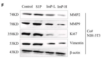
Application: WB    Species: Mouse    Sample: skin tissues

Figure 5 ImP played a role in regulating wound healing by inhibiting the downstream Rho pathway of S1P (A and B) Functions and pathways negatively enriched for ImP treated cells and positively enriched for S1P treated cells were shown. The differentially expressed proteins was identified and analyzed by GO and KEGG pathway analysis. (C) Protein-protein interaction network analysis of ImP down-regulating protein by STRING. (D and E) The effects of S1P and ImP on the expression levels of VEGF, HIF-1α, CD31, and Ki67 proteins in HUVEC cells detected by Western blot. (F and G) The effect of ImP and S1P on the protein expression levels of MMP2, MMP9, Ki67 and Vimentin in NIH3T3cells under coculture conditions. (H) The effect of ImP on the cell membrane localization of RhoA. ImP-L:500 nM, ImP-H: 1μΜ. Data are expressed as the mean ± SD (∗p < 0.05, ∗∗p < 0.01). The number of sample replicates for all experiments was 3 (n = 3).

10). Oleanolic acid inhibits epithelial-mesenchymal transition of hepatocellular carcinoma by promoting iNOS dimerization. MOLECULAR CANCER THERAPEUTICS (PubMed: 30297361) [IF=5.7]

Application: IHC    Species: human    Sample: PLC cells

Figure 5. |OA exhibits anti-EMT effect in vivo and enhances the antitumor effect of regorafenib. (F) Results of IHC assays. The expression levels of E-cadherin were significantly upregulated, whereas those of vimentin, MMP2, and MMP9 were downregulated by OA or regorafenib treatment, and OA enhanced the effects of regorafenib. The expression levels of iNOS and NT were upregulated by OA but not by regorafenib. (G) Staining indexes of IHC assays. Data are represented as mean ± standard error of the mean (*P < 0.05, **P < 0.01).

Load more

Restrictive clause


Affinity Biosciences tests all products strictly. Citations are provided as a resource for additional applications that have not been validated by Affinity Biosciences. Please choose the appropriate format for each application and consult Materials and Methods sections for additional details about the use of any product in these publications.

For Research Use Only.
Not for use in diagnostic or therapeutic procedures. Not for resale. Not for distribution without written consent. Affinity Biosciences will not be held responsible for patent infringement or other violations that may occur with the use of our products. Affinity Biosciences, Affinity Biosciences Logo and all other trademarks are the property of Affinity Biosciences LTD.