PERK Antibody - #AF5304

| Product: | PERK Antibody |
| Catalog: | AF5304 |
| Description: | Rabbit polyclonal antibody to PERK |
| Application: | WB IHC IF/ICC |
| Reactivity: | Human, Mouse, Rat |
| Prediction: | Pig, Bovine, Horse, Sheep, Rabbit, Dog, Chicken, Xenopus |
| Mol.Wt.: | 125~140kDa; 125kD(Calculated). |
| Uniprot: | Q9NZJ5 |
| RRID: | AB_2837789 |
Related Downloads
Protocols
Product Info
*The optimal dilutions should be determined by the end user.
*Tips:
WB: For western blot detection of denatured protein samples. IHC: For immunohistochemical detection of paraffin sections (IHC-p) or frozen sections (IHC-f) of tissue samples. IF/ICC: For immunofluorescence detection of cell samples. ELISA(peptide): For ELISA detection of antigenic peptide.
Cite Format: Affinity Biosciences Cat# AF5304, RRID:AB_2837789.
Fold/Unfold
DKFZp781H1925; E2AK3_HUMAN; EC 2.7.11.1; Eif2ak3; Eukaryotic translation initiation factor 2 alpha kinase 3; Eukaryotic translation initiation factor 2-alpha kinase 3; Heme regulated EIF2 alpha kinase; HRI; HsPEK; Pancreatic eIF2 alpha kinase; Pancreatic eIF2-alpha kinase; PEK; PRKR like endoplasmic reticulum kinase; PRKR-like endoplasmic reticulum kinase; WRS;
Immunogens
- Q9NZJ5 E2AK3_HUMAN:
- Protein BLAST With
- NCBI/
- ExPASy/
- Uniprot
MERAISPGLLVRALLLLLLLLGLAARTVAAGRARGLPAPTAEAAFGLGAAAAPTSATRVPAAGAVAAAEVTVEDAEALPAAAGEQEPRGPEPDDETELRPRGRSLVIISTLDGRIAALDPENHGKKQWDLDVGSGSLVSSSLSKPEVFGNKMIIPSLDGALFQWDQDRESMETVPFTVESLLESSYKFGDDVVLVGGKSLTTYGLSAYSGKVRYICSALGCRQWDSDEMEQEEDILLLQRTQKTVRAVGPRSGNEKWNFSVGHFELRYIPDMETRAGFIESTFKPNENTEESKIISDVEEQEAAIMDIVIKVSVADWKVMAFSKKGGHLEWEYQFCTPIASAWLLKDGKVIPISLFDDTSYTSNDDVLEDEEDIVEAARGATENSVYLGMYRGQLYLQSSVRISEKFPSSPKALESVTNENAIIPLPTIKWKPLIHSPSRTPVLVGSDEFDKCLSNDKFSHEEYSNGALSILQYPYDNGYYLPYYKRERNKRSTQITVRFLDNPHYNKNIRKKDPVLLLHWWKEIVATILFCIIATTFIVRRLFHPHPHRQRKESETQCQTENKYDSVSGEANDSSWNDIKNSGYISRYLTDFEPIQCLGRGGFGVVFEAKNKVDDCNYAIKRIRLPNRELAREKVMREVKALAKLEHPGIVRYFNAWLEAPPEKWQEKMDEIWLKDESTDWPLSSPSPMDAPSVKIRRMDPFATKEHIEIIAPSPQRSRSFSVGISCDQTSSSESQFSPLEFSGMDHEDISESVDAAYNLQDSCLTDCDVEDGTMDGNDEGHSFELCPSEASPYVRSRERTSSSIVFEDSGCDNASSKEEPKTNRLHIGNHCANKLTAFKPTSSKSSSEATLSISPPRPTTLSLDLTKNTTEKLQPSSPKVYLYIQMQLCRKENLKDWMNGRCTIEERERSVCLHIFLQIAEAVEFLHSKGLMHRDLKPSNIFFTMDDVVKVGDFGLVTAMDQDEEEQTVLTPMPAYARHTGQVGTKLYMSPEQIHGNSYSHKVDIFSLGLILFELLYPFSTQMERVRTLTDVRNLKFPPLFTQKYPCEYVMVQDMLSPSPMERPEAINIIENAVFEDLDFPGKTVLRQRSRSLSSSGTKHSRQSNNSHSPLPSN
Predictions
Score>80(red) has high confidence and is suggested to be used for WB detection. *The prediction model is mainly based on the alignment of immunogen sequences, the results are for reference only, not as the basis of quality assurance.
High(score>80) Medium(80>score>50) Low(score<50) No confidence
PTMs - Q9NZJ5 As Substrate
| Site | PTM Type | Enzyme | Source |
|---|---|---|---|
| T177 | Phosphorylation | Uniprot | |
| Y268 | Phosphorylation | Uniprot | |
| T441 | Phosphorylation | Uniprot | |
| S447 | Phosphorylation | Uniprot | |
| K452 | Ubiquitination | Uniprot | |
| S455 | Phosphorylation | Uniprot | |
| Y464 | Phosphorylation | Uniprot | |
| Y474 | Phosphorylation | Uniprot | |
| Y480 | Phosphorylation | Uniprot | |
| Y481 | Phosphorylation | Uniprot | |
| Y484 | Phosphorylation | Uniprot | |
| Y485 | Phosphorylation | Uniprot | |
| S555 | Phosphorylation | Uniprot | |
| T557 | Phosphorylation | Uniprot | |
| T561 | Phosphorylation | Uniprot | |
| S567 | Phosphorylation | Uniprot | |
| K581 | Ubiquitination | Uniprot | |
| Y585 | Phosphorylation | Uniprot | |
| Y619 | Phosphorylation | Q9NZJ5 (EIF2AK3) | Uniprot |
| K622 | Ubiquitination | Uniprot | |
| K669 | Ubiquitination | Uniprot | |
| S688 | Phosphorylation | Uniprot | |
| S694 | Phosphorylation | Uniprot | |
| T705 | Phosphorylation | Uniprot | |
| S715 | Phosphorylation | Uniprot | |
| S719 | Phosphorylation | Uniprot | |
| T802 | Phosphorylation | P31751 (AKT2) , P31749 (AKT1) | Uniprot |
| S803 | Phosphorylation | Uniprot | |
| S804 | Phosphorylation | Uniprot | |
| S811 | Phosphorylation | Uniprot | |
| S844 | Phosphorylation | Uniprot | |
| S845 | Phosphorylation | Uniprot | |
| S854 | Phosphorylation | Uniprot | |
| S856 | Phosphorylation | Uniprot | |
| T861 | Phosphorylation | Uniprot | |
| T862 | Phosphorylation | Uniprot | |
| T982 | Phosphorylation | Uniprot | |
| S1094 | Phosphorylation | Uniprot | |
| S1096 | Phosphorylation | Uniprot | |
| S1109 | Phosphorylation | Uniprot | |
| S1111 | Phosphorylation | Uniprot |
PTMs - Q9NZJ5 As Enzyme
Research Backgrounds
Metabolic-stress sensing protein kinase that phosphorylates the alpha subunit of eukaryotic translation initiation factor 2 (eIF-2-alpha/EIF2S1) on 'Ser-52' during the unfolded protein response (UPR) and in response to low amino acid availability. Converts phosphorylated eIF-2-alpha/EIF2S1 either in a global protein synthesis inhibitor, leading to a reduced overall utilization of amino acids, or to a translation initiation activator of specific mRNAs, such as the transcriptional activator ATF4, and hence allowing ATF4-mediated reprogramming of amino acid biosynthetic gene expression to alleviate nutrient depletion. Serves as a critical effector of unfolded protein response (UPR)-induced G1 growth arrest due to the loss of cyclin-D1 (CCND1). Involved in control of mitochondrial morphology and function.
Oligomerization of the N-terminal ER luminal domain by ER stress promotes PERK trans-autophosphorylation of the C-terminal cytoplasmic kinase domain at multiple residues including Thr-982 on the kinase activation loop (By similarity). Autophosphorylated. Phosphorylated at Tyr-619 following endoplasmic reticulum stress, leading to activate its tyrosine-protein kinase activity. Dephosphorylated by PTPN1/TP1B, leading to inactivate its enzyme activity.
N-glycosylated.
ADP-ribosylated by PARP16 upon ER stress, which increases kinase activity.
Endoplasmic reticulum membrane>Single-pass type I membrane protein.
Ubiquitous. A high level expression is seen in secretory tissues.
Forms dimers with HSPA5/BIP in resting cells (By similarity). Oligomerizes in ER-stressed cells (By similarity). Interacts with DNAJC3 and MFN2 (By similarity). Interacts with TMEM33. Interacts with PDIA6.
The lumenal domain senses perturbations in protein folding in the ER, probably through reversible interaction with HSPA5/BIP.
Belongs to the protein kinase superfamily. Ser/Thr protein kinase family. GCN2 subfamily.
Research Fields
· Cellular Processes > Transport and catabolism > Autophagy - animal. (View pathway)
· Cellular Processes > Cell growth and death > Apoptosis. (View pathway)
· Genetic Information Processing > Folding, sorting and degradation > Protein processing in endoplasmic reticulum. (View pathway)
· Human Diseases > Endocrine and metabolic diseases > Non-alcoholic fatty liver disease (NAFLD).
· Human Diseases > Neurodegenerative diseases > Alzheimer's disease.
· Human Diseases > Infectious diseases: Viral > Hepatitis C.
· Human Diseases > Infectious diseases: Viral > Measles.
· Human Diseases > Infectious diseases: Viral > Influenza A.
· Human Diseases > Infectious diseases: Viral > Herpes simplex infection.
· Human Diseases > Infectious diseases: Viral > Epstein-Barr virus infection.
References
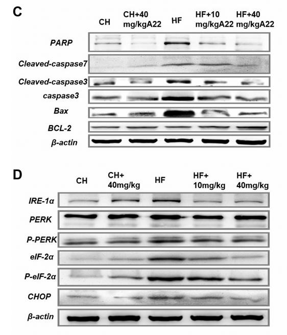
Application: WB Species: mouse Sample: liver
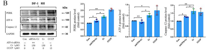
Application: WB Species: chicken Sample: DF-1 cells
Application: WB Species: Human Sample: HaCaT cells
Application: WB Species: Human Sample: HaCaT cells
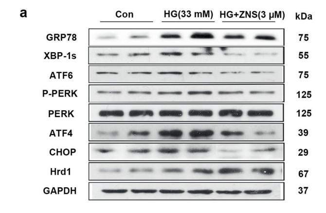
Application: WB Species: mice Sample: NRCMs
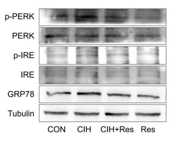
Application: WB Species: Rat Sample: heart tissue

Application: WB Species: human Sample: MDA-MB-231 cells

Application: WB Species: rat Sample: liver
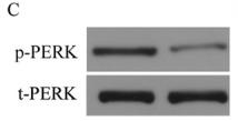
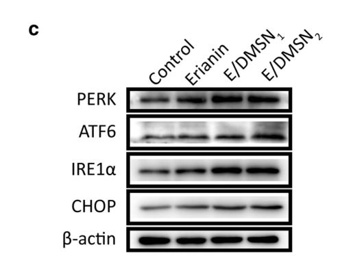
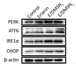
Application: WB Species: Human Sample:
Restrictive clause
Affinity Biosciences tests all products strictly. Citations are provided as a resource for additional applications that have not been validated by Affinity Biosciences. Please choose the appropriate format for each application and consult Materials and Methods sections for additional details about the use of any product in these publications.
For Research Use Only.
Not for use in diagnostic or therapeutic procedures. Not for resale. Not for distribution without written consent. Affinity Biosciences will not be held responsible for patent infringement or other violations that may occur with the use of our products. Affinity Biosciences, Affinity Biosciences Logo and all other trademarks are the property of Affinity Biosciences LTD.