Cleaved-Caspase 8 (Asp384) Antibody - #AF5267
 expression in 293 cells, treated with etoposide,The lane on the left was treated with the antigen-specific peptide.")
Related Downloads
Protocols
Product Info
*The optimal dilutions should be determined by the end user.
*Tips:
WB: For western blot detection of denatured protein samples. IHC: For immunohistochemical detection of paraffin sections (IHC-p) or frozen sections (IHC-f) of tissue samples. IF/ICC: For immunofluorescence detection of cell samples. ELISA(peptide): For ELISA detection of antigenic peptide.
Cite Format: Affinity Biosciences Cat# AF5267, RRID:AB_2837753.
Fold/Unfold
ALPS2B; Amyotrophic lateral sclerosis 2 chromosomal region candidate gene 12 protein; Apoptotic cysteine protease; Apoptotic protease Mch-5; Apoptotic protease Mch5; CAP4; CASP-8; CASP8; CASP8_HUMAN; Caspase 8; Caspase 8 apoptosis related cysteine peptidase; Caspase-8 subunit p10; CED 3; FADD Like ICE; FADD-homologous ICE/CED-3-like protease; FADD-like ICE; FLICE; FLJ17672; ICE-like apoptotic protease 5; MACH alpha 1/2/3 protein; MACH; MACH beta 1/2/3/4 protein; MCH5; MGC78473; MORT1 associated ced 3 homolog; MORT1-associated CED-3 homolog; OTTHUMP00000163717; OTTHUMP00000163720; OTTHUMP00000163724; OTTHUMP00000163725; OTTHUMP00000165062; OTTHUMP00000165063; OTTHUMP00000165064; OTTHUMP00000206552; OTTHUMP00000206582;
Immunogens
Isoform 1, isoform 5 and isoform 7 are expressed in a wide variety of tissues. Highest expression in peripheral blood leukocytes, spleen, thymus and liver. Barely detectable in brain, testis and skeletal muscle.
- Q14790 CASP8_HUMAN:
- Protein BLAST With
- NCBI/
- ExPASy/
- Uniprot
MDFSRNLYDIGEQLDSEDLASLKFLSLDYIPQRKQEPIKDALMLFQRLQEKRMLEESNLSFLKELLFRINRLDLLITYLNTRKEEMERELQTPGRAQISAYRVMLYQISEEVSRSELRSFKFLLQEEISKCKLDDDMNLLDIFIEMEKRVILGEGKLDILKRVCAQINKSLLKIINDYEEFSKERSSSLEGSPDEFSNGEELCGVMTISDSPREQDSESQTLDKVYQMKSKPRGYCLIINNHNFAKAREKVPKLHSIRDRNGTHLDAGALTTTFEELHFEIKPHDDCTVEQIYEILKIYQLMDHSNMDCFICCILSHGDKGIIYGTDGQEAPIYELTSQFTGLKCPSLAGKPKVFFIQACQGDNYQKGIPVETDSEEQPYLEMDLSSPQTRYIPDEADFLLGMATVNNCVSYRNPAEGTWYIQSLCQSLRERCPRGDDILTILTEVNYEVSNKDDKKNMGKQMPQPTFTLRKKLVFPSD
PTMs - Q14790 As Substrate
| Site | PTM Type | Enzyme | Source |
|---|---|---|---|
| S16 | Phosphorylation | Uniprot | |
| K23 | Ubiquitination | Uniprot | |
| K34 | Ubiquitination | Uniprot | |
| K39 | Ubiquitination | Uniprot | |
| K63 | Ubiquitination | Uniprot | |
| S113 | Phosphorylation | Uniprot | |
| K121 | Ubiquitination | Uniprot | |
| K130 | Ubiquitination | Uniprot | |
| K148 | Ubiquitination | Uniprot | |
| K156 | Acetylation | Uniprot | |
| K156 | Ubiquitination | Uniprot | |
| K161 | Ubiquitination | Uniprot | |
| K169 | Ubiquitination | Uniprot | |
| K173 | Ubiquitination | Uniprot | |
| Y178 | Phosphorylation | Uniprot | |
| K183 | Ubiquitination | Uniprot | |
| S192 | Phosphorylation | Uniprot | |
| S209 | Phosphorylation | Uniprot | |
| S211 | Phosphorylation | Uniprot | |
| S219 | Phosphorylation | Uniprot | |
| K224 | Ubiquitination | Uniprot | |
| K229 | Ubiquitination | Uniprot | |
| Y235 | Phosphorylation | Uniprot | |
| K253 | Ubiquitination | Uniprot | |
| T263 | Phosphorylation | P51812 (RPS6KA3) | Uniprot |
| T273 | Phosphorylation | Uniprot | |
| S305 | Phosphorylation | Uniprot | |
| Y334 | Phosphorylation | Uniprot | |
| K344 | Ubiquitination | Uniprot | |
| S347 | Phosphorylation | Q16539 (MAPK14) | Uniprot |
| K351 | Ubiquitination | Uniprot | |
| K353 | Ubiquitination | Uniprot | |
| T373 | Phosphorylation | Uniprot | |
| S375 | Phosphorylation | Uniprot | |
| Y380 | Phosphorylation | P07948 (LYN) , P12931 (SRC) | Uniprot |
| S387 | Phosphorylation | P28482 (MAPK1) , P06493 (CDK1) , P27361 (MAPK3) | Uniprot |
| Y448 | Phosphorylation | P07948 (LYN) | Uniprot |
| K461 | Ubiquitination | Uniprot | |
| K473 | Ubiquitination | Uniprot |
Research Backgrounds
Most upstream protease of the activation cascade of caspases responsible for the TNFRSF6/FAS mediated and TNFRSF1A induced cell death. Binding to the adapter molecule FADD recruits it to either receptor. The resulting aggregate called death-inducing signaling complex (DISC) performs CASP8 proteolytic activation. The active dimeric enzyme is then liberated from the DISC and free to activate downstream apoptotic proteases. Proteolytic fragments of the N-terminal propeptide (termed CAP3, CAP5 and CAP6) are likely retained in the DISC. Cleaves and activates CASP3, CASP4, CASP6, CASP7, CASP9 and CASP10. May participate in the GZMB apoptotic pathways. Cleaves ADPRT. Hydrolyzes the small-molecule substrate, Ac-Asp-Glu-Val-Asp-|-AMC. Likely target for the cowpox virus CRMA death inhibitory protein. Isoform 5, isoform 6, isoform 7 and isoform 8 lack the catalytic site and may interfere with the pro-apoptotic activity of the complex. Cleaves RIPK1 at 'Asp-325' which is crucial for limiting apoptosis and necroptosis during embryonic development (By similarity).
Generation of the subunits requires association with the death-inducing signaling complex (DISC), whereas additional processing is likely due to the autocatalytic activity of the activated protease. GZMB and CASP10 can be involved in these processing events.
Phosphorylation on Ser-387 during mitosis by CDK1 inhibits activation by proteolysis and prevents apoptosis. This phosphorylation occurs in cancer cell lines, as well as in primary breast tissues and lymphocytes.
Cytoplasm.
Isoform 1, isoform 5 and isoform 7 are expressed in a wide variety of tissues. Highest expression in peripheral blood leukocytes, spleen, thymus and liver. Barely detectable in brain, testis and skeletal muscle.
Heterotetramer that consists of two anti-parallel arranged heterodimers, each one formed by a 18 kDa (p18) and a 10 kDa (p10) subunit. Interacts with FADD, CFLAR and PEA15. Isoform 9 interacts at the endoplasmic reticulum with a complex containing BCAP31, BAP29, BCL2 and/or BCL2L1. Interacts with TNFAIP8L2 (By similarity). Interacts with CASP8AP2. Interacts with RFFL and RNF34; negatively regulate CASP8 through proteasomal degradation. Interacts with NOL3; decreases CASP8 activity in a mitochondria localization- and phosphorylation-dependent manner and this interaction is dissociated by calcium. Interacts with UBR2. Interacts with RIPK1 (By similarity). Interacts with stimulated TNFRSF10B; this interaction is followed by CASP8 proteolytic cleavage and activation.
(Microbial infection) Interacts with human cytomegalovirus/HHV-5 protein vICA/UL36; this interaction inhibits CASP8 activation.
(Microbial infection) Interacts with NleF from pathogenic E.coli.
(Microbial infection) Interacts with molluscum contagiosum virus protein MC160.
Isoform 9 contains a N-terminal extension that is required for interaction with the BCAP31 complex.
Belongs to the peptidase C14A family.
Research Fields
· Cellular Processes > Cell growth and death > p53 signaling pathway. (View pathway)
· Cellular Processes > Cell growth and death > Apoptosis. (View pathway)
· Cellular Processes > Cell growth and death > Apoptosis - multiple species. (View pathway)
· Cellular Processes > Cell growth and death > Necroptosis. (View pathway)
· Environmental Information Processing > Signal transduction > TNF signaling pathway. (View pathway)
· Human Diseases > Drug resistance: Antineoplastic > Platinum drug resistance.
· Human Diseases > Endocrine and metabolic diseases > Non-alcoholic fatty liver disease (NAFLD).
· Human Diseases > Neurodegenerative diseases > Alzheimer's disease.
· Human Diseases > Neurodegenerative diseases > Huntington's disease.
· Human Diseases > Infectious diseases: Bacterial > Legionellosis.
· Human Diseases > Infectious diseases: Parasitic > Chagas disease (American trypanosomiasis).
· Human Diseases > Infectious diseases: Parasitic > Toxoplasmosis.
· Human Diseases > Infectious diseases: Bacterial > Tuberculosis.
· Human Diseases > Infectious diseases: Viral > Hepatitis B.
· Human Diseases > Infectious diseases: Viral > Human papillomavirus infection.
· Human Diseases > Infectious diseases: Viral > Herpes simplex infection.
· Human Diseases > Cancers: Overview > Pathways in cancer. (View pathway)
· Human Diseases > Cancers: Overview > Viral carcinogenesis.
· Human Diseases > Cardiovascular diseases > Viral myocarditis.
· Organismal Systems > Immune system > Toll-like receptor signaling pathway. (View pathway)
· Organismal Systems > Immune system > NOD-like receptor signaling pathway. (View pathway)
· Organismal Systems > Immune system > RIG-I-like receptor signaling pathway. (View pathway)
· Organismal Systems > Immune system > IL-17 signaling pathway. (View pathway)
References
Application: WB Species: human Sample: HT-29 cells
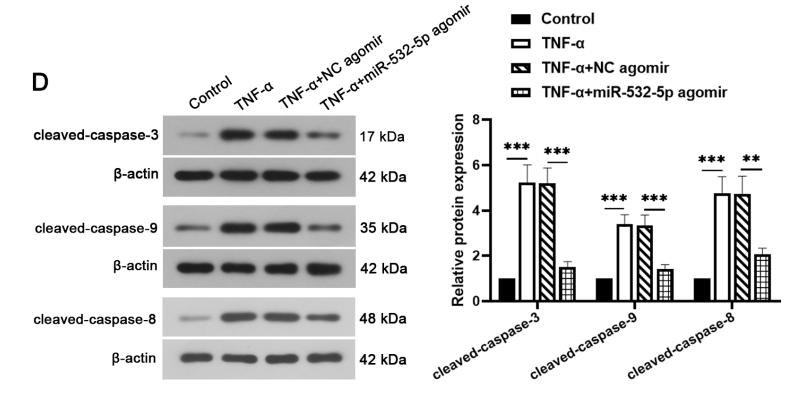
Application: WB Species: rat Sample: NP cells
Application: WB Species: human Sample: LS174T cells

Application: WB Species: Human Sample: HepG2 cells
Application: WB Species: Human Sample: HepG2 cells
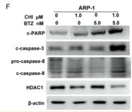
Application: WB Species: Human Sample: MM cells
Application: WB Species: human Sample: HepG2 cells
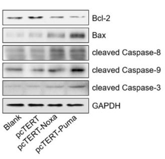
Application: WB Species: Human Sample: bone marrow mesenchymal stem cells (BMSCs)
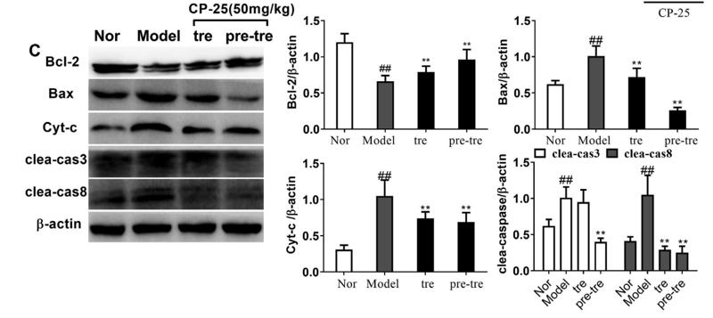
Application: WB Species: Rat Sample: renal tissue
Application: WB Species: human Sample: HepG2
Restrictive clause
Affinity Biosciences tests all products strictly. Citations are provided as a resource for additional applications that have not been validated by Affinity Biosciences. Please choose the appropriate format for each application and consult Materials and Methods sections for additional details about the use of any product in these publications.
For Research Use Only.
Not for use in diagnostic or therapeutic procedures. Not for resale. Not for distribution without written consent. Affinity Biosciences will not be held responsible for patent infringement or other violations that may occur with the use of our products. Affinity Biosciences, Affinity Biosciences Logo and all other trademarks are the property of Affinity Biosciences LTD.