NGF Antibody - #AF5172

| Product: | NGF Antibody |
| Catalog: | AF5172 |
| Description: | Rabbit polyclonal antibody to NGF |
| Application: | WB IHC |
| Reactivity: | Human, Mouse, Rat |
| Prediction: | Pig, Zebrafish, Bovine, Horse, Sheep, Rabbit, Dog |
| Mol.Wt.: | 26 kDa; 27kD(Calculated). |
| Uniprot: | P01138 |
| RRID: | AB_2837658 |
Related Downloads
Protocols
Product Info
*The optimal dilutions should be determined by the end user.
*Tips:
WB: For western blot detection of denatured protein samples. IHC: For immunohistochemical detection of paraffin sections (IHC-p) or frozen sections (IHC-f) of tissue samples. IF/ICC: For immunofluorescence detection of cell samples. ELISA(peptide): For ELISA detection of antigenic peptide.
Cite Format: Affinity Biosciences Cat# AF5172, RRID:AB_2837658.
Fold/Unfold
Beta nerve growth factor; Beta NGF; Beta-nerve growth factor; Beta-NGF; HSAN5; MGC161426; MGC161428; Nerve growth factor (beta polypeptide); Nerve growth factor; Nerve growth factor beta; Nerve growth factor beta polypeptide; Nerve growth factor beta subunit; NGF; NGF_HUMAN; NGFB; NID67;
Immunogens
- P01138 NGF_HUMAN:
- Protein BLAST With
- NCBI/
- ExPASy/
- Uniprot
MSMLFYTLITAFLIGIQAEPHSESNVPAGHTIPQAHWTKLQHSLDTALRRARSAPAAAIAARVAGQTRNITVDPRLFKKRRLRSPRVLFSTQPPREAADTQDLDFEVGGAAPFNRTHRSKRSSSHPIFHRGEFSVCDSVSVWVGDKTTATDIKGKEVMVLGEVNINNSVFKQYFFETKCRDPNPVDSGCRGIDSKHWNSYCTTTHTFVKALTMDGKQAAWRFIRIDTACVCVLSRKAVRRA
Predictions
Score>80(red) has high confidence and is suggested to be used for WB detection. *The prediction model is mainly based on the alignment of immunogen sequences, the results are for reference only, not as the basis of quality assurance.
High(score>80) Medium(80>score>50) Low(score<50) No confidence
PTMs - P01138 As Substrate
| Site | PTM Type | Enzyme | Source |
|---|---|---|---|
| S43 | O-Glycosylation | Uniprot | |
| T46 | O-Glycosylation | Uniprot | |
| S84 | Phosphorylation | Uniprot | |
| K155 | Acetylation | Uniprot |
Research Backgrounds
Nerve growth factor is important for the development and maintenance of the sympathetic and sensory nervous systems. Extracellular ligand for the NTRK1 and NGFR receptors, activates cellular signaling cascades to regulate neuronal proliferation, differentiation and survival (Probable). The immature NGF precursor (proNGF) functions as ligand for the heterodimeric receptor formed by SORCS2 and NGFR, and activates cellular signaling cascades that lead to inactivation of RAC1 and/or RAC2, reorganization of the actin cytoskeleton and neuronal growth cone collapse. In contrast to mature NGF, the precursor form (proNGF) promotes neuronal apoptosis (in vitro) (By similarity). Inhibits metalloproteinase-dependent proteolysis of platelet glycoprotein VI. Binds lysophosphatidylinositol and lysophosphatidylserine between the two chains of the homodimer. The lipid-bound form promotes histamine relase from mast cells, contrary to the lipid-free form (By similarity).
Secreted. Endosome lumen.
Note: ProNGF is endocytosed after binding to the cell surface receptor formed by SORT1 and NGFR.
Homodimer. The homodimer interacts with a single NTRK1 chain. The homodimer interacts with a single NGFR chain. The NGF dimer interacts with a single SORCS2 chain (via extracellular domain) (By similarity). The NGF precursor (proNGF) binds to a receptor complex formed by SORT1 and NGFR, which leads to NGF endocytosis. Both mature NGF and the immature NGF precursor (proNGF) interact with SORCS2 and with the heterodimer formed by SORCS2 and NGFR (via extracellular domains) (By similarity). The NGF precursor (proNGF) has much higher affinity for SORCS2 than mature NGF. The NGF precursor (proNGF) has much higher affinity for SORT1 than mature NGF (By similarity). Interacts with ADAM10 in a divalent cation-dependent manner.
Belongs to the NGF-beta family.
Research Fields
· Cellular Processes > Cell growth and death > Apoptosis. (View pathway)
· Environmental Information Processing > Signal transduction > MAPK signaling pathway. (View pathway)
· Environmental Information Processing > Signal transduction > Ras signaling pathway. (View pathway)
· Environmental Information Processing > Signal transduction > Rap1 signaling pathway. (View pathway)
· Environmental Information Processing > Signal transduction > PI3K-Akt signaling pathway. (View pathway)
· Organismal Systems > Nervous system > Neurotrophin signaling pathway. (View pathway)
· Organismal Systems > Sensory system > Inflammatory mediator regulation of TRP channels. (View pathway)
References
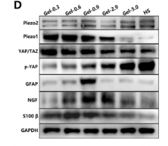
Application: WB Species: Rat Sample: MSCs

Application: WB Species: human Sample: MCF‑7 cells
Restrictive clause
Affinity Biosciences tests all products strictly. Citations are provided as a resource for additional applications that have not been validated by Affinity Biosciences. Please choose the appropriate format for each application and consult Materials and Methods sections for additional details about the use of any product in these publications.
For Research Use Only.
Not for use in diagnostic or therapeutic procedures. Not for resale. Not for distribution without written consent. Affinity Biosciences will not be held responsible for patent infringement or other violations that may occur with the use of our products. Affinity Biosciences, Affinity Biosciences Logo and all other trademarks are the property of Affinity Biosciences LTD.