Collagen IV alpha 2 Antibody - #DF3550

| Product: | Collagen IV alpha 2 Antibody |
| Catalog: | DF3550 |
| Description: | Rabbit polyclonal antibody to Collagen IV alpha 2 |
| Application: | WB IHC IF/ICC |
| Reactivity: | Human, Mouse, Rat |
| Prediction: | Pig, Horse, Sheep, Rabbit |
| Mol.Wt.: | 150 KD; 168kD(Calculated). |
| Uniprot: | P08572 |
| RRID: | AB_2835923 |
Product Info
*The optimal dilutions should be determined by the end user.
*Tips:
WB: For western blot detection of denatured protein samples. IHC: For immunohistochemical detection of paraffin sections (IHC-p) or frozen sections (IHC-f) of tissue samples. IF/ICC: For immunofluorescence detection of cell samples. ELISA(peptide): For ELISA detection of antigenic peptide.
Cite Format: Affinity Biosciences Cat# DF3550, RRID:AB_2835923.
Fold/Unfold
Canstatin; CO4A2_HUMAN; COL4A 2; Col4a2; Collagen alpha 2(IV) chain; Collagen, type IV, alpha 2;
Immunogens
- P08572 CO4A2_HUMAN:
- Protein BLAST With
- NCBI/
- ExPASy/
- Uniprot
MGRDQRAVAGPALRRWLLLGTVTVGFLAQSVLAGVKKFDVPCGGRDCSGGCQCYPEKGGRGQPGPVGPQGYNGPPGLQGFPGLQGRKGDKGERGAPGVTGPKGDVGARGVSGFPGADGIPGHPGQGGPRGRPGYDGCNGTQGDSGPQGPPGSEGFTGPPGPQGPKGQKGEPYALPKEERDRYRGEPGEPGLVGFQGPPGRPGHVGQMGPVGAPGRPGPPGPPGPKGQQGNRGLGFYGVKGEKGDVGQPGPNGIPSDTLHPIIAPTGVTFHPDQYKGEKGSEGEPGIRGISLKGEEGIMGFPGLRGYPGLSGEKGSPGQKGSRGLDGYQGPDGPRGPKGEAGDPGPPGLPAYSPHPSLAKGARGDPGFPGAQGEPGSQGEPGDPGLPGPPGLSIGDGDQRRGLPGEMGPKGFIGDPGIPALYGGPPGPDGKRGPPGPPGLPGPPGPDGFLFGLKGAKGRAGFPGLPGSPGARGPKGWKGDAGECRCTEGDEAIKGLPGLPGPKGFAGINGEPGRKGDRGDPGQHGLPGFPGLKGVPGNIGAPGPKGAKGDSRTITTKGERGQPGVPGVPGMKGDDGSPGRDGLDGFPGLPGPPGDGIKGPPGDPGYPGIPGTKGTPGEMGPPGLGLPGLKGQRGFPGDAGLPGPPGFLGPPGPAGTPGQIDCDTDVKRAVGGDRQEAIQPGCIGGPKGLPGLPGPPGPTGAKGLRGIPGFAGADGGPGPRGLPGDAGREGFPGPPGFIGPRGSKGAVGLPGPDGSPGPIGLPGPDGPPGERGLPGEVLGAQPGPRGDAGVPGQPGLKGLPGDRGPPGFRGSQGMPGMPGLKGQPGLPGPSGQPGLYGPPGLHGFPGAPGQEGPLGLPGIPGREGLPGDRGDPGDTGAPGPVGMKGLSGDRGDAGFTGEQGHPGSPGFKGIDGMPGTPGLKGDRGSPGMDGFQGMPGLKGRPGFPGSKGEAGFFGIPGLKGLAGEPGFKGSRGDPGPPGPPPVILPGMKDIKGEKGDEGPMGLKGYLGAKGIQGMPGIPGLSGIPGLPGRPGHIKGVKGDIGVPGIPGLPGFPGVAGPPGITGFPGFIGSRGDKGAPGRAGLYGEIGATGDFGDIGDTINLPGRPGLKGERGTTGIPGLKGFFGEKGTEGDIGFPGITGVTGVQGPPGLKGQTGFPGLTGPPGSQGELGRIGLPGGKGDDGWPGAPGLPGFPGLRGIRGLHGLPGTKGFPGSPGSDIHGDPGFPGPPGERGDPGEANTLPGPVGVPGQKGDQGAPGERGPPGSPGLQGFPGITPPSNISGAPGDKGAPGIFGLKGYRGPPGPPGSAALPGSKGDTGNPGAPGTPGTKGWAGDSGPQGRPGVFGLPGEKGPRGEQGFMGNTGPTGAVGDRGPKGPKGDPGFPGAPGTVGAPGIAGIPQKIAVQPGTVGPQGRRGPPGAPGEMGPQGPPGEPGFRGAPGKAGPQGRGGVSAVPGFRGDEGPIGHQGPIGQEGAPGRPGSPGLPGMPGRSVSIGYLLVKHSQTDQEPMCPVGMNKLWSGYSLLYFEGQEKAHNQDLGLAGSCLARFSTMPFLYCNPGDVCYYASRNDKSYWLSTTAPLPMMPVAEDEIKPYISRCSVCEAPAIAIAVHSQDVSIPHCPAGWRSLWIGYSFLMHTAAGDEGGGQSLVSPGSCLEDFRATPFIECNGGRGTCHYYANKYSFWLTTIPEQSFQGSPSADTLKAGLIRTHISRCQVCMKNL
Predictions
Score>80(red) has high confidence and is suggested to be used for WB detection. *The prediction model is mainly based on the alignment of immunogen sequences, the results are for reference only, not as the basis of quality assurance.
High(score>80) Medium(80>score>50) Low(score<50) No confidence
PTMs - P08572 As Substrate
| Site | PTM Type | Enzyme | Source |
|---|---|---|---|
| S111 | O-Glycosylation | Uniprot | |
| N138 | N-Glycosylation | Uniprot | |
| Y306 | Phosphorylation | Uniprot | |
| S321 | Phosphorylation | Uniprot | |
| Y327 | Phosphorylation | Uniprot | |
| K453 | Acetylation | Uniprot | |
| K456 | Acetylation | Uniprot | |
| K1002 | Acetylation | Uniprot | |
| T1204 | Phosphorylation | Uniprot | |
| S1303 | Phosphorylation | Uniprot | |
| S1309 | Phosphorylation | Uniprot | |
| T1313 | Phosphorylation | Uniprot | |
| T1361 | O-Glycosylation | Uniprot | |
| T1403 | O-Glycosylation | Uniprot | |
| T1403 | Phosphorylation | Uniprot | |
| S1446 | O-Glycosylation | Uniprot | |
| S1446 | Phosphorylation | Uniprot | |
| S1485 | Phosphorylation | Uniprot | |
| Y1586 | Phosphorylation | Uniprot | |
| S1683 | Phosphorylation | Uniprot | |
| S1687 | Phosphorylation | Uniprot |
Research Backgrounds
Type IV collagen is the major structural component of glomerular basement membranes (GBM), forming a 'chicken-wire' meshwork together with laminins, proteoglycans and entactin/nidogen.
Canstatin, a cleavage product corresponding to the collagen alpha 2(IV) NC1 domain, possesses both anti-angiogenic and anti-tumor cell activity. It inhibits proliferation and migration of endothelial cells, reduces mitochondrial membrane potential, and induces apoptosis. Specifically induces Fas-dependent apoptosis and activates procaspase-8 and -9 activity. Ligand for alphavbeta3 and alphavbeta5 integrins.
Prolines at the third position of the tripeptide repeating unit (G-X-Y) are hydroxylated in some or all of the chains.
Type IV collagens contain numerous cysteine residues which are involved in inter- and intramolecular disulfide bonding. 12 of these, located in the NC1 domain, are conserved in all known type IV collagens.
The trimeric structure of the NC1 domains is stabilized by covalent bonds between Lys and Met residues.
Proteolytic processing produces the C-terminal NC1 peptide, canstatin.
Secreted>Extracellular space>Extracellular matrix>Basement membrane.
There are six type IV collagen isoforms, alpha 1(IV)-alpha 6(IV), each of which can form a triple helix structure with 2 other chains to generate type IV collagen network.
Alpha chains of type IV collagen have a non-collagenous domain (NC1) at their C-terminus, frequent interruptions of the G-X-Y repeats in the long central triple-helical domain (which may cause flexibility in the triple helix), and a short N-terminal triple-helical 7S domain.
Belongs to the type IV collagen family.
Research Fields
· Cellular Processes > Cellular community - eukaryotes > Focal adhesion. (View pathway)
· Environmental Information Processing > Signal transduction > PI3K-Akt signaling pathway. (View pathway)
· Environmental Information Processing > Signaling molecules and interaction > ECM-receptor interaction. (View pathway)
· Human Diseases > Infectious diseases: Parasitic > Amoebiasis.
· Human Diseases > Infectious diseases: Viral > Human papillomavirus infection.
· Human Diseases > Cancers: Overview > Pathways in cancer. (View pathway)
· Human Diseases > Cancers: Specific types > Small cell lung cancer. (View pathway)
· Organismal Systems > Endocrine system > Relaxin signaling pathway.
· Organismal Systems > Digestive system > Protein digestion and absorption.
References
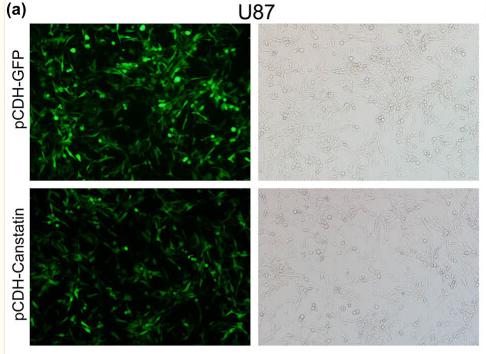
Application: IF/ICC Species: Mice Sample: U87 cell
Restrictive clause
Affinity Biosciences tests all products strictly. Citations are provided as a resource for additional applications that have not been validated by Affinity Biosciences. Please choose the appropriate format for each application and consult Materials and Methods sections for additional details about the use of any product in these publications.
For Research Use Only.
Not for use in diagnostic or therapeutic procedures. Not for resale. Not for distribution without written consent. Affinity Biosciences will not be held responsible for patent infringement or other violations that may occur with the use of our products. Affinity Biosciences, Affinity Biosciences Logo and all other trademarks are the property of Affinity Biosciences LTD.